Skip to main page content
U.S. flag

An official website of the United States government

Dot gov

The . gov means it’s official. Federal government websites often end in . gov or VSports app下载. mil. Before sharing sensitive information, make sure you’re on a federal government site. .

Https

The site is secure. The https:// ensures that you are connecting to the official website and that any information you provide is encrypted and transmitted securely V体育官网. .

. 2017 Jan;23(1):79-90.
doi: 10.1038/nm.4252. Epub 2016 Dec 12.

Fasting selectively blocks development of acute lymphoblastic leukemia via leptin-receptor upregulation

Affiliations

Fasting selectively blocks development of acute lymphoblastic leukemia via leptin-receptor upregulation

Zhigang Lu et al. Nat Med. 2017 Jan.

Abstract

New therapeutic approaches are needed to treat leukemia effectively. Dietary restriction regimens, including fasting, have been considered for the prevention and treatment of certain solid tumor types. However, whether and how dietary restriction affects hematopoietic malignancies is unknown. Here we report that fasting alone robustly inhibits the initiation and reverses the leukemic progression of both B cell and T cell acute lymphoblastic leukemia (B-ALL and T-ALL, respectively), but not acute myeloid leukemia (AML), in mouse models of these tumors. Mechanistically, we found that attenuated leptin-receptor (LEPR) expression is essential for the development and maintenance of ALL, and that fasting inhibits ALL development by upregulation of LEPR and its downstream signaling through the protein PR/SET domain 1 (PRDM1). The expression of LEPR signaling-related genes correlated with the prognosis of pediatric patients with pre-B-ALL, and fasting effectively inhibited B-ALL growth in a human xenograft model. Our results indicate that the effects of fasting on tumor growth are cancer-type dependent, and they suggest new avenues for the development of treatment strategies for leukemia VSports手机版. .


Conflict of interest statement

COMPETING FINANCIAL INTERESTS

The authors declare no competing financial interests.

Figures

Figure 1
Figure 1
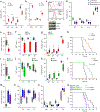
Fasting selectively inhibits ALL development. (a) Leukemia induction and fasting scheme. Lin cells were infected with N-Myc-IRES-GFP (B-ALL), Notch1-IRES-GFP (T-ALL), or MLL-AF9-IRES-YFP (AML) expressing retrovirus and transplanted into irradiated mice. Mice were fed normally or fasted with six cycles of 1-d fasting/1-d feeding at day 2 after transplantation. (b–i) N-Myc-infected Lin cells were transplanted into lethally irradiated recipient mice to induce B-ALL. Mice were either placed on a standard diet or fasted (six cycles of 1-d fast/1-d fed), initiated at day 2 after transplantation. PB was analyzed at 3, 5 and 7 weeks after transplantation; BM and SP were collected at 7 weeks. (b) Representative flow cytometry plot of GFP+ leukemic BM cells in fed and fasted mice. (c) Percentage of cells expressing GFP in PB at the indicated time points and in BM and SP (n = 5 mice per group). (d) WBC numbers in the PB at week 7 (n = 5 per group). (e) Representative flow cytometry plot of Mac-1 and B220 staining in GFP+ BM cells in fed and fasted mice. (f) Percentages of B220 and Mac-1 cells in GFP+ cells from PB, BM and SP (n = 5 per group). (g) Analysis of B-ALL surface markers B220 (pan B cell marker), CD19 (B cell marker) and IgM (B cell maturation marker) in GFP+ cells from PB, BM and SP (n = 5 per group). (h) Photographs of representative spleens and lymph nodes (left; scale bars, 1 cm) and quantitation of spleen weight (right) from fed and fasted mice with B-ALL and nonleukemic (healthy) mice (n = 3 per group). (i) Combined survival analysis of fed and fasted mice with N-Myc-driven B-ALL from four independent experiments (n = 20 per group). (j–m) Notch1-infected fetal liver Lin cells were transplanted into lethally irradiated recipient mice to induce T-ALL. Mice were placed on a standard diet or a six-cycle fasting regimen initiated at 2 d after transplantation. (j) Percentage of GFP+ cells in PB at 3, 5 and 7 weeks and in BM and SP at 7 weeks post-transplantation (n = 5 per group). (k) WBC numbers in the PB at 7 weeks in fed and fasted mice (n = 5 per group). (l) Levels of surface markers CD3 and Mac-1 in GFP+ cells of PB, BM and SP at 7 weeks. (m) Combined survival analysis of fed and fasted mice with Notch1-driven T-ALL from three independent experiments (n = 15 per group). (n–q) MLL-AF9-infected fetal liver Lin cells were transplanted into lethally irradiated recipient mice to induce AML. Mice were either placed on a standard diet or a six-cycle fasting regimen initiated at 2 d after transplantation. (n) Percentage of YFP+ cells in PB at 3, 5 and 7 weeks and in BM and SP at 7 weeks (n = 5 per group). (o) Levels of surface markers Mac-1 and B220 in YFP+ cells of PB, BM and SP at 6 weeks (n = 5 per group). (p) Levels of the AML progenitor marker c-Kit in PB, BM and SP at 6 weeks (n = 5 per group). (q) Combined survival analysis of fed and fasted mice with MLL-AF9-driven AML from three independent experiments (n = 15 per group). Representative data from three or four independent experiments are presented as dot plots (means ± s.e.m.) in c, j and n, or as box-and-whisker plots (median values (line), 25th–75th percentiles (box outline) and minimum and maximum values (whiskers)) in d, fh, k, l, o and p. In b and e, numerals in outlined area indicate the percentage of cells in the gates or in each quantile. Statistical significance was calculated by Student’s t-test, *P < 0.05. Statistical significance for survival in i, m and q was calculated by the log–rank test.
Figure 2
Figure 2
Fasting inhibits B-ALL development in both early and late stages. (a) Leukemia induction and fasting regimens. Shown are regimens for fasting at an early stage (1 and 2) and at a mid-to-late stage (3) of primary B-ALL development, or in secondary B-ALL development (4). (b–i) N-Myc-infected Lin cells were transplanted into lethally irradiated recipient mice to induce B-ALL. One to six cycles of 1-d fasting/1-d feeding (1F) (b–e) or one to four cycles of 2-d fasting/2-d feeding (2F) (f–i) were initiated at 2 d post-transplantation. (b,f) Percentage of GFP+ cells in PB, BM and SP at 7 weeks post-transplantation (n = 5 per group). (c,g) WBC numbers in PB at 7 weeks post-transplantation (n = 5 per group). (d,h) Leukemia burden rates, defined as >20% of GFP+ cells in PB with no decrease in the percentage of GFP+ cells as compared to the prior week, at the indicated time points (n = 10 per group before and at week 7, and n = 5 per group after week 7). (e,i) Survival of mice with B-ALL (n = 5 per group). (j,g) N-Myc-infected Lin cells were transplanted into lethally irradiated recipient mice, and two and four-cycles of 1F or two and three-cycles of 2F fasting were initiated when the percentage of GFP+ cells in PB reached ~60% (at around 3–4 weeks). (j) Percentage of GFP+ cells in PB before fasting was initiated and at 2 and 3 weeks after fasting, and in BM and SP at 3 weeks after fasting (n = 5 per group). (k) WBC numbers in PB 3 weeks after fasting (n = 5 per group). (l) Leukemia burden rates at the indicated time points (n = 10 per group before and at week 7, and n = 5 per group after week 7). (m) Survival analysis of B-ALL mice (n = 5 per group). (n–q) GFP+ cells from the BM of primary N-Myc-induced B-ALL mice were transplanted into lethally irradiated recipient mice. Two and four-cycles of 1F or two and three-cycles of 2F fasting were initiated at 2 d after transplantation. (n) Percentage of GFP+ cells in PB, BM, and SP at 8 weeks post-transplantation (n = 5 per group). (o) WBC numbers in PB at 8 weeks post-transplantation (n = 5 per group). (p) Leukemia burden rates mice at the indicated weeks (n = 10 per group before and at week 7, and n = 5 per group after week 7). (q) Survival of secondary B-ALL mice (n = 5 per group). Data in b, c, f, g, j, k, n and o, are presented as box-and-whisker plots (median values (line), 25th–75th percentiles (box outline) and minimum and maximum values (whiskers)). Statistical significance was calculated by Student’s t-test, *P < 0.05. Statistical significance for survival analysis in e, i, m and q was calculated by the log–rank test, *P < 0.05, **P < 0.01.
Figure 3
Figure 3
Fasting upregulates LEPR expression and its downstream signaling. (ah) N-Myc-infected Lin cells were transplanted into lethally irradiated recipient mice, and a 48-h fast was initiated when the percentage of GFP+ cells in PB reached ~60% (at around 3–4 weeks). Analyses were conducted before initiation of fasting and at days 1, 2 and 5 after initiation of fasting. (a) Representative flow cytometry plots showing GFP and B220 staining in PB of fasted and fed mice at the indicated time points. Numerals in the outlined area indicate the percentage of cells in each quantile. (b) The percentages of GFP+ cells in PB, BM, SP and LV from fasted and fed mice at the indicated time points (n = 5 per group). (c) The percentages of B220+, Annexin V+ and Ki67+ cells in the GFP+ and GFP compartments from BM of fasted and fed mice (n = 5 per group). (d) The percentages of CD43+, IgM+, Igκ+, Igλ+ and Tdt+ cells in the GFP+B220+ B-ALL cell compartment from BM of fasted and fed mice (n = 5 per group). A representative flow cytometry plot of Tdt staining is also shown (lower right). (e,f) GFP+B220+ B-ALL cells from 1-d (D1) or 2-d (D2) fasted mice, and control mice were sorted for RNA-seq analysis. (e) Fasting induced alterations of signature transcription factors of B cell terminal differentiation, shown for mRNA levels (fasted versus fed, left) and inferred activities (fasted versus fed, right). (f) Fasting-induced alterations in pathways (top) and transcription factor activities (fasted versus fed, bottom). (g) Relative mRNA expression (log2-fold change, FC) of all 42 cytokine receptors in the KEGG pathway database in the sorted cells by qPCR (fasted versus fed, left), the log2 values of their original hazard ratios (HRs) (middle) and their mRNA fold change normalized hazard ratios (log2 HR multiplied by fold change) for survival of human pediatric patients with pre-B-ALL. Up- and downregulation of mRNA levels are indicated by red and green, respectively; HRs below and above 1 are indicated by blue and red, respectively. (h) Representative flow cytometry plots (left and middle) and quantitation (right) of LEPR staining (mean fluorescence intensity, MFI) in GFP+ and GFP BM cell populations from fasted and fed mice at the indicated time points. (n = 5 per group). (i) Ratio of Lepr mRNA isoforms Ob-Rb to Ob-Ra in GFP+ and GFP PB cells of fed B-ALL mice, by qPCR (n = 4 per group). (j) Fold change of Ob-Rb mRNA in BM GFP+ cells of fasted mice at the indicat ed time points, by qPCR (n = 5 per group). (k) Quantitation of phospho-STAT3 staining in BM GFP+ cells from fasted mice at the indicated time points (n = 5 per group). (l) Fold change of surface LEPR in the indicated populations of BM cells relative to pre-fast levels (n = 5 per group). (m) Comparison of surface LEPR expression on N-Myc B-ALL cells, Notch1 T-ALL cells and MLL-AF9 AML cells with normal BM B, T and myeloid cell subpopulations: pro-B (B220+IgMCD43+), Pre-B (B220+IgMCD43), immature-B (B220+IgM+IgD), mature-B (B220+IgM+IgD+) and total B220+ cells (B cell subpopulations); DN (CD3+CD4CD8), CD3+CD4, CD3+CD8, DP (CD3+CD4+CD8+) and total CD3+ cells (T cell subpopulations); and Mac1+cKit+ myeloid progenitors and total Mac1+ cells (myeloid cell subpopulations) (n = 5 per group). Representative data from three independent experiments are presented as means ± s.e.m. in bd, h and l, or as box-and-whisker plots (median values (line), 25th–75th percentiles (box outline) and minimum and maximum values (whiskers)) in ik and m. In statistical significance was calculated by Student’s t-test, *P < 0.05.
Figure 4
Figure 4
Attenuated LEPR signaling is essential for ALL development, and fasting does not inhibit B-ALL development in the absence of LEPR. (a) B-ALL mice were fasted for 48 h with administration of leptin (5 μg/15 g body weight) or PBS when the percentage of GFP+ cells in PB reached ~60%. The percentage of GFP+ cells and surface LEPR expression on GFP+B220+ B-ALL cells were measured by flow cytometry and compared with pre-fast levels (n = 3 per group). (bf) N-Myc-infected Lin BM cells from wild-type (wt) or Leprdb/db mice were transplanted into lethally irradiated wild-type recipient mice. (b) Percentage of GFP+ cells in PB at 5, 7, and 10 weeks post-transplantation and in BM and SP at 10 weeks (n = 5 per group). (c) WBC numbers at 10 weeks (n = 5 per group). (d) Percentage of B220+ cells in the GFP+ compartment in PB at the indicated time points (n = 5 per group). (e) Percentage of cells positive for the surface markers B220, CD19, CD43 and IgM in the GFP+ compartment in PB, BM and SP at 10 weeks post-transplantation. (f) Survival analysis of wild-type (wt) and Leprdb/db mice with N-Myc-induced B-ALL (wt, n = 6; db/db, n = 5). (g) Schematic of leukemia initiation and the two fasting regimens. N-Myc-infected Lin- BM cells from wild-type (wt) or Leprdb/db mice were transplanted into lethally irradiated wild-type recipient mice. Approximately 104 GFP+ B-ALL cells from these primary B-ALL mice were sorted and transplanted into lethally irradiated secondary recipient mice. The mice were fed normally or subjected to either of two fasting regimens: a 48-h fast was initiated when the percentage of GFP+ cells in PB reached ~60%, or two cycles of 2-d fasting/2-d feeding were performed beginning at 2 d after transplantation. (gi) 48-h fast regimen, as shown in g. (h) Percentage of GFP+ cells in PB at the indicated time points after fasting was initiated (n = 5 per group). (i) Percentage of Mac-1+/B220 myeloid cells and B220+/IgM+ differentiated cells in GFP+ cells from PB at the indicated time points after fasting was initiated (n = 5 per group). (jm) Two-cycle fast regimen, as shown in g. (j) Percentage of GFP+ cells in PB, BM and SP at 5 weeks post-transplantation (n = 5 per group). (k) WBC numbers in PB at 5 weeks post-transplantation (n = 5 per group). (l) Surface marker expression, including B220, CD19, CD43 and IgM, on BM GFP+ cells at 5 weeks post-transplantation (n = 5 per group). (m) Survival analysis (n = 5 per group). Representative data from three independent experiments are presented as means ± s.d. in a, d and h, or as dot plots (means ± s.e.m.) in b and j, or as box-and-whisker plots (median values (line), 25th–75th percentiles (box outline) and minimum and maximum values (whiskers)) in c, e, i, k and l. Statistical significance was calculated by Student’s t-test, *P < 0.05. n.s., not significantly different. Statistical significance for survival analysis in f and m was calculated by the log–rank test.
Figure 5
Figure 5
LEPR inhibits B-ALL development by promoting leukemic cell differentiation through PRDM1. (ac) Primary mouse B-ALL cells from BM were infected with LEPR- or DsRed control (Ctrl)-expressing retrovirus and cultured for 2 d. (a) Percentages of IgM+, Igκ+, Igλ+ and Tdt+ cells in the GFP+ compartment, by flow cytometry (n = 3 per group). (b) Relative mRNA levels of Prdm1, Irf4 and Xbp1 in the GFP+ compartment, by qPCR (n = 3 per group). (c) Representative flow cytometry plot showing LEPR staining (top) and PRDM1 intracellular staining (middle), and a representative western blot for PRDM1 and XBP1 (bottom) in the GFP+ compartment. (d) BM cells from primary B-ALL mice were infected with LEPR-expressing retrovirus together with shCtrl- or shPRDM1-expressing lentivirus (with a DsRed reporter) and cultured for 2 d. The percentages of IgM+, Igκ+, Igλ+ and Tdt+ cells in Lepr+DsRed+GFP+ B-ALL cells were measured by flow cytometry (n = 3 per group). (eh) BM cells from primary B-ALL mice were infected with LEPR- or DsRed (Ctrl) expressing retrovirus. GFP+Lepr+ or GFP+DsRed+ cells were sorted, and 1 × 10⁴ double-positive cells per mouse were transplanted into lethally irradiated mice together with normal cell competitors. (e) WBC numbers in PB at week 4 (n = 5 per group). (f) The percentage of GFP cells in PB, BM and SP at week 4, by flow cytometry (n = 5 per group). (g) Expression of Tdt, surface IgM, Igκ and Igλ on BM GFP+ cells at week 4, by flow cytometry (n = 5 per group). (h) Survival analysis (Ctrl group, n = 5; LEPR group, n = 10). (il) BM cells from mice with B-ALL were infected with PRDM1- or DsRed (Ctrl)-expressing retrovirus. GFP+DsRed+ cells were sorted, and 1 × 10⁴ double-positive cells per mouse were transplanted into lethally irradiated mice together with normal cell competitors. (i) WBC numbers in PB at week 4 (n = 5 per group). (j) The percentage of GFP+ cells in PB, BM and SP at week 4, by flow cytometry (n = 5 per group). (k) Expression of Tdt, surface IgM, Igκ and Igλ on BM GFP+ cells at week 4, by flow cytometry (n = 5 per group). (l) Survival analysis (n = 5 per group). (mp) GFP+ BM cells from B-ALL mice were infected with LEPR-expressing retrovirus together with shCtrl- or shPRDM1-expressing lentivirus (with a DsRed reporter). GFP+LEPR+DsRed+ cells were sorted, and 1 × 10⁴ sorted cells were transplanted per mouse into lethally irradiated mice together with normal cell competitors. (m) WBC numbers in PB at week 4 (n = 5 per group). (n) The percentage of GFP+ cells in PB, BM and SP at week 4, by flow cytometry (n = 5 per group). (o) Expression of Tdt, surface IgM, Igκ and Igλ on GFP+ BM cells at week 4, by flow cytometry (n = 5). (p) Survival analysis (shCtrl, n = 6; shPRDM1, n = 5 per group). Colors in mo as in p. Representative data from three independent experiments are presented as dot plots (means ± s.e.m.) in a, b and d, or as box-and-whisker plots (median values (line), 25th–75th percentiles (box outline) and minimum and maximum values (whiskers)) plots in eg, ik, and mo. Statistical significance was calculated by Student’s t-test, *P < 0.05. Statistical significance for survival analysis in h, i and p was calculated by the log–rank test.
Figure 6
Figure 6
Fasting and LEPR signaling inhibit human ALL development. (a) LEPR mRNA levels in the indicated types of human lymphoid leukemia and myeloid leukemia samples relative to healthy bone marrow samples (GEO data set GSE13159, n = 2096). (b) Flow cytometry analysis showing surface LEPR on human patient T-ALL (CD3+, n = 5 patients), B-ALL (CD19+, n = 8 patients) and AML (CD33+, n = 52 patients) cells; on human cord blood-derived normal T cell subpopulations including DN (CD3+CD4CD8), CD3+CD4, CD3+CD8, DP (CD3+CD4+CD8+) and total CD3+ T cells; normal B cell subpopulations including Pro-B (CD19+CD34+CD38+), Pre-B (CD19+CD34+CD40+); immature-B (CD19+CD40+IgM+), mature-B (CD19+IgM+IgD+) and total CD19+ B cells; and myeloid subpopulations including CD33+CD34+ myeloid progenitors and CD33+ myeloid cells (for all normal cell populations, n = 5 healthy donors per group). (c) Overall survival of pediatric patients with pre-B-ALL patients (COG P9906, n = 206) and patients with AML (TCGA, n = 186) relative to LEPR mRNA expression levels (above (high) or below (low) the 50th percentile). (d) Correlation analysis of the expression of LEPR-signaling-related genes (118) and nonrelated genes (20933) with overall survival (OS) and event-free survival (EFS) of patients with B-ALL, or with the overall survival of patients with AML. (e) Correlation analysis of the expression of 29 leptin/LEPR-signaling related genes with OS of patients with AML, or OS and EFS of patients with B-ALL. Positive and negative correlations with patient survival are indicated by red and blue, respectively; genes that are negatively or positively associated with LEPR signaling are indicated in the bar on the far right as green or pink, respectively. (f) Expression of surface LEPR on human BM CD19+ NALM-6 cells, by flow cytometry, in scid mice xenografted with 5 × 10⁶ human B-ALL NALM-6 cells, which were fed or subjected to a 48-h fasting regimen initiated at day 10 after cell injection (n = 5 per group). (gi) Xenografted mice as in f were fed or subjected to three cycles of 2-d fasting/2-d feeding initiated on day 2 after cell injection, and analyses were performed on day 25. (g) Paraplegia occurrence (n = 5 per group). (h) Percentage of human CD19+ NALM-6 cells in PB, BM and SP, by flow cytometry (n = 5 per group). (i) Survival analysis (n = 5 per group). Data are presented as means ± s.e.m. in a, or as dot plot (means ± s.e.m.) in g, or as box-and-whisker plots (median values (line), 25th–75th percentiles (box outline) and minimum and maximum values (whiskers)) in b, f and h. Statistical significance was calculated by Student’s t-test, *P < 0.05. Statistical significance in d was calculated by the χ-squared test. Statistical significance for survival analysis in c, e and i was calculated by the log–rank test.

Comment in

References

    1. Hursting SD, Lavigne JA, Berrigan D, Perkins SN & Barrett JC Calorie restriction, aging, and cancer prevention: mechanisms of action and applicability to humans. Annu. Rev. Med 54, 131–152 (2003). - PubMed
    1. Fontana L & Partridge L Promoting health and longevity through diet: from model organisms to humans. Cell 161, 106–118 (2015). - PMC - PubMed
    1. Mihaylova MM, Sabatini DM & Yilmaz OH Dietary and metabolic control of stem cell function in physiology and cancer. Cell Stem Cell 14, 292–305 (2014). - PMC - PubMed
    1. Bordone L & Guarente L Calorie restriction, SIRT1 and metabolism: understanding longevity. Nat. Rev. Mol. Cell Biol 6, 298–305 (2005). - PubMed
    1. Longo VD & Mattson MP Fasting: molecular mechanisms and clinical applications. Cell Metab. 19, 181–192 (2014). - PMC - PubMed

MeSH terms (VSports注册入口)

Substances