Skip to main page content (V体育ios版)
U.S. flag

An official website of the United States government

Dot gov

The . gov means it’s official. Federal government websites often end in . gov or . mil VSports app下载. Before sharing sensitive information, make sure you’re on a federal government site. .

Https

The site is secure V体育官网. The https:// ensures that you are connecting to the official website and that any information you provide is encrypted and transmitted securely. .

. 2017 Jan;18(1):45-53.
doi: 10.1038/ni.3630. Epub 2016 Nov 21.

TET proteins regulate the lineage specification and TCR-mediated expansion of iNKT cells

Affiliations

TET proteins regulate the lineage specification and TCR-mediated expansion of iNKT cells (V体育官网入口)

Ageliki Tsagaratou et al. Nat Immunol. 2017 Jan.

Abstract

TET proteins oxidize 5-methylcytosine in DNA to 5-hydroxymethylcytosine and other oxidation products. We found that simultaneous deletion of Tet2 and Tet3 in mouse CD4+CD8+ double-positive thymocytes resulted in dysregulated development and proliferation of invariant natural killer T cells (iNKT cells) VSports手机版. Tet2-Tet3 double-knockout (DKO) iNKT cells displayed pronounced skewing toward the NKT17 lineage, with increased DNA methylation and impaired expression of genes encoding the key lineage-specifying factors T-bet and ThPOK. Transfer of purified Tet2-Tet3 DKO iNKT cells into immunocompetent recipient mice resulted in an uncontrolled expansion that was dependent on the nonclassical major histocompatibility complex (MHC) protein CD1d, which presents lipid antigens to iNKT cells. Our data indicate that TET proteins regulate iNKT cell fate by ensuring their proper development and maturation and by suppressing aberrant proliferation mediated by the T cell antigen receptor (TCR). .


Conflict of interest statement

COMPETING FINANCIAL INTERESTS

The authors declare no competing financial interests.

Figures

Figure 1
Figure 1
Simultaneous deletion of Tet2 and Tet3 severely compromises T cell homeostasis, leading to iNKT cell expansion and disease. (a) Quantification of total cells in the spleen of 5- to 7-week-old wild-type mice (WT) (n = 5) and Tet2-Tet3 DKO mice (DKO) (n = 6). (b) Disease-free survival of wild-type mice (n = 10) and Tet2-Tet3 DKO mice (n = 10) (Kaplan-Meier curve). (c) iNKT cells in spleens of 4-week-old wild-type (n = 6) versus Tet2-Tet3 DKO (n = 3) mice. (d) Quantification (left) and frequency (right) of iNKT cells in spleens isolated from 4-week-old Tet2-Tet3 DKO mice (n = 3) and wild-type mice (n = 6). (e) iNKT cells in the spleen of 3- to 4-week-old wild-type mice (n = 5), Tet2−/− (Tet2 KO) mice (n = 3), Tet3 KO mice (n = 3) and Tet2-Tet3 DKO mice (n = 6). (f) Frequency of iNKT cells in wild-type mice (n = 5), Tet2 KO mice (n = 3), Tet3 KO mice (n = 4) and Tet2-Tet3 DKO mice (n = 6) (left), and number of iNKT cells in wild-type mice (n = 3), Tet2 KO mice (n = 3), Tet3 KO mice (n = 3) and Tet2-Tet3 DKO mice (n = 6) (right). Numbers adjacent to outlined areas (c,e) indicate percent tetrameter+TCRβ+ cells. Each symbol (a,d,f) represents an individual mouse; horizontal lines indicate the mean (± s.e.m.). *P < 0.05, **P < 0.01, ***P < 0.001 and ****P < 0.0001 (unpaired t-test). Data are representative of 5 experiments (a), 10 experiments (b), 3 experiments (d), 3 experiments (e) or 3 experiments (f) or are from three independent experiments (c).
Figure 2
Figure 2
Tet2-Tet3 DKO iNKT cells in the thymus of young (3- to 4-week-old) mice are skewed toward the NKT17 cell lineage. (a) iNKT cells in the thymus of 20-day-old wild-type and Tet2-Tet3 DKO mice. Numbers adjacent to outlined areas indicate percent tetrameter+TCRβ+ cells. (b,c) Frequency (b) and number (c) of iNKT cells in the thymus of Tet2-Tet3 DKO mice (n = 22) and wild-type mice (n = 15). (d) Flow cytometry analysis of the expression of PLZF and RORγt in tetramer+CD24TCRβ+ thymocytes. (d,e) Flow cytometry analysis of the expression of PLZF and RORγt (d) or of PLZF and T-bet (e) in tetramer+CD24TCRβ+ thymocytes. (f) Frequency of iNKT cell subsets among total iNKT cells. (g) Results in f, presented as frequency among total thymocytes. (h) Flow cytometry of tetramer+CD24TCRβ+ thymocytes from wild-type mice (n = 9) (top) and Tet2-Tet3 DKO mice (n = 12) (bottom), analyzing the expression of NK1.1 and binding of tetramer (left), expression of CD4 and CD27 in the tetramer+NK1.1 subset (arrow) (middle), and expression of CCR6 and CD27 in the tetramer+NK1.1+ subset (arrow) (right). (i) Frequency of the NK1.1 and NK1.1+ subsets (as defined in h) among total iNKT cells. (j) Quantification of the NK1.1 and NK1.1+ subsets (as defined in h) among total iNKT cells. Numbers adjacent to outlined areas (a,d,e,h) indicate percent cells in each. Each symbol (b,c,f,g,i,j) represents an individual mouse; horizontal lines indicate the mean (± s.e.m.). *P < 0.05, **P < 0.01, ***P < 0.001 and ****P < 0.0001 (unpaired t-test). Data are representative of 15 experiments (a), 5 experiments (i), 5 experiments (i), are from one experiment representative of four experiments (be) or are from four independent experiments with five wild-type mice and seven Tet2-Tet3 DKO mice (f,g) or five independent experiments (h).
Figure 3
Figure 3
Gene-expression profiles of thymic wild-type and Tet2-Tet3 DKO iNKT cells. (a) Mean average (MA) plot of genes expressed differentially in thymic iNKT cells isolated from Tet2-Tet3 DKO relative to their expression in such cells from wild-type mice. (b) Expression of genes encoding selected transcriptional regulators (left) and cytokines and their receptors (right) in Tet2-Tet3 DKO, Tet3 KO and wild-type thymic iNKT cells, presented as row-wise z-scores (red, higher expression, and blue, lower expression, relative to other conditions). (c) Expression of selected genes of interest in stage 0, NK1.1 and NKT1 iNKT subsets. (d) Flow cytometry analysis of BrdU incorporation in tetramer+TCRβ+ thymocytes. Numbers adjacent to outlined areas indicate percent cells with BrdU incorporation. (e) Frequency of tetramer+TCRβ+ iNKT cells that incorporated BrdU. Each symbol represents an individual mouse; horizontal lines indicate the mean (± s.e.m.). **P < 0.01, (unpaired t test). Data are representative of one experiment with three biological replicates per genotype (a), one experiment with two biological replicates per genotype (b), one experiment with two biological replicates per genotype for stage 0, three biological replicates per genotype for NK1.1, two biological replicates wild-type NKT1, and three biological replicates per genotype for DKO NKT1 (c), are from one experiment representative of two experiments with five mice per genotype (d) or from two independent experiments with five mice per genotype (e).
Figure 4
Figure 4
Antigen stimulation promotes the expansion of Tet2-Tet3 DKO iNKT cells and disease development in fully immunocompetent recipients. (a) Disease-free survival of wild-type (n = 6) and CD1dKO recipients (n = 5) of Tet2-Tet3 DKO iNKT cells (Kaplan-Meier curve). **P < 0.01 (log-rank (Mantel-Cox) test and Gehan-Brenslow-Wilcoxon test). (b) Cellularity of the spleen (left) and lymph nodes (right) of wild-type recipients (n = 12) and CD1dKO recipients (n = 6) of Tet2-Tet3 DKO iNKT cells. (c) Frequency (top) and numbers (bottom) of iNKT cells in the spleen of wild-type recipients (n = 12) and CD1dKO recipients (n = 6) of Tet2-Tet3 DKO iNKT cells. (d) MA plot of genes expressed differentially in transferred and expanded Tet2-Tet3 DKO iNKT cells isolated from the spleen of congenic recipient mice relative to their expression in iNKT cells isolated from the spleen of healthy wild-type mice. (e) Expression (log2 fold values) of selected genes encoding transcriptional regulators with differential expression (higher (red) or lower (blue)) in Tet2-Tet3 DKO iNKT cells than in wild-type iNKT cells. (f) Pathway-enrichment analysis of differentially expressed genes in transferred Tet2-Tet3 DKO iNKT cells: purple, categories related to DNA replication, cell cycle and DNA repair; green, categories related to T cell function. Each symbol (b,c) represents an individual mouse; horizontal lines indicate the mean (± s.e.m.). **P < 0.01, ***P < 0.001 (unpaired t test). Data are from two independent experiments (a), three independent experiments (b,c), or one experiment with three biological replicates per genotype (df).
Figure 5
Figure 5
5hmC shows enrichment in the bodies of genes with high expression in iNKT cells and those expressing key iNKT-cell-lineage-specifying factors. (a) CMS-IP analysis of the enrichment for 5hmC over the gene body, categorized on the basis of gene expression (RNA-seq analysis), in wild-type thymic iNKT cells. (b) Genome browser views of intragenic 5hmC (CMS-IP analysis) and gene expression (RNA-seq analysis) in Tbx21, Zbtb7b and Rorc in wild-type thymocytes. (c) Average enrichment for 5mC+5hmC over the gene body, categorized on the basis of gene expression (WGBS and RNA-seq analysis), in wild-type thymic iNKT cells. (d) Average enrichment of 5hmC in wild-type iNKT cells from tissue-specific enhancers in the thymus (orange), mouse embryonic stem cells (mESC; blue) and heart (green). (e) Modification of 5mC+5hmC in wild-type and Tet2-Tet3 DKO iNKT cells, plotted against position relative to DMR. Data are representative of one experiment with two biological replicates for CMS-IP seq, two biological replicates for WGBS and three biological replicates for RNA-seq.
Figure 6
Figure 6
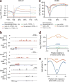
TET proteins regulate DNA modification in iNKT cells. (a) DNA modification in CLPs and wild-type and Tet2-Tet3 DKO iNKT cells, showing more (red) or less (blue) modification. (b) RNA-seq analysis of Tet1, Tet2 and Tet3 in iNKT cells and CLPs. (c) Quantification of DMRs relative to their distance to the TSS. Data are representative of one experiment with two technical replicates for CLPs and two biological replicates per genotype for iNKT cells (a,c), and three biological replicates for iNKT cells and one sample for CLPs (b).
Figure 7
Figure 7
Characterization of chromatin regions with differential accessibility in wild-type iNKT cells versus Tet2-Tet3 DKO iNKT cells. (a) ATAC-seq analysis of DARs (red) in wild-type iNKT cells versus Tet2-Tet3 DKO iNKT cells: black, DARs that overlap 5hmC (CMS-IP peaks). (c) Motif-enrichment analysis (HOMER) of the 3,162 DARs more accessible in Tet2-Tet3 DKO iNKT cells than in wild-type iNKT cells (top) and of the 2,711 DARs less accessible in Tet2-Tet3 DKO iNKT cells than in wild-type cells (bottom). (b) Distance of DARs to the nearest TSS, among DARs less (blue) or more (red) accessible in Tet2-Tet3 DKO iNKT cells than in wild-type iNKT cells or commonly accessible in both wild-type and Tet2-Tet3 DKO iNKT cells (gray). Data are representative of two experiments and three biological replicates per genotype in total for ATAC seq. For CMS-IP, data are representative of one experiment with two biological replicates.
Figure 8
Figure 8
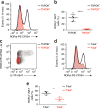
Ectopic expression of ThPOK or T-bet in Tet2-Tet3 DKO iNKT cells can suppress the aberrant increase in RORγt expression. (a) Flow cytometry analysis of RORγt expression in Tet2-Tet3 DKO iNKT cells transduced to express ThPOK (ThPOK+) or not (ThPOK). (b) Frequency of RORγt-expressing Tet2-Tet3 DKO iNKT cells as in a. (c) Flow cytometry analysis of Rorγt expression and IL-17F production in Tet2-Tet3 DKO iNKT cells as in a. (d) Flow cytometry analysis of RORγt expression in Tet2-Tet3 DKO iNKT cells transduced to express T-bet (T-bet+) or not (T-bet). (e) Frequency of RORγt-expressing Tet2-Tet3 DKO iNKT cells as in d. Each symbol (b,e) represents an individual mouse; horizontal lines indicate the mean (± s.e.m.). *P < 0.05, ****P < 0.0001 (unpaired t test). Data are representative of four experiments (a) or three experiments (d) or are from four independent experiments with a total of five mice (b), one experiment representative of two experiments with three mice (c) or one experiment representative of three experiments (e).

References

    1. Yui MA, Rothenberg EV. Developmental gene networks: a triathlon on the course to T cell identity. Nat Rev Immunol. 2014;14:529–545. - PMC - PubMed
    1. Carpenter AC, Bosselut R. Decision checkpoints in the thymus. Nat Immunol. 2010;11:666–673. - PMC - PubMed
    1. Bendelac A, Savage PB, Teyton L. The biology of NKT cells. Annu Rev Immunol. 2007;25:297–336. - PubMed (V体育安卓版)
    1. Vahedi G, et al. Helper T-cell identity and evolution of differential transcriptomes and epigenomes. Immunol Rev. 2013;252:24–40. - PMC - PubMed
    1. Hogquist KA, Jameson SC. The self-obsession of T cells: how TCR signaling thresholds affect fate ‘decisions’ and effector function. Nat Immunol. 2014;15:815–823. - PMC - PubMed

VSports手机版 - MeSH terms