Skip to main page content
U.S. flag

An official website of the United States government

Dot gov

The . gov means it’s official. Federal government websites often end in . gov or . mil. Before sharing sensitive information, make sure you’re on a federal government site VSports app下载. .

Https

The site is secure V体育官网. The https:// ensures that you are connecting to the official website and that any information you provide is encrypted and transmitted securely. .

. 2016 Nov 29;113(48):E7710-E7719.
doi: 10.1073/pnas.1612335113. Epub 2016 Nov 14.

"V体育ios版" Notch1 controls development of the extravillous trophoblast lineage in the human placenta

Affiliations

Notch1 controls development of the extravillous trophoblast lineage in the human placenta

Sandra Haider et al. Proc Natl Acad Sci U S A. .

Abstract

Development of the human placenta and its different epithelial trophoblasts is crucial for a successful pregnancy. Besides fusing into a multinuclear syncytium, the exchange surface between mother and fetus, progenitors develop into extravillous trophoblasts invading the maternal uterus and its spiral arteries. Migration into these vessels promotes remodelling and, as a consequence, adaption of blood flow to the fetal-placental unit. Defects in remodelling and trophoblast differentiation are associated with severe gestational diseases, such as preeclampsia. However, mechanisms controlling human trophoblast development are largely unknown. Herein, we show that Notch1 is one such critical regulator, programming primary trophoblasts into progenitors of the invasive differentiation pathway. At the 12th wk of gestation, Notch1 is exclusively detected in precursors of the extravillous trophoblast lineage, forming cell columns anchored to the uterine stroma. At the 6th wk, Notch1 is additionally expressed in clusters of villous trophoblasts underlying the syncytium, suggesting that the receptor initiates the invasive differentiation program in distal regions of the developing placental epithelium VSports手机版. Manipulation of Notch1 in primary trophoblast models demonstrated that the receptor promotes proliferation and survival of extravillous trophoblast progenitors. Notch1 intracellular domain induced genes associated with stemness of cell columns, myc and VE-cadherin, in Notch1- fusogenic precursors, and bound to the myc promoter and enhancer region at RBPJκ cognate sequences. In contrast, Notch1 repressed syncytialization and expression of TEAD4 and p63, two regulators controlling self-renewal of villous cytotrophoblasts. Our results revealed Notch1 as a key factor promoting development of progenitors of the extravillous trophoblast lineage in the human placenta. .

Keywords: Notch1; cell fusion; extravillous trophoblast; human placenta; trophoblast progenitors. V体育安卓版.


"V体育官网入口" Conflict of interest statement

The authors declare no conflict of interest.

"V体育平台登录" Figures

Fig. 1.
Fig. 1.
Notch1 marks a subset of cycling CCTs and decreases during the first trimester of pregnancy. (AD) Notch1 IF in first trimester placenta. Stars mark proliferative, Notch1 CCTs or vCTBs underlying the syncytium (S). Nuclei were stained with DAPI. Representative images of cell columns and placental villi from 5th–7th wk of gestation (n = 7) and 10th–12th wk of gestation (n = 5) are shown. VS, villous stroma. (Scale bars, 50 µm.) Tissue sections of 6th (A)- or 12th (B)-wk placentae were immunostained with antibodies against Notch1 and PCNA. In negative controls (Inset pictures) Notch1 primary antibody was replaced by rabbit monoclonal isotype IgG (mAB IgG). Arrowheads indicate clusters of Notch1+ vCTBs. (C) IF of different regions of an 11th-wk villous tree using Notch1 and vimentin (VIM) antibodies. Images are representative for (1) the proximal region close to the chorionic plate, (2) the intermediate portion, and (3) the distal part anchoring to the maternal decidua (n = 4). Picture at Left shows the Alcian blue-stained placental villus embedded in paraffin of which different regions (1–3) have been analyzed. (D) IF detecting Notch1 at the cell membrane and in nuclei (arrow) of CCTs. Stippled line denotes boundary between VS and the cell column. (E) Western blot analyses detecting Notch1 in MAC-sorted EGFR+ and HLA-G+ CTBs at the time of isolation from 7th- and 12th-wk placentae, respectively. Antibodies against EGFR and HLA-G were used to determine purity of CTB cell pools. GAPDH served as loading control. Bar graph denotes mean values ± SD of Notch1 protein levels measured by densitometry in three (12th wk) and four (7th wk) different CTB pools. *P < 0.05.
Fig. 2.
Fig. 2.
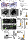
Notch1 promotes survival of extravillous trophoblast progenitors in anchoring villi. Experiments were performed with placentae between the 6th and 8th wk. (A) IHC analysis of Notch1 in CCTs after treatment of floating explant cultures with Notch1-IgG1 or ctrl-IgG1. Sections were stained with Notch1 antibodies (brown color) and hematoxylin (blue) to mark nuclear DNA. Representative cell columns of each 40 explants (derived from four different placentae) analyzed are shown. Stippled line demarcates the cell column from the underlying villous stroma (VS). (Scale bars, 50 μm.) (B) Western blot showing down-regulation of the full-length Notch1 receptor (transmembrane intracellular domain, 120 kDa) in the presence of Notch1-blocking antibody or control. A representative example (10 pooled explants per treatment and placenta, n = 4 placentae analyzed) is shown. (C) Representative pictures of collagen I-attached anchoring villi (arrows) after incubation with Notch1-blocking antibody. Explants (a total of 86 explants per condition, derived from five different placentae) were pretreated with Notch1-IgG1 or controls for 12 h and seeded onto collagen I. (Scale bars, 500 µm.) (D) After 36 h, numbers of anchoring tips were counted. Bar graph shows mean values ± SD (n = 5). *P < 0.05. (E) Representative images (Scale bar, 50 µm.) showing cytokeratin 18 neoepitope (K18-ne) or p53 IF in Notch1-IgG1–treated floating explant cultures. CC, cell column; VS, villous stroma. (F) Box plots depict median and interquartile range (IQR) values of K18-ne+ or p53+ CCTs after Notch1- or ctrl-IgG1 treatment of each of the 90 explants (n = 5 placentae). (G) Western blot and quantification of cleaved caspase-3 expression after Notch1 inhibition. For each treatment a total of 40 explants (10 pooled explants per placenta, n = 4) were analyzed. Mean values ± SD (n = 4) normalized to GAPDH are shown. *P < 0.05.
Fig. 3.
Fig. 3.
N1ICD increases proliferation and survival of purified CCTs and inhibits extravillous trophoblasts differentiation. For each preparation three to five pooled placentae of the 9th–10th wk were used. (A) Schematic representation showing protein domains of full-length Notch1, FLAG-tagged N1ICD, and N1ICD-∆RAM. (B) Notch1 IF detecting localization of N1ICD and its mutant variant after transfection into purified CCTs. (Scale bars, 50 µm.) (C) Representative Western blot and quantification of cyclin A and cleaved caspase-3 after overexpression of Notch1 constructs in CCT preparations (n = 3). Topoisomerase IIβ (TOPOIIβ) was used as loading control. (D) Cyclin D1 mRNA expression (n = 4) and (E) EdU labeling (n = 4) after ectopic expression of wild-type or mutant N1ICD in CCTs. (F) Western blot showing inhibition of HLA-G expression in differentiating cell column progenitors upon N1ICD expression. Mean values ± SD normalized to GAPDH are depicted (n = 3). *P < 0.05; ns, not significant compared with mock control.
Fig. 4.
Fig. 4.
N1ICD increases proliferation and inhibits cell fusion of purified vCTBs. FLAG-tagged N1ICD or N1ICD-∆RAM were overexpressed in vCTBs isolated from three to five pooled placentae between the 9th and 10th wk. (A) Representative Western blot showing up-regulation of cyclins and p-Histone H3, a marker of mitosis. As loading control, α-tubulin was used. (B) Percentage of EdU+ vCTBs (n = 5) and (C) relative cyclin D1 mRNA levels (n = 3) after expression of N1ICD or its mutant. AU, arbitrary units. (D) Western blot and quantification (n = 5) of markers of undifferentiated (E-cadherin, HAI-1) and differentiated (CGβ) vCTBs after N1ICD transfection. Mean values ± SD normalized to GAPDH are depicted. *P < 0.05; ns, not significant. (E) Representative IF pictures showing E-cadherin syncytial areas (marked by stippled line) in Notch1-transfected vCTBs at 120 h of in vitro differentiation. (Scale bars, 100 µm.) (F) Box plots depict median and IQR values of the percentage of multinucleated cells (n = 3). *P < 0.05; ns, not significant.
Fig. 5.
Fig. 5.
Expression pattern of markers of cytotrophoblast self-renewal and their regulation by N1ICD and IRF6. Representative pictures of first trimester placentae (A and B) showing coimmunofluorescence of TEAD4, ∆Np63, Notch1, and myc with VIM in serial sections. VE-cadherin expression in trophoblasts of the proximal cell column (CCT) partly overlapped with Notch1. Nuclei were counterstained with DAPI. (Scale bars, 50 µm.) (C) qPCR detecting mRNA expression in purified CCT and vCTB pools (6th–8th wk, three to five placentae per preparation). Mean values ± SD (n = 6) measured in duplicates are shown. AU, arbitrary units. *P < 0.05. Representative Western blots showing (D) ∆Np63, myc, and IRF6 and (E) VE-cadherin protein expression in N1ICD or N1ICD-∆RAM transfected vCTBs. GAPDH was used as loading control. (F) Quantification of Western blots. Mean values ± SD (n = 4 vCTB pools, each consisting of three to five 9th- to 10th-wk placentae) normalized to GAPDH are depicted. *P < 0.05; ns, not significant compared with mock control. Interaction of N1ICD with genomic regions in the (G) myc and (H) IRF6 gene. N1ICD and N1ICD-∆RAM were overexpressed in vCTBs and ChIP was performed using Notch1 antibody. (G) Schematic depiction of the myc gene showing localization of a putative RBPJκ binding site in the promoter (PROM) region at −3.060 bp and the previously identified Notch-dependent myc enhancer (NDME) at +1.4 Mb. (H) Representation of the proximal IRF6 promoter region delineating RBPJκ cognate sequences at −2.4 kb (PROM-1) and −3.6 kb (PROM-2). Bar graphs (G and H) represent PCR signals (mean values ± SD) obtained after ChIP (n = 3) of CTB pools (n = 11 placentae, 6th–8th wk) *P < 0.05; (I) Representative Western blot showing elevation of ∆Np63 expression after silencing of IRF6 in N1ICD-overexpressing vCTBs. Bar graph at Right delineates mean values ± SD (n = 3 vCTB pools, each consisting of three to four 6th- to 8th-wk placentae) normalized to GAPDH. *P < 0.05.
Fig. 6.
Fig. 6.
Model system depicting the presumptive role of Notch1 in human trophoblast development. N1ICD programs vCTBs into CCTs, expressing the stemness markers myc and VE-cadherin and prevents EVT differentiation by maintaining proliferation and survival of these cells. N1ICD also suppresses TEAD4 and p63, the latter by inducing its repressor IRF6, thereby alleviating self-renewal and cell fusion of vCTBs.

References

    1. Hamilton WJ, Boyd JD. Development of the human placenta in the first three months of gestation. J Anat. 1960;94:297–328. - PMC - PubMed
    1. Burton GJ, Jauniaux E, Charnock-Jones DS. The influence of the intrauterine environment on human placental development. Int J Dev Biol. 2010;54(2–3):303–312. - PubMed
    1. Evain-Brion D, Malassine A. Human placenta as an endocrine organ. Growth Horm IGF Res. 2003;13(Suppl A):S34–S37. - "V体育官网入口" PubMed
    1. Aplin JD. Developmental cell biology of human villous trophoblast: current research problems. Int J Dev Biol. 2010;54(2–3):323–329. - PubMed
    1. Pijnenborg R, Dixon G, Robertson WB, Brosens I. Trophoblastic invasion of human decidua from 8 to 18 weeks of pregnancy. Placenta. 1980;1(1):3–19. - PubMed (VSports注册入口)

Publication types