Skip to main page content (V体育官网)
U.S. flag

An official website of the United States government

Dot gov

The . gov means it’s official. Federal government websites often end in . gov or . mil. Before sharing sensitive information, make sure you’re on a federal government site. VSports app下载.

Https

The site is secure. The https:// ensures that you are connecting to the official website and that any information you provide is encrypted and transmitted securely. V体育官网.

. 2016 Dec 29;85(1):e00733-16.
doi: 10.1128/IAI.00733-16. Print 2017 Jan.

Listeria monocytogenes-Induced Cell Death Inhibits the Generation of Cell-Mediated Immunity

Affiliations

Listeria monocytogenes-Induced Cell Death Inhibits the Generation of Cell-Mediated Immunity (V体育安卓版)

Erin Theisen et al. Infect Immun. .

Abstract

The influence of cell death on adaptive immunity has been studied for decades. Despite these efforts, the intricacies of how various cell death pathways shape immune responses in the context of infection remain unclear, particularly with regard to more recently discovered pathways such as pyroptosis. The emergence of Listeria monocytogenes as a promising immunotherapeutic platform demands a thorough understanding of how cell death induced in the context of infection influences the generation of CD8+ T-cell-mediated immune responses. To begin to address this question, we designed strains of L. monocytogenes that robustly activate necrosis, apoptosis, or pyroptosis VSports手机版. We hypothesized that proinflammatory cell death such as necrosis would be proimmunogenic while apoptosis would be detrimental, as has previously been reported in the context of sterile cell death. Surprisingly, we found that the activation of any host cell death in the context of L. monocytogenes infection inhibited the generation of protective immunity and specifically the activation of antigen-specific CD8+ T cells. Importantly, the mechanism of attenuation was unique for each type of cell death, ranging from deficits in costimulation in the context of necrosis to a suboptimal inflammatory milieu in the case of pyroptosis. Our results suggest that cell death in the context of infection is different from sterile-environment-induced cell death and that inhibition of cell death or its downstream consequences is necessary for developing effective cell-mediated immune responses using L. monocytogenes-based immunotherapeutic platforms. .

Keywords: Listeria monocytogenes; apoptosis; cell death; immunotherapy; inflammasome V体育安卓版. .


Figures

FIG 1
FIG 1
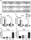
Characterization of L. monocytogenes strains that trigger cell death. (A) Unique strains of L. monocytogenes that hyperinduce the indicated form of cell death were developed. Bone marrow-derived macrophages (B, D) or bone marrow-derived dendritic cells derived from GM-CSF stimulation (C, E) were infected with the indicated strains of L. monocytogenes at an MOI of 2 and assayed for lactate dehydrogenase release (B, C) at 4 h postinfection or caspase 3/7 activation (D, E) at 2 h postinfection. Data shown are the means ± standard deviations (SD) from 3 independent experiments and are normalized to uninfected controls; ****, P < 0.0001.
FIG 2
FIG 2
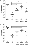
L. monocytogenes strains that induce cell death fail to induce protective immunity. Thirty days postimmunization with 1 × 10³ CFU of the indicated strain, mice were challenged with 2 LD50 of virulent L. monocytogenes expressing B8R20–27 and full-length OVA. Spleens (A) and livers (B) were harvested and analyzed for CFU burden 68 to 72 h postchallenge. Data are representative of at least three independent experiments with 4 to 5 mice per group. *, P < 0.05 (determined by Mann-Whitney U-test).
FIG 3
FIG 3
CD8+ T-cell responses are impaired following immunization with cell death-inducing strains of L. monocytogenes. Mice were immunized with 1 × 10³ CFU of the indicated strain, and splenocytes were examined by ex vivo B8R peptide stimulation 7 days postimmunization. Percentage (A) and total number (B) of IFN-γ+ B8R-specific CD8+ T cells. Percentage (C) and total number (D) of multifunctional B8R-specific CD8+ T cells expressing IFN-γ, TNF-α, and IL-2. Splenocytes were first gated for a single-cell population, followed by expression of CD8α. CD8α+ cells were then selected for expression of IFN-γ, followed by TNF-α and IL-2. Data are representative of at least 2 independent experiments with 5 mice per group; ***, P < 0.001; ****, P < 0.0001.
FIG 4
FIG 4
Recall CD8+ T-cell responses are impaired following lethal challenge with L. monocytogenes. Thirty days postimmunization with 1 × 10³ CFU of the indicated strain of L. monocytogenes, mice were challenged with a lethal dose of virulent L. monocytogenes expressing B8R20–27 and full-length OVA. Percentage (A) and total number (B) of IFN-γ+ B8R-specific CD8+ T cells at 72 h postchallenge, as examined by ex vivo peptide stimulation. Percentage (C) and total number (D) of multifunctional B8R-specific CD8+ T cells expressing IFN-γ, TNF-α, and IL-2. Splenocytes were first gated for a single-cell population, followed by expression of CD8+. CD8+ cells were then selected for expression of IFN-γ, followed by TNF-α and IL-2. Data are representative of at least 2 independent experiments with 5 mice per group. **, P < 0.01; ***, P < 0.001; ****, P < 0.0001.
FIG 5
FIG 5
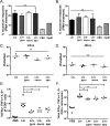
L. monocytogenes-stimulated cell death does not impair antigen presentation. (A, B) Antigen presentation of the SIINFEKL peptide was assessed using the B3Z hybridoma cell line at 24 (A) or 48 (B) hours postinfection. (C to F) To normalize burdens, mice were immunized with 1 dose of 1 × 10³ CFU Lm, 1 dose of 1 × 10⁵ CFU Lm-pyro or Lm-apo, or 2 doses of 1 × 10⁷ CFU Lm-necro dosed 12 h apart. Bacterial burdens in spleens were examined at 24 (C) or 48 (D) hours following the first immunization. Seven days postimmunization, the percentage (E) and total number (F) of multifunctional B8R-specific CD8+ T cells expressing IFN-γ, TNF-α, and IL-2 were examined. Splenocytes were first gated for a single-cell population, followed by expression of CD8α. CD8α+ cells were then selected for expression of IFN-γ, followed by TNF-α and IL-2 (E, F). Data shown are the averages ± SD and are representative of at least 2 independent experiments with 3 mice per group (A to D) or 5 mice per group (E, F). ns, not significant; *, P < 0.05; **, P < 0.01.
FIG 6
FIG 6
Dendritic cell costimulation is impaired following induction of cell death. Mice were immunized with the normalized burden-dosing scheme, and spleens were harvested at the indicated time points and analyzed for total numbers that were CD11c+ (A), CD11c+ CD86+ (B), CD8α+ CD11c+ (C), or CD8α+ CD86+ CD11c+ (D). Splenocytes were first gated for a single-cell population followed by gating on CD11c+. CD11c+ cells were then examined for expression of either CD86 or CD8α (B, C). CD11c+ CD8α+ cells were gated for expression of CD86 (D). Data are the combination of results from 2 independent experiments with 3 mice per group. *, P < 0.05; **, P < 0.01; ***, P < 0.001; ****, P < 0.0001.
FIG 7
FIG 7
Cell death-induced inflammation influences T-cell responses. Mice were immunized with the normalized burden-dosing scheme, and serum was examined at the indicated time point by cytokine bead array for levels of IFN-γ (A), IL-12p70 (B), TNF-α (C), MCP-1 (D), IL-6 (E), or IL-10 (F). Mice were immunized with 5 × 10⁵ LPS-matured DCs from wild-type mice loaded with B8R20–27; 24 h after immunization, mice were boosted with 1 × 10³ CFU of the indicated strain lacking B8R (G, H). Percentages (G) and total numbers (H) of IFN-γ-expressing CD8+ cells specific for B8R. Splenocytes were first gated for a single-cell population, followed by expression of CD8+. CD8+ cells were then selected for expression of IFN-γ. Data are the combination of results from 2 independent experiments with 3 mice per group (A to F), or data are representative of 2 independent experiments with 5 mice per group (G, H). Significance in comparison to Lm: *, P < 0.05; **, P < 0.01; ***, P < 0.001; ****, P < 0.0001 compared to Lm, Significance in comparison to PBS: #, P < 0.05; ###, P < 0.001; ####, P < 0.0001.

References

    1. Le DT, Dubensky TW, Brockstedt DG. 2012. Clinical development of Listeria monocytogenes-based immunotherapies. Semin Oncol 39:311–322. doi:10.1053/j.seminoncol.2012.02.008. - "VSports手机版" DOI - PMC - PubMed
    1. Le DT, Wang-Gillam A, Picozzi V, Greten TF, Crocenzi T, Springett G, Morse M, Zeh H, Cohen D, Fine RL, Onners B, Uram JN, Laheru DA, Lutz ER, Solt S, Murphy AL, Skoble J, Lemmens E, Grous J, Dubensky T, Brockstedt DG, Jaffee EM. 2015. Safety and survival with GVAX pancreas prime and Listeria monocytogenes-expressing mesothelin (CRS-207) boost vaccines for metastatic pancreatic cancer. J Clin Oncol 33:1325–1333. doi:10.1200/JCO.2014.57.4244. - DOI - PMC - PubMed
    1. Witte CE, Archer KA, Rae CS, Sauer J-D, Woodward JJ, Portnoy DA. 2012. Innate immune pathways triggered by Listeria monocytogenes and their role in the induction of cell-mediated immunity. Adv Immunol 113:135–156. doi:10.1016/B978-0-12-394590-7.00002-6. - DOI - PubMed
    1. Janda J, Schöneberger P, Skoberne M, Messerle M, Rüssmann H, Geginat G. 2004. Cross-presentation of Listeria-derived CD8 T cell epitopes requires unstable bacterial translation products. J Immunol 173:5644–5651. doi:10.4049/jimmunol.173.9.5644. - DOI - PubMed
    1. Reinicke AT, Omilusik KD, Basha G, Jefferies WA. 2009. Dendritic cell cross-priming is essential for immune responses to Listeria monocytogenes. PLoS One 4:e7210. doi:10.1371/journal.pone.0007210. - DOI - PMC - PubMed

Substances