Skip to main page content
U.S. flag

An official website of the United States government

Dot gov

The . gov means it’s official. Federal government websites often end in VSports app下载. gov or . mil. Before sharing sensitive information, make sure you’re on a federal government site. .

Https

The site is secure. The https:// ensures that you are connecting to the official website and that any information you provide is encrypted and transmitted securely V体育官网. .

. 2017 May 1;23(9):2255-2266.
doi: 10.1158/1078-0432.CCR-16-1300. Epub 2016 Nov 4.

V体育官网 - Multiplex Genome Editing to Generate Universal CAR T Cells Resistant to PD1 Inhibition

Affiliations

"V体育官网入口" Multiplex Genome Editing to Generate Universal CAR T Cells Resistant to PD1 Inhibition

Jiangtao Ren et al. Clin Cancer Res. .

"VSports app下载" Abstract

Purpose: Using gene-disrupted allogeneic T cells as universal effector cells provides an alternative and potentially improves current chimeric antigen receptor (CAR) T-cell therapy against cancers and infectious diseases. Experimental Design: The CRISPR/Cas9 system has recently emerged as a simple and efficient way for multiplex genome engineering. By combining lentiviral delivery of CAR and electro-transfer of Cas9 mRNA and gRNAs targeting endogenous TCR, β-2 microglobulin (B2M) and PD1 simultaneously, to generate gene-disrupted allogeneic CAR T cells deficient of TCR, HLA class I molecule and PD1. Results: The CRISPR gene-edited CAR T cells showed potent antitumor activities, both in vitro and in animal models and were as potent as non-gene-edited CAR T cells VSports手机版. In addition, the TCR and HLA class I double deficient T cells had reduced alloreactivity and did not cause graft-versus-host disease. Finally, simultaneous triple genome editing by adding the disruption of PD1 led to enhanced in vivo antitumor activity of the gene-disrupted CAR T cells. Conclusions: Gene-disrupted allogeneic CAR and TCR T cells could provide an alternative as a universal donor to autologous T cells, which carry difficulties and high production costs. Gene-disrupted CAR and TCR T cells with disabled checkpoint molecules may be potent effector cells against cancers and infectious diseases. Clin Cancer Res; 23(9); 2255-66. ©2016 AACR. .


Conflict of interest statement

COMPETING FINANCIALINTERESTS

X V体育ios版. L. , C. H. J. and Y. Z. have financial interests due to intellectual property and patents in the field of cell and gene therapy. Conflicts of interest are managed in accordance with University of Pennsylvania policy and oversight. The other authors declare that they have no competing interests.

Figures (V体育官网入口)

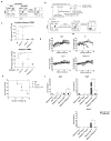
Figure 1
Figure 1. CRISPR/Cas9 mediates efficient TCR disruption in T cells
(a) CD3 expression of T cells after sequential CRISPR RNA electroporation with Cas9 and eSpCas9 (1.1). (Healthy donors, n=5) (b) Amount of TCR-targeted gene disruption measured by a mismatch-selective T7E1 surveyor nuclease assay on DNA amplified from the cells shown. The calculated amount of targeted gene disruption in TRAC and TRBC is shown at the bottom. Arrows indicate expected bands. (c) A diagram of the human locus encoding the TCR α and β CRISPR gRNA targeting sites within the genomic locus of the TCR α and β constant region. Multiple peaks in the Sanger sequencing results show the CRISPR-mediated events of NHEJ at the TRAC and TRBC genomic loci. Each exon is shown by a block. Red arrow: sense strand gRNA targeting site; blue arrow: anti-sense strand gRNA targeting site. (d) Indels and insertions observed by clonal sequence analysis of PCR amplicons after CRISPR-mediated recombination of the TCR α and β locus. Red arrow indicates putative cleavage site. (e) CD3 expression on purified TCRneg population (n=3). (f) Off-target mutagenesis measurement of TRAC and TRBC. Indel frequencies were measured by TIDE analysis (n=3). All TIDE analyses below the detection sensitivity of 1.5% were set to 0%. Bars, SE, n = 3;T: target; OT: Off-target.
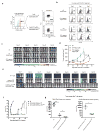
Figure 2
Figure 2. CRISPR/Cas9 editing does not impair antitumor efficacy of primary T cells
(a) Relative CD19-CAR expression after the electrotransfer of CD19-CAR RNA into Cas9 MOCK and TCR/CD3neg cells. No significant functional difference was observed between CD19-CAR redirected Cas9 MOCK and TCR/CD3neg cells as confirmed by (b) CD107 release assay, (c) cytotoxicity assay and (d) IL2 and IFNγ secretion when incubated with the Nalm6 target cell line. Representative data from 3 independent experiments are shown. Bars, SE. (e) NSG mice (n=12) were injected with 1×10⁶ Nalm6 tumor cells (i.v.), and the mice were randomly sorted into three groups. Cas9 MOCK and TCR/CD3neg T cells (10×106) expressing the CD19-CAR after electroporation were injected i.v. every 4 days for a total of three injections (arrows); mice treated with no RNA electroporated T cells from the same donor served as the control. Images were obtained from the surviving animals as indicated. Imaging commenced 1 day before the start of T cell treatment. Bars, SE; E:T, effector to tumor ratio; arrow, time point of T cell infusion; ns, not significant. ****P<0.001, ns, by comparison of the slopes with linear regression.
Figure 3
Figure 3. Multiple gene ablation by CRISPR/Cas9 to generate universal effector cells
(a) HLA-I disruption with gRNA targeting B2M. (b) Flow chart of the protocol to generate universal effector cells as described in methods. (c) The allo-reactivity of TCR and TCR/HLA disrupted was tested with an IFNγ Elispot assay by challenging the gene-ablated T cells with irradiated allogenic PBMCs (left panel) or co-culturing allogenic PBMCs with irradiated gene-ablated T cells. Specific spots are shown on the y axis as the spots produced in the presence of stimulators minus the spots produced by the effectors alone. **P<0.01 by Mann-Whitney test. (d) Survival without severe GVHD and (e) weight loss in mice after infusion of PBS (n = 5), Cas9 Mock wild type (Cas9 Mock) T cell (n = 5), TCR ablated (TCRneg) cells (n = 5) or TCR/HLA-I double ablated (TCR/HLA-Ineg) (n = 5). ***P<0.005 by the log-rank Mantel-Cox test. (f) Abolishment of target recognition of allogeneic T cells by disrupting MHC-I on target T cells. Allogeneic T cells were primed by dendritic cells of the same donor with gene-disrupted T cells and infused into NSG mice with TCRneg or TCR/HLA-Ineg target T cells. Significant prolonged survival of HLA-I ablated T cells was observed by the presence of CD3neg T cells, which is also confirmed by the failed expansion of allogeneic effector T cells (n=3). ***P<0.001**P<0.01, *P<0.05, by Mann-Whitney test.
Figure 4
Figure 4. Generation of universal CART cells with a combination of lentiviral gene transfer and CRISPR/Cas9 electroporation
(a) Flow chart of the generation of universal CD19-CART cells. T cells were transduced with lentiviral CD19-CAR on day 1 after stimulation, and Cas9 mRNA and gRNAs targeting the TCR β chain and B2M were electroporated in the T cells on day 3 followed by a second delivery of gRNAs on day 4. The TCR and HLA-I double-negative cell population was enriched by negative selection using microbeads on day9. (b) CD19-CAR expression of gene-modified lenti-CD19-CAR T cells. (c) Phenotype of universal CD19-CAR T cells. Function of TCR-negative and TCR/HLA-I double-negative CD19-CAR T cells tested by (d) CD107a release, (e) cytokine secretion and (f) tumor lytic capability. Representative data from 3 independent experiments are shown. Bars, SE. (g) CFSE-labeled CD19-CAR and non-transduced T cells were incubated with K562 and target K562-CD19 tumor cells at the indicated E:T ratio for 72 hours. (h) Survival curve of mice receiving gene-edited CD19-CART cells. Tumors were established in NSG mice (n=5 per group) by i.v. injection of 1×10⁶ Nalm6 cells. Beginning on day 7, T cells (5×10⁶) expressing lentiviral (LV) transduced CD19-CAR were infused with a single injection. T cells expressing LV GFP protein were injected as controls. ns, no difference by the log-rank Mantel-Cox test. (i) Peripheral blood from Nalm6-bearing NSG mice treated with CD19 CART cells was obtained on day 21 and quantified for the presence of CD45 T cells by a FACS Trucount assay. Results are expressed as the mean absolute count per μl of peripheral blood±SD with n=5 for all groups. ns, ****P<0.001 by Mann-Whitney test.
Figure 5
Figure 5. PD1 ablation enhances the therapeutic effect of CART cells
(a) Generation of PD1-negative PSCA-CAR T cells. T cell PD1 ablation was confirmed by flow cytometry after stimulation. PD1 deficient CART cells were sorted. (b) Co-culture of PD1 disrupted CART cells with PC3-PDL1 tumor cells. PD1 and CD137 expression were measured on the CRISPR/Cas9 edited CART cells. (c) PC3-PSCA-PDL1 tumors were established in the flank of NSG mice by inoculating 1×10⁶ tumor cells/mouse (s.c. with Matrigel, n=4). After 3 weeks, the mice were treated with 2×10⁶ PSCA CAR transduced WT (PSCA CAR) or PD1neg (PSCA CAR PD1neg) T cells (i.v.); mice treated with non-transduced T cells (NT) served as the control. BLI conducted before (day 21) and after the mice treated with a single T cell injection. (d) Tumor volume of mice. Results are expressed as the mean tumor volume (mm3±SE) with n=4 for all groups. **P<0.01, ****P<0.001 by comparison of the slopes with linear regression. (e) PD1 ablated universal CD19-CART cells were generated by co-delivery of Cas9 mRNA and gRNAs targeting TRBC, B2M and PD1 after transduction with lenti-CD19-CAR. TCR and HLA-I disruption was confirmed by flow cytometry, PD1 disruption was confirmed Sanger sequencing (Supplementary Figure 6a,b). TCR and HLA-I double negative cells were sorted at day 9. Nalm6-PDL1 tumor were established in NSG mice (n=4 or 5 per group) by i.v. injection of 1×10⁶ cells. Beginning on day 7, T cells (5×10⁶) expressing lentiviral transduced CD19-CAR were infused with a single injection. T cells expressing LV GFP protein were injected as controls. (f) Bioluminescence of mice receiving different treatment. Imaging commenced 1 day before the start of T cell treatment. P<0.05, by comparison of the slopes with liner regression.(g) Tumor burden of different groups were compared at day 2 and day 59 after T cell treatment (n= 4 or 5). *P<0.05, **P<0.01, ****P<0.001 by Mann-Whitney test.

References

    1. Brentjens RJ, Davila ML, Riviere I, Park J, Wang X, Cowell LG, et al. CD19-targeted T cells rapidly induce molecular remissions in adults with chemotherapy-refractory acute lymphoblastic leukemia. Science translational medicine. 2013;5(177):177ra38. doi: 10.1126/scitranslmed.3005930. - V体育官网 - DOI - PMC - PubMed
    1. Maude SL, Frey N, Shaw PA, Aplenc R, Barrett DM, Bunin NJ, et al. Chimeric antigen receptor T cells for sustained remissions in leukemia. N Engl J Med. 2014;371(16):1507–17. doi: 10.1056/NEJMoa1407222. - DOI (VSports) - PMC - PubMed
    1. Porter DL, Levine BL, Kalos M, Bagg A, June CH. Chimeric antigen receptor-modified T cells in chronic lymphoid leukemia. N Engl J Med. 2011;365(8):725–33. doi: 10.1056/NEJMoa1103849. - DOI - PMC - PubMed
    1. Robbins PF, Morgan RA, Feldman SA, Yang JC, Sherry RM, Dudley ME, et al. Tumor regression in patients with metastatic synovial cell sarcoma and melanoma using genetically engineered lymphocytes reactive with NY-ESO-1. Journal of clinical oncology : official journal of the American Society of Clinical Oncology. 2011;29(7):917–24. doi: 10.1200/JCO.2010.32.2537. - "V体育官网" DOI - PMC - PubMed
    1. Grupp SA, Kalos M, Barrett D, Aplenc R, Porter DL, Rheingold SR, et al. Chimeric antigen receptor-modified T cells for acute lymphoid leukemia. N Engl J Med. 2013;368(16):1509–18. doi: 10.1056/NEJMoa1215134. - DOI - PMC - PubMed

MeSH terms

Substances

V体育ios版 - Supplementary concepts