Skip to main page content
U.S. flag

An official website of the United States government

Dot gov

The . gov means it’s official VSports app下载. Federal government websites often end in . gov or . mil. Before sharing sensitive information, make sure you’re on a federal government site. .

Https

The site is secure. The https:// ensures that you are connecting to the official website and that any information you provide is encrypted and transmitted securely. V体育官网.

. 2016 Dec 23;291(52):26816-26836.
doi: 10.1074/jbc.M116.719823. Epub 2016 Oct 31.

Anoctamin 6 Contributes to Cl- Secretion in Accessory Cholera Enterotoxin (Ace)-stimulated Diarrhea: AN ESSENTIAL ROLE FOR PHOSPHATIDYLINOSITOL 4,5-BISPHOSPHATE (PIP2) SIGNALING IN CHOLERA (VSports)

Affiliations

Anoctamin 6 Contributes to Cl- Secretion in Accessory Cholera Enterotoxin (Ace)-stimulated Diarrhea: AN ESSENTIAL ROLE FOR PHOSPHATIDYLINOSITOL 4,5-BISPHOSPHATE (PIP2) SIGNALING IN CHOLERA

Joydeep Aoun et al. J Biol Chem. .

Abstract

Accessory cholera enterotoxin (Ace) of Vibrio cholerae has been shown to contribute to diarrhea. However, the signaling mechanism and specific type of Cl- channel activated by Ace are still unknown. We have shown here that the recombinant Ace protein induced ICl of apical plasma membrane, which was inhibited by classical CaCC blockers. Surprisingly, an Ace-elicited rise of current was neither affected by ANO1 (TMEM16A)-specific inhibitor T16A(inh)-AO1(TAO1) nor by the cystic fibrosis transmembrane conductance regulator (CFTR) blocker, CFTR inh-172. Ace stimulated whole-cell current in Caco-2 cells. However, the apical ICl was attenuated by knockdown of ANO6 (TMEM16F). This impaired phenotype was restored by re-expression of ANO6 in Caco-2 cells. Whole-cell patch clamp recordings of ANO currents in HEK293 cells transiently expressing mouse ANO1-mCherry or ANO6-GFP confirmed that Ace induced Cl- secretion. Application of Ace produced ANO6 but not the ANO1 currents. Ace was not able to induce a [Ca2+]i rise in Caco-2 cells, but cellular abundance of phosphatidylinositol 4,5-bisphosphate (PIP2) increased. Identification of the PIP2-binding motif at the N-terminal sequence among human and mouse ANO6 variants along with binding of PIP2 directly to ANO6 in HEK293 cells indicate likely PIP2 regulation of ANO6. The biophysical and pharmacological properties of Ace stimulated Cl- current along with intestinal fluid accumulation, and binding of PIP2 to the proximal KR motif of channel proteins, whose mutagenesis correlates with altered binding of PIP2, is comparable with ANO6 stimulation VSports手机版. We conclude that ANO6 is predominantly expressed in intestinal epithelia, where it contributes secretory diarrhea by Ace stimulation in a calcium-independent mechanism of RhoA-ROCK-PIP2 signaling. .

Keywords: epithelial cell; epithelial cell adhesion molecule (EpCAM); intestinal epithelium; ion channel; membrane; pathogenesis; phosphatidylinositol; signaling V体育安卓版. .


"VSports在线直播" Figures

FIGURE 1.
FIGURE 1.
Summarized effects of recombinant Ace stimulation on Cl current in Caco-2 cell monolayers. A, representative time course of changes in Isc and the effect of different doses of apically applied Ace on the changes in Isc (insets). Each concentration of Ace was added as a single dose to separate monolayers. Values are means ± S.E. n = 3–5. NS, statistically not significant. B, effects of basolateral bumetanide (100 μm) on basal and Ace (1 μm)-stimulated Isc. Summary of data showed average ΔIsc in response to Ace in the absence (−) or presence (+) of bumetanide. Values are means ± S.E. n = 4. C, effects of Ace on apical ICl. After establishment of a basolateral-to-apical Cl gradient in the high K+ solution by depolarizing the basolateral membrane, addition of Ace (1 μm) evoked an outward current consistent with a secretory Cl flow from the basolateral-to-apical side. Summary data show the change in ΔICl induced by Ace stimulation. Values are means ± S.E.; n = 5. Error bars represent S.E. *, significant difference compared with control (unpaired t test).
FIGURE 2.
FIGURE 2.
Ace-induced Isc and ICl response to CFTR inhibitor and classical CaCC inhibitors NPPB and NFA and ANO inhibitors AO1 and TAO1 in Caco-2 cells. A, representative trace showing the effects of FSK (10 μm) and CFTR inhibitor CFTRinh-172 (20 μm) on Ace-stimulated Isc. CFTRinh-172 inhibited FSK- but not Ace-stimulated Isc in Caco-2 cells (inset). Values are means ± S.E.; n = 3–6. B, effects of cumulative addition of CaCC blockers DIDS (500 μm), TFN (10 μm), AO1 (10 μm), and TAO1 (10 μm) on Ace-induced Isc. AO1 was most effective to inhibit Ace-induced Isc than cumulative addition of different CaCC blockers. C, summary of the inhibitory effects on ICl by various CaCC inhibitors NPPB (100 μm), NFA (100 μm), AO1 (10 μm), TAO1 (10 μm), TFN (10 μm), and TA (100 μm) in Caco-2 cell monolayers. D, mouse ileum. Values are means ± S.E.; n = 5–8. Error bars represent S.E. *, p < 0.01, significant inhibition by blockers (ANOVA). NS, statistically not significant; ND, not determined.
FIGURE 3.
FIGURE 3.
ANO6 is expressed abundantly in Caco-2 cells and mouse ileum. Quantitative PCR analysis of ANO1 and ANO6 in human colonic cells T84 and Caco-2 (A) and different parts of mouse intestine as indicated (B and C), which are shown as mRNA levels relative to levels of GAPDH and determined using the difference of PCR cycles to reach a threshold amplification (ΔCT), and the relative amount of the target mRNA are given as 2−ΔCT. CT, cycle threshold. Values are means ± S.E. of triplicate experiments. D, protein from total lysates of T84 and Caco-2 cell monolayers was resolved by 10% SDS-PAGE and immunoblotted with ANO6 antibody. ANO6 protein migrated in all cases with an apparent molecular mass of 106 kDa (top). Paraffin sections of mouse ileum were fixed and stained for ANO6 (green), and xy images were collected by confocal microscopy as described under “Experimental Procedures.” Image is representative of results for three experiments. Significant difference at p < 0.001 was compared between ANO1 and ANO6 (paired t test). NS, statistically not significant; ND, not determined.
FIGURE 4.
FIGURE 4.
ANO6 expression correlates with Ace-stimulated Isc change in mouse intestinal epithelium. A, concentration-response showing the increase in Isc following apical treatment with various concentrations of Ace in mouse ileum. The maximum effect was achieved with 5–6 μm Ace. Dose-response curve was created by a non-cumulative single addition to individual tissues. Results are shown as means ± S.E. n = 6–10 tissues from 10 mice were used. B, effect of anion substitution on maximal Isc response to Ace in mouse ileum. Replacement of Cl in both apical and basolateral solution completely inhibited the maximal Isc response to Ace. Data are means ± S.E., n = 5. C, summary of the effects of Ace on change in Isc of different parts of mouse intestine. Ileum had the highest response to Ace than duodenum, jejunum, and colon. Values are means ± S.E.; n = 4–7. D, representative of Ace-induced Isc after addition of Ace to the apical solution showing the level of recovery after washing out the Ace in mouse ileum. *, significant difference when compared with control to Ace stimulation (paired t test).
FIGURE 5.
FIGURE 5.
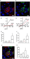
ANO6 is activated by Ace but not the ANO1. HEK293 cells transiently transfected with mcherry-tagged mouse ANO1 (A) and GFP-tagged mouse ANO6 (D) showing membrane expression of ANO1 and ANO6 in these cells. The images were taken using ×63 objective in LSM510 confocal microscope. Bars, 20 μm. Representative whole-cell current traces for ANO1 (B) and ANO6 (E) measured in transfected HEK293 cells treated with either Ace or A23187. Ace stimulated ANO6 but not ANO1 currents and A23187 evoked both of these currents. Average current amplitudes of ANO1 (C) and ANO6 (F) in transiently transfected HEK293 cells in response to Ace and A23187 are shown. ANO6 co-localized with ANO1 at the membrane of transiently transfected HEK293 cells (G). Co-localization augmented the whole-cell current amplitude in these cells (H). Data are presented as mean ± S.E. and tested by one-way ANOVA; *, p < 0.05. For transfection with individual ANO constructs, 2 μg of DNA was used; co-transfection was performed using 1 μg of each ANO constructs.
FIGURE 6.
FIGURE 6.
Ace stimulated calcium-independent Cl current and ATP but not Ace increased intracellular calcium in mouse ileum and Caco-2 cells. A, effects of BAPTA-AM on (left panel) and removal of extracellular calcium (right panel) on Ace-stimulated Isc of mouse ileum. BAPTA-AM was applied serosally as indicated. AO1 was applied to ensure that current was ANO-mediated. Traces are representative of four independent experiments. B, time course for Ace + ATP induced increase in [Ca2+]i in Caco-2 cells, as detected by ratiometric Fura2 fluorescence. Stimulation with ATP activates [Ca2+]i rise. Cells were treated with 1 μm Ace followed by the addition of 100 μm ATP. Ace was not able to increase [Ca2+]i, but subsequent addition of ATP evoked a [Ca2+]i rise in these cells. Resting [Ca2+]i of these cells was around 100 nm when measured in the solution contained 10 mm glucose.
FIGURE 7.
FIGURE 7.
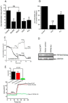
Inhibition of PIP2 synthesis by lipid kinase inhibitors affects Ace-stimulated ICl. A, pre-incubation with two different concentrations of Wort50 (50 μm), Wort1 (1 μm), PAO (10 μm), and PLL (25 μg/ml) significantly inhibited Isc in Caco-2 cells. NS, statistically not significant. B, following a 30-min incubation in the presence of PAO (20 μm) and PLL (25 μg/ml), Isc was reduced in response to Ace (5 μm) in mouse ileum. C, PIP2 speeds Isc recovery by subsequent washout of PAO. PIP2 modulators were added to both sides of the solutions. Ace was added only on the apical side. D, Western blot (with PIP5K antibody) detection of proteins in total lysate of wild-type Caco-2, vector control, and PIP5K knock out cells. Anti-GAPDH was used to confirm equal loading (n = 3). E, representative time course on the effect of Ace on Isc in PIP5K KO Caco-2, WT, and vector control cells. PIP5K knock-out cells had very little or no effect in response to Ace stimulated Isc. The inset shows the summary of the data from six such monolayers. Values represent means ± S.E. significant difference at *, p < 0.05, or **, p < 0.01when compared with the corresponding control value by ANOVA.
FIGURE 8.
FIGURE 8.
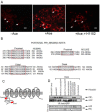
Cellular abundance of PIP2 and its potential binding sites in the N terminus among the human and mouse ANO6 variants. A, Caco-2 wild-type cells were stimulated with Ace. Shown are representative images of fixed cells labeled for PIP2 in non-stimulated cells (control, left panel), Ace-stimulated cells (middle panel), and for cells that are pre-treated with H1152 and stimulated with Ace (right panel). Red color represents PIP2 abundance. Scale bars, 10 μm. B, sequence alignments at a comparable position in ANO6 variants showing two similar PIP2-binding motifs are conserved beginning immediately after the putative first membrane spanning domain of human (left) and mouse (right).Typical PIP2-binding motifs consisting of a cluster of basic residues are indicated by the red box. Sequences were obtained from the NCBI protein database. Accession numbers are as follows: variant 1 isoform a, NM_001025356.2 > NP_001020527.2; variant 2 isoform b, NM_001142678.1>NP_001136150.1; variant 3 isoform c, NM_001142679.1 > NP_001136151.1; and variant 4 isoform d, NM_001204803.1>NP_001191732.1. Mouse variant accession numbers are as follows: variant 1 isoform a, NM_001253813.1 > NP_0012407421.1, and variant 2 isoform b NM_175344.4 > NP_780553.2. C, schematic diagram illustrating the topology of human ANO6, highlighting two potential PIP2-binding sites (red box) of the channel N terminus. D, PIP2 co-immunoprecipitates with ANO6 in mANO6-GFP-transfected HEK293 cells whereas truncation mutants led to a significant decrease in the interaction. The cell lysates were incubated with anti-GFP-protein G-Sepharose beads, and inputs and immunoprecipitation eluate fractions were analyzed by Western blotting with either anti-PIP2 with or without Ace stimulation. PIP2 antibodies recognize GFP-tagged ANO6 protein in transfected HEK293 cells. No signal is detected in non-transfected HEK293 cells. This experiment was performed twice. The GFP blot for this experiment was presented to indicate the level of ANO6-GFP precipitation. D and P in truncated protein designate the deletion of distal and both distal and proximal KR motifs. Introduction of four mutations in the proximal and distal KR motifs are as indicated. IB, immunoblot; IP, immunoprecipitation.
FIGURE 9.
FIGURE 9.
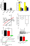
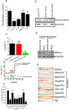
Quantitative mRNA expression and activation of ANO6 whole-cell current in Caco-2 cells. A, relative expression (normalized by expression of GAPDH) of ANO6 variants in Caco-2 cells; B, mouse intestinal tissues in log scale. Data are means ± S.E. from triplicate experiments. C, whole-cell current response to Ace showing representative current-voltage relationship with gramicidin-perforated patch recording in Caco-2 cells. D, summary graph of current amplitude in response to Ace stimulation. * indicates significant difference at p < 0.05 compared with control (paired t test). E, knocking out of ANO6 mostly abolished Ace-stimulated Isc. Shown are representative time courses on the effects of Ace on Isc in WT, vector control, and ANO6 KO Caco-2 cells (bottom panel); top panel shows the summary of the data from four such monolayers. Values are means ± S.E. F, rescue experiments for ANO6 in Caco-2 cells by re-expression of ANO6 using retroviral expression system described under “Experimental Procedures.” Top panel, Western blotting analysis of proteins in total lysate of wild type, ANO6 KO, ANO6 KI, and in vector control cells. Anti-GAPDH was used as loading control. Bottom panel, summary of the data on Ace-stimulated Isc in ANO6 KO and ANO6 KI cells with WT and vector control. ** indicates significant difference at p < 0.001compared with WT or vector control (paired t test). NS, statistically not significant.
FIGURE 10.
FIGURE 10.
RhoA, ROCK, and lipid kinase inhibitors attenuate Ace-induced diarrhea in vivo. A, summary graph of Ace-induced Isc in Caco-2 cell monolayer pre-treatments with C3 toxin (at 1 μg/ml for 4 h in the 6-well plate), H1152 (1 μm), PD98059 (10 μm), and Wort (1 μm) on both sides of the chamber for 20 min before addition of Ace. B, immunoblot analysis of RhoA in WT, vector control, and RhoA KO Caco-2 cells total lysate. Probe for GAPDH was used to ensure equal loading. C, knock-out of RhoA expression abolished Ace-stimulated Isc. Top panel, summary; bottom panel, representative traces of time course experiments showing that RhoA KO inhibited the increase in Isc in response to Ace. Mean ± S.E. were calculated from n = 5 experiments. ** indicates significant difference at p < 0.01 compared with WT or vector control (paired t test). ND, not determined. D, representative immunoblot from three independent experiments demonstrate Ace stimulation increases membrane amount of RhoA. Typical Western blotting probed with anti-RhoA antibody containing the plasma membrane fraction from Caco-2 cells. E, fluid accumulation ratio in closed mouse loops by Ace stimulation in the presence of ROCK inhibitor H1152 (1 μm), lipid kinase inhibitors PAO (100 μm), PLL (25 μg/ml), and CaCC blockers. F, photographs of representative mouse ileal loops at 6 h after luminal installation of Ace with or without inhibitors. Data represent the means ± S.E. n = 10 mice per group.* indicates significant difference at p < 0.05 when compared with control or with Ace stimulation by ANOVA; NS, statistically not significant.
FIGURE 11.
FIGURE 11.
Model for the role and regulation of ANO6 to induce diarrhea by Ace stimulation. Ace-mediated activation of RhoA-ROCK influences function of PIP5K, the enzyme that catalyzes the formation of PIP2, which after subsequent interaction to the N-terminal proximal KR motif became the key mechanism to acutely activate ANO6 for Cl secretion to induce diarrhea.

V体育ios版 - References

    1. Walker C. L., Rudan I., Liu L., Nair H., Theodoratou E., Bhutta Z. A., O'Brien K. L., Campbell H., and Black R. E. (2013) Global burden of childhood pneumonia and diarrhoea. Lancet 381, 1405–1416 - PMC - PubMed
    1. Sears C. L., and Kaper J. B. (1996) Enteric bacterial toxins: mechanisms of action and linkage to intestinal secretion. Microbiol. Rev. 60, 167–215 - PMC - PubMed
    1. Fasano A., Baudry B., Pumplin D. W., Wasserman S. S., Tall B. D., Ketley J. M., and Kaper J. B. (1991) Vibrio cholerae produces a second enterotoxin, which affects intestinal tight junctions. Proc. Natl. Acad. Sci. U.S.A. 88, 5242–5246 - PMC - PubMed
    1. Fasano A., and Uzzau S. (1997) Modulation of intestinal tight junctions by zonula occludens toxin permits enteral administration of insulin and other macromolecules in an animal model. J. Clin. Invest. 99, 1158–1164 - PMC - PubMed
    1. Trucksis M., Galen J. E., Michalski J., Fasano A., and Kaper J. B. (1993) Accessory cholera enterotoxin (Ace), the third toxin of a Vibrio cholerae virulence cassette. Proc. Natl. Acad. Sci. U.S.A. 90, 5267–5271 - PMC - PubMed

MeSH terms