Skip to main page content (VSports手机版)
U.S. flag

An official website of the United States government

Dot gov

The . gov means it’s official. Federal government websites often end in . gov or . mil VSports app下载. Before sharing sensitive information, make sure you’re on a federal government site. .

Https

The site is secure V体育官网. The https:// ensures that you are connecting to the official website and that any information you provide is encrypted and transmitted securely. .

. 2016 Oct 19;8(361):361ra138.
doi: 10.1126/scitranslmed.aag1711.

"V体育2025版" Cancer cells induce metastasis-supporting neutrophil extracellular DNA traps

Affiliations

Cancer cells induce metastasis-supporting neutrophil extracellular DNA traps

Juwon Park et al. Sci Transl Med. .

Abstract

Neutrophils, the most abundant type of leukocytes in blood, can form neutrophil extracellular traps (NETs). These are pathogen-trapping structures generated by expulsion of the neutrophil's DNA with associated proteolytic enzymes. NETs produced by infection can promote cancer metastasis. We show that metastatic breast cancer cells can induce neutrophils to form metastasis-supporting NETs in the absence of infection. Using intravital imaging, we observed NET-like structures around metastatic 4T1 cancer cells that had reached the lungs of mice. We also found NETs in clinical samples of triple-negative human breast cancer. The formation of NETs stimulated the invasion and migration of breast cancer cells in vitro. Inhibiting NET formation or digesting NETs with deoxyribonuclease I (DNase I) blocked these processes. Treatment with NET-digesting, DNase I-coated nanoparticles markedly reduced lung metastases in mice VSports手机版. Our data suggest that induction of NETs by cancer cells is a previously unidentified metastasis-promoting tumor-host interaction and a potential therapeutic target. .


Figures

Figure 1
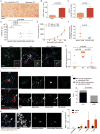
Figure 1. Neutrophil extracellular traps (NETs) form during metastasis of breast cancer
(A, B) More neutrophils infiltrated metastatic 4T1 tumors than non-metastatic 4T07 tumors (Ly6G immunostaining, mean ± SEM; n=4 mice, t-test). Scale bar: 50 μm. (C) CXCL1 protein level was higher in 4T1 than in 4T07 tumors (mean ± SEM, n=5 mice, t-test). (D) Cancer cell-derived CXCL1 promoted metastatic seeding after intravenous injection of luciferase-expressing cells (bioluminescence radiance 19 days after cell injection; individual mice and means indicated; one-way ANOVA p=0.0008, Dunnett's multiple comparison: p<0.01). (E) CXCL1 secretion by 4T1 cells did not affect primary tumor growth in nude mice (mean ± SEM, n=5 mice). (F) Knockdown of Cxcl1 in 4T1 cells reduced lung metastasis in nude mice (individual mice and means ± SD indicated; t-test). (G, H) NET-like structures of extracellular DNA, sensitive to intravenous DNase I, were found around 4T1-cancer cells in LysM-EGFP mice using CILI. Grey scale insert shows DNA channel (shown and quantified 30-60 minutes after cancer cell injection, values from individual mice and mean ± SD are indicated; one-way ANOVA p=0.002, Sidak's multiple comparison: p<0.01). Scale bar: 50 μm. (I, J) Extracellular DNA and neutrophil elastase activity co-localized near 4T1, but not 4T07, cancer cells (shown and quantified 30-60 minutes after cancer cell injection, Fisher's exact test). Scale bar: 50 μm. (K, L) The number of NETs in the lungs was higher after 4T1 cell injection than in controls (co-localized myeloperoxidase and citrullinated histone H3 immune staining). White arrow: NET; yellow arrow: intact neutrophil (mean ± SEM, n=3 mice). Scale bar: 10 μm.
Figure 2
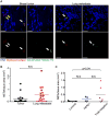
Figure 2. NETs are present in metastatic, triple-negative human breast cancer
(A) Detection of NETs by immunofluorescence in human breast tumors and lung metastases. White arrows point to NETs (defined as co-localized myeloperoxidase, citrullinated histone H3, and DNA), and yellow arrows point to intact neutrophils. Scale bars: 20 μm. (B) Number of NETs in matched primary tumors and lung metastases (paired t-test). (C) Number of NETs in primary tumor of different breast cancer subtypes (ANOVA, Tukey's multiple comparisons test).
Figure 3
Figure 3. Formation of NETs by metastatic 4T1 breast cancer cells is associated with cancer cell invasion
(A, B) 4T1 but not 4T07 cells increased the formation of NETs (immunostaining for histone H3 and neutrophil elastase, mean ± SEM, n=3, t-test). Scale bar: 50 μm. (C) Neutrophils promoted invasion of 4T1 but not 4T07 cells (mean ± SEM, n=5, t-test). (D, E) DNase I (1.5 U) digested NETs (mean ± SEM, n=3, t-test). Scale bar: 50 μm. (F) DNase I treatment inhibited neutrophil-stimulated invasion of 4T1 cells (mean ± SEM, n=5-7, t-test). (G) Primary C3(1)-Tag cancer cells induced NETs. Scale bar: 50 μm. (H) DNase I treatment inhibited neutrophil-stimulated migration of C3(1)-Tag cancer cells (mean ± SEM, n=3, t-test). (I, J) Human BT-549 breast cancer cells promoted NET formation (immunostaining for histone H3 and myeloperoxidase, mean ± SEM, n=3, t-test). Scale bar: 50 μm. (K) DNase I (1.5 U) treatment blocked neutrophil-stimulated invasion of BT-549 breast cancer cells (mean ± SEM; BT-549 cells only or BT-549 cells with neutrophils and vehicle or DNase I: n=5; BT-549 cells and vehicle or DNase I in 10% FCS: n=2).
Figure 4
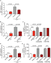
Figure 4. Cancer cells induce NET formation through G-CSF, and neutrophil-stimulated invasion requires NADPH oxidase and PAD4 activity
(A) Blocking anti-G-CSF antibodies (1.6 μg/mL) decreased 4T1-induced NET extension (mean ± SEM, neutrophils with vehicle or anti-G-CSF: n=3; neutrophils with 4T1 cells and vehicle or anti-G-CSF: n=5, t-test). (B) The NADPH oxidase inhibitor apocynin (10 μM) inhibited NET formation (mean ± SEM, neutrophils and vehicle or NAPDH oxidase inhibitor: n=3, neutrophils with cancer cells and vehicle or apocynin: n=5, t-test). (C) Apocynin (10 μM) inhibited neutrophil-stimulated invasion (mean ± SEM, 4T1 cells only or 4T1 cells with neutrophils and vehicle or apocynin: n=4; 4T1 cells only and vehicle or apocynin in 10% FCS: n=1, t-test). (D) PAD4 inhibition (200 μM Cl-amidine) reduced cancer cell-induced NET formation (mean ± SEM; neutrophils and vehicle or PAD4 inhibitor: n=6; neutrophils with 4T1 cells and vehicle or PAD4 inhibitor: n=4, t-test). (E) PAD4 inhibition (200 μM Cl-amidine) blocked neutrophil-stimulated cancer cell invasion (mean ± SEM; n=3, t-test).
Figure 5
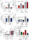
Figure 5. NET formation and neutrophil-stimulated invasion require neutrophil protease activity
(A) Cathepsin G inhibitor I (2 μM) reduced cancer cell-induced NET formation (mean ± SEM, n=4, t-test). (B) Cathepsin G inhibitor I (2 μM) inhibited neutrophil-stimulated invasion (mean ± SEM; 4T1 cells only, 4T1 cells with neutrophils: n=4; 4T1 cells in 10% FCS: n=3, t-test). (C) Neutrophil elastase inhibitor sivelestat (10 μM) reduced cancer cell-induced NET formation (mean ± SEM; n=3, t-test). (D) Neutrophil elastase inhibitor sivelestat (10 μM) weakly reduced neutrophil-stimulated invasion of 4T1 cells (mean ± SEM; 4T1 cells only or 4T1 cells with neutrophils and vehicle or sivelestat: n=5; 4T1 cells and vehicle or sivelestat in 10% FCS: n=2). (E) NADPH oxidase or neutrophil elastase inhibition (10 μM apocynin or sivelestat, respectively) blocked neutrophil-stimulated invasion of human breast cancer cells (mean ± SEM, BT-549 cells only or BT-549 cells with neutrophils and vehicle or apocynin or sivelestat: n=4, BT-549 cells and apocynin or sivelestat in 10% FCS: n=2). (F) CM from neutrophils induced to form NETs by culturing with cancer cells promoted invasion, but not when NET induction occurred in the presence of NADPH oxidase inhibition (10 μM apocynin, mean ± SEM, n=3-6, t-tests).
Figure 6
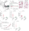
Figure 6. Targeting NETs in vivo reduces metastasis
(A) DNase I-coated nanoparticles reduced neutrophil-stimulated cancer cell invasion in vitro (mean ± SEM, n=4, t-test). (B) Injection of DNase I-coated nanoparticles results in higher plasma nuclease activity than injection of free DNase I (mice injected with 75 U free DNase I, 75 U nanoparticle bound DNase I, or equivalent vehicle, mean ± SEM; n=3 mice). (C, D) DNase I-coated nanoparticles (75 U/mouse) reduced experimental lung metastasis of 4T1 cells (mean ± SEM; n=9-10 mice; t-test). Arrows point to metastases. Scale bars: 4 mm. (E, F) DNase I-coated nanoparticles (75 U/mouse) reduced the number and the size of metastatic foci arising after injection of 4T1 cells (mean ± SEM; n=9-10 mice; t-test. The data were transformed by taking the square root before performing the t-test due to significantly [p=0.0003] different variances [untransformed data graphed]). (G) DNase I-coated nanoparticles did not affect primary tumor growth. Nanoparticle treatment was initiated 7 days after tumor cell transplantation (mean ± SEM; n=6 mice). (H) DNase I-coated nanoparticles (75 U/mouse) reduced spontaneous metastasis of 4T1 cells (mean ± SEM; n=6 mice; t-test. The data were transformed by taking the square root before performing the t-test due to significantly [p=0.007] different variances).

"VSports注册入口" Comment in

  • Cancer: Blocking metastasis.
    Crunkhorn S. Crunkhorn S. Nat Rev Drug Discov. 2016 Dec;15(12):822. doi: 10.1038/nrd.2016.239. Epub 2016 Nov 18. Nat Rev Drug Discov. 2016. PMID: 27857143 No abstract available.

References (VSports手机版)

    1. Nguyen DX, Bos PD, Massague J. Metastasis: from dissemination to organ-specific colonization. Nat Rev Cancer. 2009;9:274–284. - "VSports最新版本" PubMed
    1. Sethi N, Kang Y. Unravelling the complexity of metastasis - molecular understanding and targeted therapies. Nat Rev Cancer. 2011;11:735–748. - "V体育官网入口" PMC - PubMed
    1. Hanahan D, Coussens LM. Accessories to the crime: functions of cells recruited to the tumor microenvironment. Cancer Cell. 2012;21:309–322. - PubMed
    1. Tazawa H, Okada F, Kobayashi T, Tada M, Mori Y, Une Y, Sendo F, Kobayashi M, Hosokawa M. Infiltration of neutrophils is required for acquisition of metastatic phenotype of benign murine fibrosarcoma cells: implication of inflammation-associated carcinogenesis and tumor progression. Am J Pathol. 2003;163:2221–2232. - PMC - PubMed
    1. Cools-Lartigue J, Spicer J, McDonald B, Gowing S, Chow S, Giannias B, Bourdeau F, Kubes P, Ferri L. Neutrophil extracellular traps sequester circulating tumor cells and promote metastasis. J Clin Invest. 2013;123:3446–3458. - PMC - PubMed

Publication types

VSports在线直播 - MeSH terms

Substances