Skip to main page content
U.S. flag

An official website of the United States government

Dot gov

The . gov means it’s official VSports app下载. Federal government websites often end in . gov or . mil. Before sharing sensitive information, make sure you’re on a federal government site. .

Https

The site is secure. The https:// ensures that you are connecting to the official website and that any information you provide is encrypted and transmitted securely V体育官网. .

. 2016 Dec 1;143(23):4462-4473.
doi: 10.1242/dev.132142. Epub 2016 Oct 21.

Six3 dosage mediates the pathogenesis of holoprosencephaly

Affiliations

"V体育2025版" Six3 dosage mediates the pathogenesis of holoprosencephaly

Xin Geng et al. Development. .

Abstract

Holoprosencephaly (HPE) is defined as the incomplete separation of the two cerebral hemispheres. The pathology of HPE is variable and, based on the severity of the defect, HPE is divided into alobar, semilobar, and lobar VSports手机版. Using a novel hypomorphic Six3 allele, we demonstrate in mice that variability in Six3 dosage results in different HPE phenotypes. Furthermore, we show that whereas the semilobar phenotype results from severe downregulation of Shh expression in the rostral diencephalon ventral midline, the alobar phenotype is caused by downregulation of Foxg1 expression in the anterior neural ectoderm. Consistent with these results, in vivo activation of the Shh signaling pathway rescued the semilobar phenotype but not the alobar phenotype. Our findings show that variations in Six3 dosage result in different forms of HPE. .

Keywords: Forebrain; Gene dosage; Holoprosencephaly; Mouse; Six3; Transcription factor. V体育安卓版.


V体育安卓版 - Conflict of interest statement

The authors declare no competing or financial interests.

"V体育平台登录" Figures

Fig. 1.
Fig. 1.
Changes in Six3 levels lead to different types of HPE-like phenotypes in mice. Scanning electron microscopy analysis was performed on E10.5 wild-type (A), Six3neo/neo (E) and Six3neo/– (I) embryos. The telencephalic vesicles are pseudocolored in magenta, medial nasal prominences (MNPs) in yellow, and lateral nasal prominences (LNPs) in green. (A) In control embryos, both the telencephalic vesicles and the MNPs are well separated (n=3). (E) In Six3neo/neo embryos, the LNPs are well formed; however, a single telencephalic vesicle is present and the MNP is not separated (n=2). (I) In Six3neo/– embryos, the single telencephalic vesicle is small, the MNPs are absent and the LNPs are not separated (n=2). Coronal sections [rostral (R) to caudal (C)] of P0 control (B-D), Six3neo/neo (F-H) and Six3neo/– (J-L) pups stained with Hematoxylin and Eosin. At the most rostral level, the cartilage nasal septum is seen in controls (B, arrow), but this structure is severely defective in Six3neo/neo pups (F, arrow). The nasal structures are absent in Six3neo/– pups (J, arrow). At the mid level, the corpus callosum (C,G,K, red arrows) and the septum (C,G,K, black arrows) are absent in Six3neo/neo and Six3neo/– embryos. More caudally, the cerebral hemispheres are well formed and separated by the diencephalon and the hippocampus in wild-type and in Six3neo/neo pups (D,H, arrows). However, the hippocampus is relatively enlarged and misplaced on the surface of the brain (L, arrow). n=3 control; n=4 Six3neo/neo; n=3 Six3neo/−.
Fig. 2.
Fig. 2.
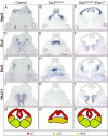
Activation of the Shh signaling pathway rescues dorsoventral patterning defects in the Six3neo/neo telencephalon. E12.5 wild-type, Six3neo/neo and Six3neo/neo;Ptch1+/– embryos were coronally sectioned and analyzed for possible alterations in dorsoventral patterning of the telencephalon. Ngn2 expression is restricted to the dorsal telencephalon (DT) in control (A), Six3neo/neo (B) and Six3neo/neo;Ptch1+/– (C) embryos. (D) Dlx2, a marker for the ventral telencephalon, is expressed in both the lateral ganglionic eminence (LGE) and medial ganglionic eminence (MGE) of control embryos. A single Dlx2-positive lobe is seen in Six3neo/neo embryos (E). Normal separation of the ganglionic eminences is restored in Six3neo/neo;Ptch1+/– embryos (F). Ebf1 expression labels the LGEs in control embryos (G). The single ganglionic eminence in Six3neo/neo embryos is positive for Ebf1 (H). Proper separation of the LGE lobes is rescued in Six3neo/neo;Ptch1+/– embryos (I). Nkx2.1 is expressed in the two MGEs of control embryos (J) but is absent in the Six3neo/neo embryos (K). Normal expression of Nkx2.1 is restored in Six3neo/neo;Ptch1+/– embryos (L). n=4 control; n=5 Six3neo/neo; n=4 Six3neo/neo;Ptch1+/−. Schematic representation of the various telencephalic domains in control (M), Six3neo/neo (N) and Six3neo/neo; Ptch1+/− (O) embryos.
Fig. 3.
Fig. 3.
Morphogenesis and dorsoventral patterning are defective in the Six3neo/– telencephalon. E14.5 control and Six3neo/– brains were coronally sectioned and in situ hybridization was performed for various markers at the rostral (A-H) and caudal (K-R) levels. At the rostral level, two well-separated cerebral vesicles are present in control embryos (A). Ngn2 expression is also restricted to the ventricle of the cerebral cortex (CC) (A), and Gad67, Ebf1 and Nkx2.1 are expressed in the ventral region (C,E,G). However, a single vesicle is observed in Six3neo/– embryos, and it is much smaller than that of the control telencephalon. In these mutant embryos, Ngn2 expression also expands ventrally to cover the entire ventricle (B) at the expense of Gad67, Ebf1 and Nkx2.1, which are no longer expressed in this region (D,F,H). These results suggest that the forebrain of Six3neo/– embryos is completely dorsalized, as schematically summarized (I,J). (K) In control embryos at the caudal level, Ngn2 is expressed along the ventricle of the CC and the dorsal thalamus, but excluded from the caudal ganglionic eminence (CGE). (L) In Six3neo/– embryos, Ngn2 expression is abnormally expanded ventrally. In control (M) and Six3neo/– (N) embryos, Prox1 expression is seen in the dentate gyrus neuroepithelium (DNE) of the hippocampal region (black arrows), and the dorsal thalamus (red arrows). Wnt8b expression labels the fimbria neuroepithelium (FNE) of the hippocampal region (O,P, black arrows) and eminentia thalami (EmT) (O,P, red arrows). Ttr is expressed in the choroid plexus (CP) (Q,R, arrows). The DNE, FNE and CP are localized to the surface of the brain (N,P,R, respectively). (S,T) Summary of the expression analysis results. Di, diencephalon; ST, septum. n=3 control; n=5 Six3neo/−.
Fig. 4.
Fig. 4.
Elevated Shh signaling fails to rescue brain defects in Six3neo/– embryos. Coronal sections of E12.5 control and Six3neo/–;Ptch1+/– forebrains. In controls, the dorsal telencephalic marker Ngn2 is excluded from the caudal ganglionic eminence (CGE) (A,C, arrows), which is identified by Dlx2 expression (C). By contrast, Ngn2 is expressed throughout the telencephalic neural epithelium in Six3neo/–;Ptch1+/– embryos (B, arrows). Rostral-expanded diencephalon separates the telencephalic vesicles (B, red arrow). Wnt8b labels the dorsal midline (DM) of the telencephalon and the eminentia thalami (EmT) in control and mutant samples (E,F, arrows). The presence of the EmT is confirmed by the expression of Lim1 (G,H, black arrows). Lim1 is also a marker for the zona limitans intrathalamica (ZLI), the ventral thalamus (VTh) and the hypothalamus (HTh). Compared with that in wild-type embryos (G, red arrows), the region of Lim1 expression is slightly smaller and located more ventrally in Six3neo/–;Ptch1+/– embryos (H, red arrows). Similarly, Dlx2, another marker for the VTh and HTh, is expressed more ventrally and in a smaller area in the mutant embryos (D, red arrows) than in controls (C, red arrows). The presence of the HTh is marked by Nkx2.1 expression in control and mutant embryos (I,J). Ngn2 and Gbx2 are expressed in the dorsal thalamus (DTh), and their expression domain is enlarged in the mutant brain (B,L) compared with that in controls (A,K). (M,N) Summary of the expression analysis results. DT, dorsal telencephalon; PT, pretectum. n=3 control; n=3 Six3neo/−;Ptch1+/−.
Fig. 5.
Fig. 5.
Six3 regulates Foxg1 and Wnt8b expression in a dosage-dependent manner. (A-C) Frontal view of E8.5 control (A), Six3neo/neo (B) and Six3neo/– (C) embryos showing Wnt8b expression. Wnt8b labels the forebrain and midbrain boundary in control and Six3neo/neo embryos (A,B, arrowheads). By contrast, Wnt8b expression expands into the rostral end of the anterior neural plate in Six3neo/– embryos (C, arrowheads). (D-F) Lateral view of E9.0 control (D), Six3neo/neo (E) and Six3neo/– (F) embryos showing Foxg1 expression. Foxg1 is expressed in the telencephalon of control and Six3neo/neo embryos (D,E, arrowhead). By contrast, Foxg1 expression is dramatically downregulated in Six3neo/– embryos (F, arrowhead). n=3 for A-C,E; n=4 for D,F.
Fig. 6.
Fig. 6.
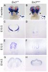
Foxg1 expression is downregulated in Six3 mutant embryos. Foxg1 expression during development (A-H). Frontal view of 7-somite embryos (A,B). Foxg1 is expressed in the anterior neuroectoderm (ANE) (black arrowheads) and the surface ectoderm beneath the neural plate (red arrowheads) of control embryos (A). Foxg1 expression in the ANE is significantly reduced in Six3neo/– embryos (black arrowheads); however, Foxg1 levels in the surface ectoderm are comparable to that of controls (B, red arrowheads). Insets illustrate the transverse sections of the stained embryos at the level indicated by the white dotted line. (C-F) Foxg1 expression in the frontal telencephalon at E10.5 and E12.5 is lower in Six3neo/– embryos than in Six3+/+ littermates. (G,H) Foxg1 expression levels appear normal in coronal sections of telencephalon of E14.5 Six3neo/– embryos.
Fig. 7.
Fig. 7.
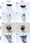
Shh is not necessary to regulate Foxg1 expression in the telencephalon. E8.5 control (A,C,E,G) and Shh–/– (B,D,F,H) embryos were analyzed for the expression of Six3 (A,B) and its target genes Wnt1 (C,D), Foxg1 (E,F) and Wnt8b (G,H). Although the midline remains fused in Shh–/– embryos (F,H, arrows), no obvious changes in gene expression are observed. Six3 and Foxg1 are expressed in the ANE of Shh–/– embryos (B,F) at a comparable level to that of control embryos (A,E). Consistently, Wnt1 and Wnt8b expression is restricted to the forebrain/midbrain boundary of control (C,G, arrowheads) and mutant (D,H, arrowheads) embryos. A, n=4; B, n=3.
Fig. 8.
Fig. 8.
Six3 directly regulates Foxg1 expression. (A) Alignment of the Foxg1 DNA sequences in different vertebrates reveals several conserved regions. The putative Six3 binding site identified in one conserved region 1.5 kb upstream of the 5′ UTR is indicated by the arrow and nucleotide sequence in red. (B) DNA enrichment after Six3 immunoprecipitation of Foxg1 DNA isolated from heads and trunks of 5- to 16-somite staged embryos. Gapdh promoter primers were used as a negative control. All samples were normalized with the input. (C,D) Six3 binding to the Foxg1 enhancer element as shown by activation of luciferase (Foxg1-Luc). (C) Luciferase activity was augmented significantly (***P<0.0001, ANOVA) as the concentration of the Six3 coding vector was increased. (D) The activator form of Six3 (Six3-VP16) induced a more significant increase (3.6-fold, **P<0.001, t-test) in luciferase activity than the normal reporter. No changes in luciferase activity were observed in the presence of a repressor form of Six3 (Six3-enR). Error bars indicate s.d.
Fig. 9.
Fig. 9.
Model for the dosage effect of Six3 during normal and pathological forebrain development. (A) Six3 serves multiple roles during mammalian forebrain development, as a transcriptional activator and repressor of major signaling pathways. (B) Dosage effect of Six3 in the pathogenesis of HPE. Depending on the amount of functional Six3 in the embryo, the brain phenotype can range from normal, semilobar HPE, alobar HPE to aprosencephaly/atelencephaly.

References

    1. Aguiar D. P., Sghari S. and Creuzet S. (2014). The facial neural crest controls fore- and midbrain patterning by regulating Foxg1 expression through Smad1 activity. Development 141, 2494-2505. 10.1242/dev.101790 - DOI - PubMed
    1. Beccari L., Conte I., Cisneros E. and Bovolenta P. (2012). Sox2-mediated differential activation of Six3.2 contributes to forebrain patterning. Development 139, 151-164. 10.1242/dev.067660 - DOI - PubMed
    1. Billington C. J. Jr, Schmidt B., Marcucio R. S., Hallgrimsson B., Gopalakrishnan R. and Petryk A. (2015). Impact of retinoic acid exposure on midfacial shape variation and manifestation of holoprosencephaly in Twsg1 mutant mice. Dis. Model. Mech. 8, 139-146. 10.1242/dmm.018275 - DOI - PMC - PubMed
    1. Carl M., Loosli F. and Wittbrodt J. (2002). Six3 inactivation reveals its essential role for the formation and patterning of the vertebrate eye. Development 129, 4057-4063. - PubMed
    1. Carlin D., Sepich D., Grover V. K., Cooper M. K., Solnica-Krezel L. and Inbal A. (2012). Six3 cooperates with Hedgehog signaling to specify ventral telencephalon by promoting early expression of Foxg1a and repressing Wnt signaling. Development 139, 2614-2624. 10.1242/dev.076018 - DOI - PMC - PubMed

Publication types

MeSH terms

LinkOut - more resources