Skip to main page content
U.S. flag

An official website of the United States government

Dot gov

The VSports app下载. gov means it’s official. Federal government websites often end in . gov or . mil. Before sharing sensitive information, make sure you’re on a federal government site. .

Https

The site is secure V体育官网. The https:// ensures that you are connecting to the official website and that any information you provide is encrypted and transmitted securely. .

. 2016 Oct 13:7:13096.
doi: 10.1038/ncomms13096.

Macrophage-derived extracellular vesicle-packaged WNTs rescue intestinal stem cells and enhance survival after radiation injury

Affiliations

"VSports手机版" Macrophage-derived extracellular vesicle-packaged WNTs rescue intestinal stem cells and enhance survival after radiation injury

Subhrajit Saha et al. Nat Commun. .

Abstract

WNT/β-catenin signalling is crucial for intestinal homoeostasis. The intestinal epithelium and stroma are the major source of WNT ligands but their origin and role in intestinal stem cell (ISC) and epithelial repair remains unknown. Macrophages are a major constituent of the intestinal stroma VSports手机版. Here, we analyse the role of macrophage-derived WNT in intestinal repair in mice by inhibiting their release using a macrophage-restricted ablation of Porcupine, a gene essential for WNT synthesis. Such Porcn-depleted mice have normal intestinal morphology but are hypersensitive to radiation injury in the intestine compared with wild-type (WT) littermates. Porcn-null mice are rescued from radiation lethality by treatment with WT but not Porcn-null bone marrow macrophage-conditioned medium (CM). Depletion of extracellular vesicles (EV) from the macrophage CM removes WNT function and its ability to rescue ISCs from radiation lethality. Therefore macrophage-derived EV-packaged WNTs are essential for regenerative response of intestine against radiation. .


Figures
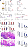
Figure 1
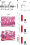
Figure 1. Deletion of Porcn in macrophages radio-sensitizes mice to lethal doses of WBI.
(a) Kaplan–Meier survival analysis of Csf1r.iCre;Porcnfl/fl and WT mice exposed to WBI (9.4–12.4 Gy). Csf1r.iCre;Porcnfl/fl mice show reduced survival following lethal doses of WBI (11.4–12.4 Gy WBI) with 100% mortality within 7–12 days of radiation exposure, compared with WT that have 60% survival beyond 15 days post WBI (P<0.0001, P<0.004 Log-rank (Mantel–Cox) test; n=15 per group). No significant survival difference between Csf1r.iCre;Porcnfl/fl or WT littermate mice was observed with the irradiation dose of 9.4–10.4 Gy WBI (n=15 per group). (b) Body weight of mice at post irradiation time points (11.4 Gy and 12.4 Gy WBI). (c) Complete blood count (CBC) analysis. CBC measurements for Csf1r.iCre;Porcnfl/fl and WT mice exposed to 0 Gy/11.4 Gy/12.4 Gy WBI. Blood samples were drawn at 5 days post irradiation (n=3 per group). (d) HE staining of jejunum section from Csf1r.iCre;Porcnfl/fl and WT mice exposed to 0 Gy/11.4 Gy WBI at day 5 post irradiation. No significant differences in crypt villus structure were noted in non-irradiated Csf1r.iCre;Porcnfl/fl mice compared with WT mice. Note, shortening of crypt depth as well as loss of crypts in Csf1r.iCre;Porcnfl/fl mice exposed to irradiation (n=3 per group) compared with WT mice. (e) Histogram demonstrating crypt depth (μM) from jejunal sections of Csf1r.iCre;Porcnfl/fl and WT mice exposed to 0 or 11.4 Gy WBI. Both WT and Csf1r.iCre;Porcnfl/fl mice exposed to irradiation showed reduction in crypt depth at day 5 post irradiation compared with un-irradiated control WT mice *P<4.66E−05, Csf1r.iCre;Porcnfl/fl mice *P<9.63E−08. However, loss of crypt depth was significantly greater in Csf1r.iCre;Porcnfl/fl mice compared with WT mice *P<8.84E−07 unpaired t-test, two-tailed. (f) Histogram demonstrating the number of crypts mm−1 from jejunal section of Csf1r.iCre;Porcnfl/fl and WT mice exposed to 0 or 11.4 Gy WBI. Both WT and Csf1r.iCre;Porcnfl/fl mice exposed to irradiation showed reduction in crypt number at day 5 post irradiation compared with un-irradiated control WT mice *P<0.0008, Csf1r.iCre;Porcnfl/fl mice *P<1E−08. However, loss of crypt was significantly greater in Csf1r.iCre;Porcnfl/fl mice compared with WT mice *P<9.79E−09 unpaired t-test, two-tailed. (g) Histogram demonstrating villi length from jejunal section of Csf1r.iCre;Porcnfl/fl and WT mice exposed to 0 or 11.4 Gy WBI. Both WT and Csf1r.iCre;Porcnfl/fl mice exposed to irradiation showed reduction in crypt number at day 5 post irradiation compared with un-irradiated control WT mice*P<0.0006, Csf1r.iCre;Porcnfl/fl mice *P<8.9E−08. However, loss of villi length was significantly greater in Csf1r.iCre;Porcnfl/fl mice compared with WT mice *P<7.98E−09 unpaired t-test, two-tailed.
Figure 2
Figure 2. Deletion of Porcn in macrophages sensitizes mice against lethal dose of AIR.
(a) Schematic diagram demonstrating the AIR exposure field for Csf1r.iCre;Porcnfl/fl and WT mice. A 2 cm area of the mice containing the GI was irradiated (irradiation field), thus shielding the upper thorax, head and neck as well as lower and upper extremities, protecting a significant portion of the bone marrow, thus inducing predominantly RIGS. (b) Kaplan–Meier survival analysis. Csf1r.iCre;Porcnfl/fl mice have reduced survival against a lethal dose (18 Gy) of abdominal radiation compared with WT mice (n=10 per group; P<0.009 Log-rank (Mantel–Cox) test). (c) HE staining of jejunum sections from Csf1r.iCre;Porcnfl/fl and WT mice exposed to 18 Gy AIR. Mice were killed and jejunum was collected at day 5 post irradiation. Csf1r.iCre;Porcnfl/fl mice showed more villi denudation and crypt loss compared with WT littermate mice at day 5 post irradiation (n=3 per group). (d) Histogram showing crypt depth (μM) in jejunal sections of Csf1r.iCre;Porcnfl/fl and WT mice exposed to 0 Gy or 18 Gy AIR. Csf1r.iCre;Porcnfl/fl mice exposed to AIR had significantly higher reduction in crypt depth compared with WT *P<9.64E−08 unpaired t-test, two-tailed. (e) Histogram showing number of crypts mm−1 in jejunal sections of Csf1r.iCre;Porcnfl/fl and WT mice exposed to 0 Gy or 18 Gy AIR. Csf1r.iCre;Porcnfl/fl mice exposed to AIR has significantly higher reduction in crypt number compared with WT *P<8.77E−09 unpaired t-test, two-tailed. (f) Histogram showing villus length in jejunal sections of Csf1r.iCre;Porcnfl/fl and WT mice exposed to 0 or 18 Gy AIR. Csf1r.iCre;Porcnfl/fl mice exposed to AIR has significantly higher reduction in villi length compared with WT *P<8E−07 unpaired t-test, two-tailed.
Figure 3
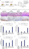
Figure 3. CM from WT but not from Porcn-null BMMΦ inhibits RIGS in Csf1r.iCre;Porcnfl/fl mice exposed to lethal dose of WBI.
(a) Experimental design and Kaplan–Meier survival analysis of Csf1r.iCre;Porcnfl/fl mice (n=10 per group) receiving CM (i.v.) derived from WT/Porcn-null BMMΦ at 1 and 48 h post WBI (11.2 Gy). Mice receiving WT BMMΦ CM showed 40% survival (P<0.003 Log-rank (Mantel–Cox) test) beyond 25 days compared with mice receiving Porcn-null BMMΦ CM or untreated mice where 100% of mice died within 12 days after irradiation. (b) Representative HE and BrdU immunohistochemistry of mice jejunal sections. Note, restitution on crypt villus structure with the increase in crypt cell proliferation in Csf1r.iCre;Porcnfl/fl mice receiving WT BMMΦ CM compared with Porcn-null BMMΦ CM treatment. (c) The proliferation rate was calculated as the percentage of BrdU-positive cells over the total number of cells in each crypt and displayed as bar diagrams. Crypt cell proliferation rate in irradiated mice: WT BMMΦ CM versus Porcn-null BMMΦ CM treatment group *P<2.21E−07 (n=5 per group), WT BMMΦ CM versus αMEM growth medium treatment group *P<2.69E−07 (n=5 per group; unpaired t-test, two-tailed). (d) Histogram showing number of crypts mm−1 in jejunal sections of Csf1r.iCre;Porcnfl/fl mice. Irradiated Csf1r.iCre;Porcnfl/fl mice receiving WT BMMΦ CM showed less crypt loss compared with mice receiving Porcn-null BMMΦ CM or αMEM growth medium (*P<6.86E−09 and *P<6.74E−08 unpaired t-test, two-tailed). (e) Histogram showing villus length in jejunal sections of Csf1r.iCre;Porcnfl/fl mice. Irradiated Csf1r.iCre;Porcnfl/fl mice receiving WT BMMΦ CM showed less reduction in villi length compared with mice receiving Porcn-null BMMΦ CM or αMEM growth medium (*P<3.74E−06 and *P<3.60E−06 unpaired t-test, two-tailed). (f) Histogram demonstrating serum dextran level in Csf1r.iCre;Porcnfl/fl mice. Mice receiving WT BMMΦ CM showed a lower serum dextran level thereby suggesting restitution of epithelial integrity compared with mice receiving Porcn-null BMMΦ CM or αMEM growth medium (*P<0.006 and *P<0.009 respectively; n=3 per group). Untreated mice also showed a lower serum dextran level compared with irradiated mice receiving αMEM growth medium (P<0.003) or Porcn-null BMMΦ CM (P<0.001; unpaired t-test, two-tailed).
Figure 4
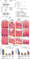
Figure 4. WNTs in BMMΦ CM are required to inhibit RIGS in Csf1r.iCre;Porcnfl/fl mice exposed to AIR.
(a) Experimental design for partial body irradiation. Mice exposed to AIR were treated with WT or Porcn-null BMMΦ CM at 1 and 24 h post exposure. (b) Kaplan–Meier survival analysis of Csf1r.iCre;Porcnfl/fl mice (n=10 per group) receiving CM (500 μl per mice i.v.) derived from WT or Porcn-null BMMΦ at 1 h and 48 h post AIR (18 Gy). Mice receiving WT BMMΦ CM showed 60% survival beyond 20 days compared with mice receiving Porcn-null BMMΦ CM or EV-depleted WT BMMΦ CM, where 100% of mice died within 12 days after irradiation (P<0.002 and P<0.003, respectively, Log-rank (Mantel–Cox) test). Reagent used for chemical depletion of EV did not confer any toxicity to normal mice. (c) HE-stained representative transverse sections of duodenum, jejunum and ileum from Csf1r.iCre;Porcnfl/fl mice (n=3 per group). Note, restitution on crypt villus structure in irradiated Csf1r.iCre;Porcnfl/fl mice receiving WT BMMΦ CM. However, treatment with Porcn-null BMMΦ CM or αMEM growth medium showed significant loss of crypts along with villi denudation. (d) Histogram demonstrating number of crypts mm−1 in duodenal, jejuna, and illeul sections of Csf1r.iCre;Porcnfl/fl mice. Irradiated Csf1r.iCre;Porcnfl/fl mice receiving WT BMMΦ CM showed less crypt loss compared with mice receiving Porcn-null BMMΦ CM (Duodenum *P<6.86E−07, Jejunum **P<7.89E−08 and Ileum ***P<8.16E−08) or αMEM growth medium (Duodenum *P<7.92E−08, Jejunum **P<8.26E−07 and Ileum ***P<8.96E−09; unpaired t-test, two-tailed). (e) Histogram demonstrating villus length in duodenal, jejunal and illeul sections of Csf1r.iCre;Porcnfl/fl mice. Irradiated Csf1r.iCre;Porcnfl/fl mice receiving WT BMMΦ CM showed less reduction in villus length compared with mice receiving Porcn-null BMMΦ CM (Duodenum *P<3.64E−06, Jejunum **P<2.86E−06 and Ileum ***P<2.16E−05) or αMEM growth medium (Duodenum *P<4.21E−06, Jejunum **P<3.16E−08 and Ileum ***P<2.88E−05; unpaired t-test, two-tailed).
Figure 5
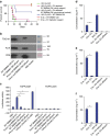
Figure 5. Presence of EV-packaged WNT in BMMΦ CM is critical for radio-mitigating function.
(a) Kaplan–Meier survival analysis of WT mice (n=10 per group) receiving CM/EV-depleted CM (500 μl per mice i.v.) derived from WT BMMΦ at 1 h and 48 h post 18.5 Gy AIR. Mice receiving WT BMMΦ CM showed 60% survival beyond 30 days compared with mice receiving EV-depleted WT BMMΦ CM (using reagent (P<0.004) or conventional method (P<0.002)) or untreated mice (P<0.0009; Log-rank (Mantel–Cox) test) where 80–100% of mice died within 12 days after irradiation (n=10 per group). Reagent (500 μl per mice i.v.) used for chemical depletion of exosome did not confer any toxicity to normal mice. (b) Immunoblot to detect exosomal markers TSG101, ALIX and CD9 in EV from WT BMMΦ CM or Porcn-null BMMΦ CM or respective effluents. EV from WT BMMΦ CM and from Porcn-null BMMΦ CM showed the presence of EV markers. However, EV markers were not detected in effluents. (c) TCF/LEF reporter assay. HEK293 cells having TCF/LEF luciferase reporter construct were treated with EV (prepared with the conventional method) from WT or Porcn-null BMMΦ CM or effluents or LiCl. Treatment with EV (100 μg ml−1) from WT BMMΦ CM showed higher Luciferase activity compared with EV (100 μg ml−1) from Porcn-null BMMΦ CM (P<0.0002) and effluent from WT BMMΦ CM or Porcn-null BMMΦ CM (P<0.0001 and P<0.0002 respectively; unpaired t-test, two-tailed). (df) ELISA to detect WNT5a, 6 and 9a in EVs from WT or Porcn-null BMMΦ CM and effluents. Presence of WNT5a, WNT6 and WNT9a were detected in EVs from WT BMMΦ CM (Cre− EV) but not in EVs from Porcn-null BMMΦ CM (Cre+ EV; P<0.0002; P<2.78E−05 and P<3.26E−06 respectively; unpaired t-test, two-tailed). WNT5a, WNT6 and WNT9a were also absent in effluents derived from WT or Porcn-null BMMΦ CM. Recombinant WNT5a, WNT6 and WNT9a were used as positive control respectively.
Figure 6
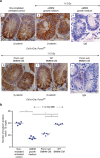
Figure 6. Macrophage-derived WNTs induce β-catenin activity in irradiated crypts.
(a) Representative microscopic images (× 60 magnification) of jejunal sections immunostained with anti β-catenin antibody to determine β-catenin nuclear localization in Csf1r.iCre;Porcnfl/fl mice. Nucleus stained with haematoxylin. Irradiated Csf1r.iCre;Porcnfl/fl mice receiving WT BMMΦ CM (i.v.) showed more nuclear β-catenin staining (dark brown; indicated with arrows) at the base of the crypt compared with mice receiving Porcn-null BMMΦ CM (i.v.) or αMEM growth medium (ii; nucleus stained blue). Fig iii and vi are representative IgG controls indicating lack of staining and thus showing specificity for the anti β-catenin antibody. (b) Nuclear β-catenin count: each data point is the average of the number of β-catenin-positive nucleus from 15 crypts per field, 5 fields per mice. Number of β-catenin-positive nucleus in irradiated Csf1r.iCre;Porcnfl/fl mice receiving WT BMMΦ CM is higher compared with Porcn-null BMMΦ CM (*P<1E−04 ) or αMEM growth medium (*P<1E−04). Treatment with Porcn-null BMMΦ CM and αMEM growth medium following irradiation showed significantly fewer β-catenin-positive nuclei than the non-irradiated control (*P<1.2E−04 and *P<1E−04 respectively; unpaired t-test, two-tailed).
Figure 7
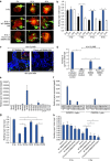
Figure 7. CM from WT BMMΦ rescued the Lgr5+ve ISC population in both in vivo and in vitro.
(a) Two-photon microscopic images of organoids from Lgr5/GFP-IRES-Cre-ERT2 knock-in mouse irradiated and then treated with CM. CM from WT BMMΦ (Cre-) rescued the LGR5+ve (GFP+ve cells) ISC population from radiation injury as indicated by arrow. GFP+ve cells disappeared in organoids treated with/without Porcn-null BMMΦ CM (Cre+) within 48 h of exposure to 8 Gy irradiation. Scale bar=50 μM. (b) Histograms demonstrating the effect of Cre±CM treatment on crypt organoid growth following irradiation. Treatment groups: 4 Gy versus 4 Gy+Cre−CM (*P<0.013), 6 Gy versus 6 Gy+Cre−CM (*P<0.001), 8 Gy versus 8 Gy+Cre−CM (*P<0.004; unpaired t-test, two-tailed). (c) Representative images of jejunal sections demonstrating the presence of GFP+ve Lgr5+ve ISCs (arrow) in Lgr5/GFP-IRES-Cre-ERT2 knock-in mice receiving WT BMMΦ CM. Note, the absence of GFP+ve cells in mice receiving Porcn-null BMMΦ CM. Nuclei are stained with DAPI. Top panel Inset: phalloidin (red) staining to show localization of cell membrane. (d) Histograms demonstrating the number of GFP+veLgr5+ve ISCs/crypt in jejunal sections from Lgr5/GFP-IRES-Cre-ERT2 knock-in mice exposed to irradiation and then treated with Porcn-null or WT BMMΦ CM. Irradiated mice receiving WT BMMΦ CM showed higher numbers of GFP+ve cells compared with mice receiving Porcn-null BMMΦ CM (*P<0.0002) or αMEM (*P<1.74858E−06; unpaired t-test, two-tailed). (e) qPCR analysis of RNA from WT BMMΦ demonstrated mRNA expression of Wnt5a, Wnt6 and Wnt9a. (f) qPCR analysis of intestinal macrophages RNA from WT mice. (g) Organoids were exposed to irradiation (6 Gy) and treated with Porcn-null BMMΦ CM supplemented with Wnt5a, Wnt6, Wnt9a and Wnt5b (1 μg ml−1). Organoid survival was improved with treatment of canonical WNT ligands WNT5a (*P<0.009), WNT6 (*P<0.0001) and WNT9a (*P<8.99662E−05) compared with irradiated control. However, treatment with non-canonical WNT5b failed to rescue organoids from radiation lethality (WNT6 versus WNT5b *P<0.0001; WNT9a versus WNT5b *P<0.0001; unpaired t-test, two-tailed). (h) Histogram demonstrating the effect of EVs from WT/Porcn-null BMMΦ CM on crypt organoid growth following irradiation. Treatment groups: 6 Gy (control) versus 6 Gy+WT BMMΦ CM EV (*P<0.009), 6 Gy+ WT BMMΦ CM EV versus 6 Gy+Porcn-null BMMΦ CM EV (*P<0.004), 6 Gy+WT BMMΦ CM EV versus 6 Gy+WT BMMΦ CM effluent (*P<0.0002), 6 Gy+WT BMMΦ CM EV versus Porcn-null BMMΦ CM effluent (*P<0.0001; unpaired t-test, two-tailed).

References

    1. Kabiri Z. et al.. Stroma provides an intestinal stem cell niche in the absence of epithelial Wnts. Development 141, 2206–2215 (2014). - PubMed
    1. Saha S. et al.. Bone marrow stromal cell transplantation mitigates radiation-induced gastrointestinal syndrome in mice. PLoS ONE 6, e24072 (2011). - "VSports最新版本" PMC - PubMed
    1. Smith P. D. et al.. Intestinal macrophages and response to microbial encroachment. Mucosal. Immunol. 4, 31–42 (2011). - PMC - PubMed
    1. Pull S. L., Doherty J. M., Mills J. C., Gordon J. I. & Stappenbeck T. S. Activated macrophages are an adaptive element of the colonic epithelial progenitor niche necessary for regenerative responses to injury. Proc. Natl Acad. Sci. USA 102, 99–104 (2005). - PMC - PubMed
    1. Saha S. et al.. TLR9 agonist protects mice from radiation-induced gastrointestinal syndrome. PLoS ONE 7, e29357 (2012). - PMC - PubMed

MeSH terms