Skip to main page content
U.S. flag

An official website of the United States government

Dot gov

The . gov means it’s official. Federal government websites often end in . gov or VSports app下载. mil. Before sharing sensitive information, make sure you’re on a federal government site. .

Https

The site is secure. The https:// ensures that you are connecting to the official website and that any information you provide is encrypted and transmitted securely V体育官网. .

. 2016 Dec 2;15(12):4436-4451.
doi: 10.1021/acs.jproteome.6b00521. Epub 2016 Oct 18.

Determining the Mitochondrial Methyl Proteome in Saccharomyces cerevisiae using Heavy Methyl SILAC

Affiliations

VSports最新版本 - Determining the Mitochondrial Methyl Proteome in Saccharomyces cerevisiae using Heavy Methyl SILAC

"V体育安卓版" Katelyn E Caslavka Zempel et al. J Proteome Res. .

Abstract

Methylation is a common and abundant post-translational modification. High-throughput proteomic investigations have reported many methylation sites from complex mixtures of proteins. The lack of consistency between parallel studies, resulting from both false positives and missed identifications, suggests problems with both over-reporting and under-reporting methylation sites. However, isotope labeling can be used effectively to address the issue of false-positives, and fractionation of proteins can increase the probability of identifying methylation sites in lower abundance. Here we have adapted heavy methyl SILAC to analyze fractions of the budding yeast Saccharomyces cerevisiae under respiratory conditions to allow for the production of mitochondria, an organelle whose proteins are often overlooked in larger methyl proteome studies. We have found 12 methylation sites on 11 mitochondrial proteins as well as an additional 14 methylation sites on 9 proteins that are nonmitochondrial. Of these methylation sites, 20 sites have not been previously reported. This study represents the first characterization of the yeast mitochondrial methyl proteome and the second proteomic investigation of global mitochondrial methylation to date in any organism VSports手机版. .

Keywords: MudPIT; heavy methyl SILAC; methyl proteome; methylation; mitochondria; protein arginine methylation; protein lysine methylation; yeast. V体育安卓版.


Figures

Figure 1
Figure 1
Improved enrichment of the deuterated methionine label into proteins of S. cerevisiae strains with a deletion in the MET6 gene encoding the cobalamin-independent methionine synthase. As described in "Materials and Methods", wild type (WT) and met6Δ strains were heavy-labeled, mitochondrial ribosomal proteins were isolated, digested with trypsin/Lys-C and analyzed by LC-MS/MS. For each peptide identified, the ratio of the areas of the extracted ion chromatograms for each labeled and unlabeled peptide pair was calculated to obtain a fractional enrichment of L-[methyl-D3]-methionine (Met-D3).
Figure 2
Figure 2
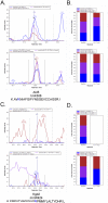
Examples of good and poor methylated peptide identifications in MS1 ion chromatograms. (A) Extracted precursor ion chromatograms for light (top) and heavy (bottom) peptides containing a candidate trimethyl K308 on Ald5. The zero, +1 and +2 isotopes are shown in blue, magenta, and red, respectively. The methylated residue is highlighted in blue in the peptide sequence. (B) Corresponding peak areas of both experimental and theoretical precursor ions for the heavy and light peptide containing this modification. (C, D) Extracted precursor ion chromatograms for light and heavy peptides containing an identified, but unlikely monomethyl R424 on Kgd2, analyzed as in panels A and B above. These chromatograms are from the mitochondrial fraction.
Figure 3
Figure 3
Overlap of methylated proteins between cytoplasmic ribosomal proteins (blue), mitochondrial ribosomal proteins (pink), and mitochondrial protein fractions (green). The four methylated proteins found in all three protein fractions are listed. Known methylated proteins are bolded and italicized.
Figure 4
Figure 4
Confirmation of known methylation sites on EF1A. Extracted precursor ion chromatograms for light (top) and heavy (bottom) peptides containing (A) monomethyl K30, (B) trimethyl K79, and (C) monomethyl K390. The zero, +1 and +2 isotopes are shown in blue, magenta, and red, respectively. The methylated residue is highlighted in blue in the peptide sequence. Chromatograms are representative of three replicates from the cytoplasmic ribosomal protein fraction.
Figure 5
Figure 5
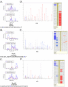
Novel methylated peptides found in mitochondrial ribosomal protein fraction. Extracted precursor ion chromatograms for light (top) and heavy (bottom) peptides containing (A) dimethyl K56 on Mnp1, (B) monomethyl R204 on Mrpl4, and (C) monomethyl K186 on Mrpl40. The zero, +1 and +2 isotopes are shown in blue, magenta, and red, respectively. The methylated residue is highlighted in blue in the peptide sequence. Fragmentation patterns of product ions for (D) Mnp1, (E) Mrpl4, and (F) Mrpl40 methyl peptides. Methylated residue is highlighted in yellow in the peptide sequence.
Figure 6
Figure 6
Novel methylation of Ssa2/4 in all three protein fractions. Extracted precursor ion chromatograms for light (top) and heavy (bottom) peptides containing (A) monomethyl K421/2 on Ssa2/4. The zero, +1 and +2 isotopes are shown in blue, magenta, and red, respectively. The methylated residue is highlighted in blue in the peptide sequence. (B) Fragmentation patterns of product ions for Ssa2/4 methyl peptide. Methylated residue is highlighted in yellow in the peptide sequence. Chromatograms and fragmentations are representative of three replicates from the three protein fractions.
Figure 7
Figure 7
Sequence similarity between identified methylated residues. (A) Logo of sequence of amino acids 10 residues before and after identified methylated lysine. (B) Zoomed in view of logo, looking at the first bit of identified methylated lysine sequences. (C) Logo of sequence of amino acids 10 residues before and after identified methylated arginine.
Figure 8
Figure 8
Overlap of methylated proteins identified from the literature and curated from Uniprot. (A) Comparison between “Our study” (red), “MILS 2015” (green), and “FIND MOD 2010” (blue). (B) Comparison between known studies “Heavy methyl SILAC 2015” (red), “MILS 2015” (green), and “FIND MOD 2010” (blue). (C) Comparison to “Uniprot Curated 2015”, “MILS 2015” (green), and “FIND MOD 2010” (blue). (D) Comparison to “Uniprot Curated 2015”, “Our study” (green), and “Heavy methyl SILAC 2015” (blue). “Our study” was candidate proteins identified in this work. “FIND MOD 2010” proteins are from Pang et al., 2010, “MILS 2015” proteins are from Wang et al., 2015, “Heavy methyl SILAC 2015” proteins are from Plank et al., 2015, and “Uniprot Curated 2015” proteins were manually curated from Uniprot in March 2015.

VSports - References

    1. Walsh C. Posttranslational Modification of Proteins: Expanding Nature’s Inventory. Roberts and Company Publishers; 2006. Protein Methylation; pp. 121–150.
    1. Clarke SG. Protein methylation at the surface and buried deep: thinking outside the histone box. Trends Biochem. Sci. 2013;38(5):243–252. - PMC - PubMed
    1. Carlson SM, Gozani O. Emerging technologies to map the protein methylome. J. Mol. Biol. 2014;426(20):3350–3362. - PMC - PubMed
    1. Lanouette S, Mongeon V, Figeys D, Couture J-F. The functional diversity of protein lysine methylation. Mol. Syst. Biol. 2014;10(4):724. - PMC - PubMed
    1. Biggar KK, Li SS-C. Non-histone protein methylation as a regulator of cellular signalling and function. Nat. Rev. Mol. Cell Biol. 2015;16(1):5–17. - PubMed