"V体育2025版" Skip to main page content
U.S. flag

An official website of the United States government

Dot gov

The . gov means it’s official. Federal government websites often end in . gov or . mil. Before sharing sensitive information, make sure you’re on a federal government site VSports app下载. .

Https

The site is secure. The https:// ensures that you are connecting to the official website and that any information you provide is encrypted and transmitted securely V体育官网. .

. 2016 Oct 4;7(40):64527-64542.
doi: 10.18632/oncotarget.11743.

VSports app下载 - Hypoxia-inducible factors regulate pluripotency factor expression by ZNF217- and ALKBH5-mediated modulation of RNA methylation in breast cancer cells

Affiliations

Hypoxia-inducible factors regulate pluripotency factor expression by ZNF217- and ALKBH5-mediated modulation of RNA methylation in breast cancer cells

Chuanzhao Zhang et al. Oncotarget. .

Abstract (V体育ios版)

Exposure of breast cancer cells to hypoxia increases the percentage of breast cancer stem cells (BCSCs), which are required for tumor initiation and metastasis, and this response is dependent on the activity of hypoxia-inducible factors (HIFs). We previously reported that exposure of breast cancer cells to hypoxia induces the ALKBH5-mediated demethylation of N6-methyladenosine (m6A) in NANOG mRNA leading to increased expression of NANOG, which is a pluripotency factor that promotes BCSC specification. Here we report that exposure of breast cancer cells to hypoxia also induces ZNF217-dependent inhibition of m6A methylation of mRNAs encoding NANOG and KLF4, which is another pluripotency factor that mediates BCSC specification. Although hypoxia induced the BCSC phenotype in all breast-cancer cell lines analyzed, it did so through variable induction of pluripotency factors and ALKBH5 or ZNF217. However, in every breast cancer line, the hypoxic induction of pluripotency factor and ALKBH5 or ZNF217 expression was HIF-dependent. Immunohistochemistry revealed that expression of HIF-1α and ALKBH5 was concordant in all human breast cancer biopsies analyzed. ALKBH5 knockdown in MDA-MB-231 breast cancer cells significantly decreased metastasis from breast to lungs in immunodeficient mice VSports手机版. Thus, HIFs stimulate pluripotency factor expression and BCSC specification by negative regulation of RNA methylation. .

Keywords: N6-methyladenosine; breast cancer stem cells; hypoxia; metastasis; pluripotency factors V体育安卓版. .


Conflict of interest statement (V体育安卓版)

CONFLICTS OF INTEREST

The authors declare no conflict of interest.

Figures
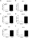
Figure 1
Figure 1. Hypoxia induces BCSC enrichment
A-F. The following breast cancer cell lines were exposed to 20% or 1% O2 for 72 h and the percentage of cells expressing aldehyde dehydrogenase (ALDH+) was determined (mean ± SEM; n = 3): MDA-MB-231 (A), MCF- 7 (B), HCC-1954 (C), SUM-149 (D), T47D (E), and ZR-75.1 (F). **P < 0.01, ***P < 0.001 vs. same cell line at 20% O2 by Student's t test.
Figure 2
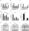
Figure 2. HIFs are required for hypoxia-induced expression of pluripotency factors
A-C. Breast cancer cell lines were exposed to 20% or 1% O2 for 24 h and NANOG (A), KLF4 (B), and SOX2 (C) mRNA levels were determined by RT-qPCR, relative to 18S rRNA, and normalized to the mean value for MDA-MB-231 cells (MDA231) at 20% O2 (mean ± SEM; n = 3). *P < 0.05, **P < 0.01, ***P < 0.001 vs. same cell line at 20% O2 by Student's t test. D and E. HCC-1954 (D) and MCF-7 (E) subclones, which were stably transfected with an expression vector encoding a non-targeting control (NTC) shRNA, or vector encoding shRNA targeting HIF-1α (sh1α) or HIF-2α (sh2α), or vectors encoding shRNAs targeting both HIF-1α and HIF-2α (DKD), were exposed to 20% or 1% O2 for 24 h and RT-qPCR was performed to determine NANOG (D) or KLF4 (E) mRNA levels relative to 18S rRNA. The results were normalized to NTC at 20% O2 (mean ± SEM; n = 3). *P < 0.05, **P < 0.01, ***P < 0.001 vs. NTC at 20% O2; #P < 0.05, ##P < 0.01, ###P < 0.001 vs. NTC at 1% O2 by ANOVA. F. ZR75.1 cells treated with vehicle or digoxin (200 nM) were exposed to 20% or 1% O2 for 24 h and SOX2 mRNA was measured (mean ± SEM; n = 3). *P < 0.05, **P < 0.01 vs. NTC at 20% O2; ###P < 0.001 vs. NTC at 1% O2 by ANOVA. G and H. NTC and DKD subclones of HCC-1954 (G) and MCF-7 (H) were exposed to 20% or 1% O2 for 48 h, whole cell lysates were prepared, and immunoblot assays were performed to analyze HIF-1α, HIF-2α, NANOG and KLF4 protein expression. Actin was also analyzed as a loading control. I. ZR75.1 cells were treated with vehicle or digoxin (200 nM), exposed to 20% or 1% O2 for 48 h, and HIF-1α, NANOG and SOX2 immunoblot assays were performed.
Figure 3
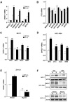
Figure 3. ZNF217, but not FTO, expression was induced by hypoxia in a HIF-dependent manner
A and B. Breast cancer cell lines were exposed to 20% or 1% O2 for 24 h and ZNF217 (A) and FTO (B) mRNA levels were determined by RT-qPCR (mean ± SEM; n = 3). The data were normalized to the mean for the NTC-20% O2 condition. *P < 0.05, **P < 0.01, ***P < 0.001 vs. same cell line at 20% O2 by Student's t test (performed prior to normalization). C and D. MCF-7 (C) and HCC-1954 (D) subclones were exposed to 20% or 1% O2 for 24 h and ZNF217 mRNA levels were determined. *P < 0.05, ***P < 0.001 vs. NTC at 20% O2; #P < 0.05, ##P < 0.01 vs. NTC at 1% O2 by ANOVA. E. ZR75.1 cells treated with vehicle or digoxin (200 nM) were exposed to 20% or 1% O2 for 24 h and ZNF217 mRNA was measured (mean ± SEM; n = 3). **P < 0.01 vs. vehicle at 20% O2; ###P < 0.001 vs. vehicle at 1% O2 by ANOVA. F. MCF-7 and HCC-1954 subclones (upper panels), and ZR75.1 cells treated with vehicle or 200 nM digoxin (lower panels), were exposed to 20% or 1% O2 for 48 h and immunoblot assays were performed.
Figure 4
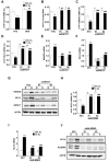
Figure 4. ZNF217 and ALKBH5 regulate NANOG and KLF4 expression via modulation of m6A levels
A. MCF-7 subclones expressing a non-targeting control shRNA (NTC) or shRNAs targeting HIF-1α and HIF-2α (double knockdown [DKD]), were exposed to 20% or 1% O2 for 48 h. Immunoprecipitation of RNA with m6A antibody and RT-qPCR were performed to determine the percentage of KLF4 mRNA with methylation (m6A+) (mean ± SEM; n = 3). **P < 0.01 vs. NTC at 20% O2; ##P < 0.01 vs. NTC at 1% O2 by two-way ANOVA. B. MCF-7 subclones, which were stably transfected with lentiviral vector expressing NTC or either of two independent shRNAs targeting different nucleotide sequences in ZNF217, were exposed to 20% or 1% O2 for 24 h. Total RNA was extracted and m6A levels were determined as a percentage of all adenosine residues (mean ± SEM; n = 3). *P < 0.05, **P < 0.01 vs. NTC at 20%; ##P < 0.01 vs. NTC at 1% O2 by two-way ANOVA. C and D. MCF-7 subclones were exposed to 20% or 1% O2 for 48 h. The percentage of m6A+ NANOG (C) and KLF4 (D) mRNA was determined (mean ± SEM; n = 3). **P < 0.01 vs. NTC at 20% O2; ##P < 0.01, ###P < 0.001 vs. NTC at 1% O2 by two-way ANOVA. E and F. MCF-7 subclones were exposed to 20% or 1% O2 for 24 h. NANOG (E) and KLF4 (F) mRNA levels were determined. *P < 0.05, **P < 0.01, ***P < 0.001 vs. NTC at 20% O2; ##P < 0.01, ###P < 0.001 vs. NTC at 1% O2 by two-way ANOVA. G. MCF-7 subclones were exposed to 20% or 1% O2 for 48 h and immunoblot assays were performed. H. MCF-7 subclones were exposed to 20% or 1% O2 for 48 h. The percentage of m6A+ KLF4 mRNA was determined. *P < 0.05, **P < 0.01 vs. NTC at 20% O2; ##P < 0.01 vs. NTC at 1% O2 by two-way ANOVA. I. MCF-7 subclones were exposed to 20% or 1% O2 for 24 h and KLF4 mRNA levels were determined. *P < 0.05, ***P < 0.001 vs. NTC at 20% O2; ###P < 0.001 vs. NTC at 1% O2 by two-way ANOVA. J. MCF-7 subclones were exposed to 20% or 1% O2 for 48 h and immunoblot assays were performed.
Figure 5
Figure 5. ZNF217 is required for hypoxia-induced BCSC enrichment
A. ZNF217 mRNA levels were determined in MCF-7 parental cells, which were cultured as adherent monolayers or as mammospheres (mean ± SEM; n= 3). ***P < 0.001 vs. adherent cells by Student's t test. B-D. Monolayer cultures of MCF-7 subclones were exposed to 20% or 1% O2 for 72 h and transferred to ultra-low attachment plates. The number of primary (B and C) and secondary (D) mammospheres per 1,000 cells initially seeded was determined (mean ± SEM; n = 3). *P < 0.05, ***P < 0.001 vs. NTC at 20% O2; ###P < 0.001, vs. NTC at 1% O2 by two-way ANOVA. Scale bar: 500 μm. E. MCF-7 subclones were exposed to 20% or 1% O2 for 72 h and the percentage of ALDH+ cells was determined (mean ± SEM; n = 3). **P < 0.01, ***P < 0.001 vs. NTC at 20% O2; ###P < 0.001 vs. NTC at 1% O2 by two-way ANOVA.
Figure 6
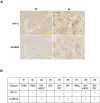
Figure 6. Immunohistochemical analysis of human breast cancer biopsies
A. Immunohistochemistry was performed on breast cancer biopsies using ALKBH5 and HIF-1α antibodies. Representative negative and positive staining (biopsies from patients #2 and #3, respectively) are shown. Scale bar: 500 μm. B. Summary of ALKBH5 and HIF-1α expression in nine human breast cancers of the indicated subtypes based on expression of ER, PR, and HER2. TNBC, triple negative breast cancer.
Figure 7
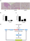
Figure 7. ALKBH5 is required for breast cancer tumorigenicity and metastasis
A-C. Analysis of lung metastasis after orthotopic transplantation of MDA-MB-231 subclones in female SCID mice. Representative hematoxylin-and-eosin stained sections showing lung metastases (arrows) in mice that received mammary fat pad injections of NTC or shALKBH5 cells; scale bar: 500 μm (A). Image analysis was performed and the percentage of lung area occupied by metastases (B) and the number of metastatic foci per lung section (C) were determined (mean + SEM, n = 3). ***P < 0.001 vs. NTC by ANOVA. D. Hypoxia induces HIF-dependent expression of ZNF217 and/or ALKBH5. ZNF217 interacts with METTL3 and inhibits METTL3-catalyzed m6A methylation of KLF4 and NANOG mRNA, whereas ALKBH5 demethylates m6A+ mRNA. m6A-free KLF4 and NANOG mRNAs are stabilized, leading to increased levels of KLF4 and NANOG protein, which specify the BCSC phenotype.

References

    1. Al-Hajj M, Wicha MS, Benito-Hernandez A, Morrison SJ, Clarke MF. Prospective identification of tumorigenic breast cancer cells. Proc Natl Acad Sci U S A. 2003;100(7):3983–3988. - PMC - PubMed
    1. Kreso A, Dick JE. Evolution of the cancer stem cell model. Cell Stem Cell. 2014;14(3):275–291. - PubMed
    1. Oskarsson T, Batlle E, Massague J. Metastatic stem cells: sources, niches, and vital pathways. Cell Stem Cell. 2014;14(3):306–321. - PMC - PubMed
    1. Li X, Lewis MT, Huang J, Gutierrez C, Osborne CK, Wu MF, Hilsenbeck SG, Pavlick A, Zhang X, Chamness GC, Wong H, Rosen J, Chang JC. Intrinsic resistance of tumorigenic breast cancer cells to chemotherapy. J Natl Cancer Inst. 2008;100(9):672–679. - PubMed (VSports)
    1. Creighton CJ, Li X, Landis M, Dixon JM, Neumeister VM, Sjolund A, Rimm DL, Wong H, Rodriguez A, Herschkowitz JI, Fan C, Zhang X, He X, et al. Residual breast cancers after conventional therapy display mesenchymal as well as tumor-initiating features. Proc Natl Acad Sci U S A. 2009;106(33):13820–13825. - PMC - PubMed

VSports手机版 - MeSH terms