Skip to main page content (V体育ios版)
U.S. flag

An official website of the United States government

Dot gov

The . gov means it’s official. Federal government websites often end in . gov or VSports app下载. mil. Before sharing sensitive information, make sure you’re on a federal government site. .

Https

The site is secure V体育官网. The https:// ensures that you are connecting to the official website and that any information you provide is encrypted and transmitted securely. .

. 2016 Sep 2:6:32320.
doi: 10.1038/srep32320.

"V体育官网" Enhanced immune response of MAIT cells in tuberculous pleural effusions depends on cytokine signaling

Affiliations

Enhanced immune response of MAIT cells in tuberculous pleural effusions depends on cytokine signaling (V体育平台登录)

Jing Jiang et al. Sci Rep. .

Abstract

The functions of MAIT cells at the site of Mycobacterium tuberculosis infection in humans are still largely unknown. In this study, the phenotypes and immune response of MAIT cells from tuberculous pleural effusions and peripheral blood were investigated. MAIT cells in tuberculous pleural effusions had greatly enhanced IFN-γ, IL-17F and granzyme B response compared with those in peripheral blood. The level of IFN-γ response in MAIT cells from tuberculous pleural effusions was inversely correlated with the extent of tuberculosis infection (p = 0. 0006). To determine whether cytokines drive the immune responses of MAIT cells at the site of tuberculosis infection, the role of IL-1β, IL-2, IL-7, IL-12, IL-15 and IL-18 was investigated. Blockade of IL-2, IL-12 or IL-18 led to significantly reduced production of IFN-γ and/or granzyme B in MAIT cells from tuberculous pleural effusions VSports手机版. Majority of IL-2-producing cells (94. 50%) in tuberculous pleural effusions had phenotype of CD3(+)CD4(+), and most IL-12p40-producing cells (91. 39%) were CD14(+) cells. MAIT cells had significantly elevated expression of γc receptor which correlated with enhanced immune responses of MAIT cells. It is concluded that MAIT cells from tuberculous pleural effusions exhibited highly elevated immune response to Mtb antigens, which are controlled by cytokines produced by innate/adaptive immune cells. .


Figures
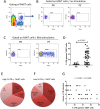
Figure 1
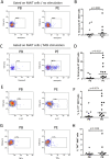
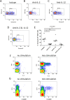
Figure 1. IFN-γ production in MAIT cells from tuberculous pleural effusions and peripheral blood.
(A) Representative flow cytometric plot showing gating of MAIT cells with phenotypes of CD3+Vα7.2+CD161high. (B) Representative flow cytometric plots showing IFN-γ production in MAIT cells from peripheral blood (PB) and from tuberculous pleural effusion (PE) of patient with tuberculous pleurisy in the absence of antigen stimulation. (C) Representative flow cytometric plots showing IFN-γ production in MAIT cells from peripheral blood (PB) and from tuberculous pleural effusion (PE) of patient with tuberculous pleurisy after Mtb antigen stimulation. (D) MAIT cells in tuberculous pleural effusions had greatly enhanced IFN-γ response to Mtb antigens compared with those in peripheral blood. Horizontal Bars in the scatter plots indicate median. (E) The ratio of patients with tuberculous pleurisy only (PE), with accompanied pulmonary TB with lesions located within one lung (PE & pTB1), with accompanied pulmonary TB with lesions located in both lungs (PE & pTB2), and with accompanied pulmonary TB and other extrapulmonary TB (PE & mTB) in the high IFN-γ-producing MAIT cell group. (F) The ratio of patients with different extent of TB infections in the low IFN-γ-producing MAIT cell group. (G) Correlation analysis showed that the frequency of IFN-γ-producing MAIT cells was inversely correlated with the extent of TB infection. The extend of TB infection was graded from 1 to 4, representing PE, PE&pTB1, PE&pTB2 and PE&mTB respectively, as described in legend in Fig. 1E. The nonparametric Mann-Whitney test was used for statistical analysis in Fig. 1D.
Figure 2
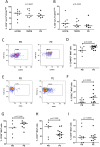
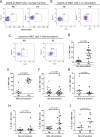
Figure 2. Expression of granzyme B, IL-17F and TNF-α in MAIT cells.
(A) Representative flow plots showing expression of granzyme B in MAIT cells from peripheral blood (PB) and tuberculous pleural effusion (PE) in the absence of antigen stimulation. (B) MAIT cells from tuberculous pleural effusions had similar expression of granzyme B compared with those from peripheral blood in the absence of antigen stimulation. (C) Representative flow plots showing expression of granzyme B in MAIT cells from peripheral blood (PB) and tuberculous pleural effusion (PE) after Mtb antigen stimulation. (D) MAIT cells from tuberculous pleural effusions had much higher expression of granzyme B than those from peripheral blood after stimulation with Mtb lysates. (E) Representative flow plots showing expression of IL-17F in MAIT cells from peripheral blood (PB) and tuberculous pleural effusion (PE) after Mtb antigen stimulation. (F) MAIT cells from tuberculous pleural effusions had significantly higher expression of IL-17F than from peripheral blood. (G) Representative flow plots showing expression of TNF-α in MAIT cells. (H) TNF-α expression in MAIT cells did not show significant difference between the two groups. Horizontal bars in the scatter plots indicate medians. The nonparametric Mann-Whitney test was used for statistical analysis between groups in Fig. 2B,D,F,H.
Figure 3
Figure 3. Phenotypic analysis of MAIT cells from tuberculous pleural effusions and peripheral blood.
(A) Frequency of CD8α+ MAIT cells in peripheral blood of healthy controls (HC/PB) and patients with TB (TB/PB), and in tuberculous pleural effusions (PE). (B) Frequency of CD4CD8α (DN) MAIT cells. (C) Representative flow plots showing expression of CD45RO in MAIT cells from peripheral blood (PB) and tuberculous pleural effusion (PE) in the absence of antigen stimulation. (D) Frequencies of CD45RO+ MAIT cells. (E) Representative flow plots showing expression of CD62L in MAIT cells in the absence of antigen stimulation. (F) Frequencies of CD62L+ MAIT cells. (GI) Frequencies of CD26+ (G), CD27+ (H), CD39+ (I) MAIT cells in peripheral blood of patients with TB (PB) and in tuberculous pleural effusions (PE) respectively. Horizontal bars in the scatter plots indicate medians. Kruskal-Wallis test was used for statistical analysis among groups in Fig. 3A,B. The nonparametric Mann-Whitney test was used for statistical analysis between groups in Fig. 3D,F,G–I.
Figure 4
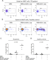
Figure 4. Influence of humoral factors on IFN-γ response of MAIT cells from PBMCs.
(A) Representative flow plots showing IFN-γ production in MAIT cells from PBMCs of patient with TB under Mtb stimulation only (Mtb) (left), or with addition of centrifuged supernatant (100 μl) of unstimulated mononuclear cells (Mtb + unstim. sup.) (middle), or supernatants of Mtb-stimulated mononuclear cells from tuberculous pleural effusion (Mtb + stim. sup.) (right). (B) Representative flow plots showing IFN-γ production in MAIT cells from PBMCs of healthy control under same stimulation conditions as stated above. (C) IFN-γ response of MAIT cells in PBMCs from patients with TB (n = 5) under condition of Mtb stimulation alone (Mtb stim.), or with addition of centrifuged supernatants (100 μl) of unstimulated (PE-unstim. supernatant) or Mtb antigen-stimulated cells (PE-stim. supernatant) from tuberculous pleural effusions. (D) Supernatants of Mtb-stimulated cells from tuberculous pleural effusions drove strong IFN-γ response of MAIT cells in PBMCs from healthy controls (n = 5). Horizontal bars in the scatter plots indicate medians. Kruskal-Wallis test was used for statistical analysis among groups.
Figure 5
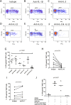
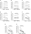
Figure 5. The role of cytokines on IFN-γ response of MAIT cells from tuberculous pleural effusions.
(AF) Representative flow plots showing IFN-γ production in Mtb-stimulated MAIT cells in the presence of isotype antibody control (A), or blocking antibody to IL-1β (B), IL-2 (C), IL-12 (D), IL-15 (E), or combined blocking antibodies of both IL-2 and IL-12 (F). (G) Reduced IFN-γ response in MAIT cells from tuberculous pleural effusions was observed when IL-2 and IL-12 were blocked with antibodies (n = 6). (H) Addition of IL-18 blocking antibody led to decreased IFN-γ production in MAIT cells (n = 8). (I) Blockade of IL-7 resulted in slightly higher IFN-γ response in MAIT cells (n = 6). (J) IFN-γ response in MAIT cells of PBMCs (PBMCs) in the presence of Mtb stimulation and centrifuged supernatants (100 μl) of Mtb antigen-stimulated cells from tuberculous pleural effusions was abolished by blocking antibodies to IL-2 and IL-12 (n = 3). Pleural effusion: mononuclear cells from tuberculous pleural effusions; Mtb stim.: Mtb antigen stimulation; PE-stim. supernatant: supernatants of Mtb antigen-stimulated cells from tuberculous pleural effusions; Anti-IL-2 & IL-12 Abs: combined blocking antibodies to IL-2 and IL-12. The Friedman’s test (p < 0.0001), followed by Dunn’s multiple comparison test was used in Fig. 5G. The non-parametric Wilcoxon signed-rank test was used for statistical analysis between groups in Fig. 5H,I. Kruskal-Wallis test was used for statistical analysis in Fig. 5J. Horizontal bars in the scatter plots indicate medians. **p < 0.01.
Figure 6
Figure 6. The role of cytokines on granzyme B production in MAIT cells from tuberculous pleural effusions.
(AD) Representative flow plots showing granzyme B production in Mtb-stimulated MAIT cells in the presence of isotype antibody control (A), or blocking antibody to IL-2 (B) and IL-12 (C), or combined blocking antibodies to both IL-2 and IL-12 (D). (E) Blockade of IL-2, but not IL-12, led to significantly reduced expression of granzyme B (n = 6). (F) Representative flow plots showing IL-2-producing cells in the tuberculous pleural effusions. Cells from tuberculous pleural effusions (n = 6) were not stimulated (left) or stimulated with Mtb lysates (right). All cells were gated and CD3+CD4+ T cells were identified as CD4+ T cells. (G) Representative flow plots showing IL-12p40-producing cells. Cells from tuberculous pleural effusions (n = 6) were not stimulated (left) or stimulated with Mtb lysates (right). All cells were gated and CD14+ cells were identified. The Friedman’s test (p < 0.0001), followed by Dunn’s multiple comparison test was used for statistical analysis among groups in Fig. 6E. Horizontal bars in the scatter plots indicate medians. *p < 0.05; **p < 0.01; ns: no significant difference.
Figure 7
Figure 7. IL-2 and IL-12 receptor expression on MAIT cells from peripheral blood and tuberculous pleural effusions.
(A) Representative flow plots showing γc (CD132) isotype control antibody staining of MAIT cells from peripheral blood (PB) and tuberculous pleural effusion (PE). (B) Representative flow plots showing expression of γc receptor (CD132) on MAIT cells in the absent of antigen stimulation. (C) Representative flow plots showing expression of γc receptor (CD132) on Mtb-stimulated MAIT cells from peripheral blood (PB) and tuberculous pleural effusion (PE). (DE) MAIT cells from tuberculous pleural effusions exhibited significantly higher expression of γc receptor (CD132) than those from peripheral blood both in the absent of antigen stimulation (D) and after Mtb stimulation (E). (FG) The expression of IL-2Rβ on MAIT cells in the absent of antigen stimulation (F) or after Mtb stimulation (G). (HI) The expression of IL-2Rα on MAIT cells in the absent of antigen stimulation (H) or after Mtb stimulation (I). (J) The expression of IL-12Rβ1 on MAIT cells in the absent of antigen stimulation. Horizontal bars in the scatter plots indicate medians. The nonparametric Mann-Whitney test was used for statistical analysis between groups.
Figure 8
Figure 8. Blockade of γc and IL-2Rβ receptors resulted in reduced immune response of MAIT cells from tuberculous pleural effusions.
(A) Blockade of IL-2Rα did not have significant effect on IFN-γ response in MAIT cells stimulated with Mtb lysates. (BC) Reduced IFN-γ response in MAIT cells was observed when IL-2Rβ (B) and γc receptors (C) were blocked with antibodies respectively. (D) Blockade of both IL-2Rβ (B) and γc receptors resulted in decreased IFN-γ response in MAIT cells. (E) Blockade of IL-2Rα did not have significant effect on granzyme B production in MAIT cells. (FG) Blockade of IL-2Rβ (F) or γc receptor (G) led to significantly reduced production of granzyme B in MAIT cells. (H) Combined blocking of both IL-2Rβ and γc receptors resulted in even more reduced production of granzyme B. The non-parametric Wilcoxon signed-rank test was used for statistical analysis.
Figure 9
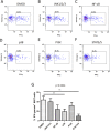
Figure 9. Blockade of signaling pathways with small molecule inhibitors.
(AF) Representative flow plots showing IFN-γ production in Mtb-stimulated MAIT cells in the presence of solvent DMSO (A), or small molecule inhibitors to JNK1/2/3 (B), NF-κB (C), p38 (D), PI3K (E), or STAT3/STAT5 (F). (G) Inhibition effect of small molecule inhibitors on IFN-γ production in Mtb-stimulated MAIT cells from tuberculous pleural effusions (n = 8). The Friedman’s test (p = 0.0004), followed by Dunn’s multiple comparison test was used for statistical analysis among groups. **p < 0.01.

References

    1. WHO. Global tuberculosis report 2014. "VSports最新版本" http://www.who.int/tb/publications/global_report/en/ (2015).
    1. Vorster M. J., Allwood B. W., Diacon A. H. & Koegelenberg C. F. Tuberculous pleural effusions: advances and controversies. J Thorac Dis 7, 981–991 (2015). - PMC - PubMed
    1. Udwadia Z. F. & Sen T. Pleural tuberculosis: an update. Curr Opin Pulm Med 16, 399–406 (2010). - V体育平台登录 - PubMed
    1. Light R. W. Update on tuberculous pleural effusion. Respirology 15, 451–458 (2010). - PubMed
    1. Le Bourhis L. et al. Antimicrobial activity of mucosal-associated invariant T cells. Nat Immunol 11, 701–708 (2010). - PubMed

"VSports在线直播" Publication types

MeSH terms

VSports手机版 - LinkOut - more resources