Skip to main page content
U.S. flag

An official website of the United States government

Dot gov

The . gov means it’s official. Federal government websites often end in VSports app下载. gov or . mil. Before sharing sensitive information, make sure you’re on a federal government site. .

Https

The site is secure. The https:// ensures that you are connecting to the official website and that any information you provide is encrypted and transmitted securely V体育官网. .

. 2016 Sep 30;291(40):20911-20923.
doi: 10.1074/jbc.M116.748251. Epub 2016 Aug 17.

The Phylogeny and Active Site Design of Eukaryotic Copper-only Superoxide Dismutases

Affiliations

The Phylogeny and Active Site Design of Eukaryotic Copper-only Superoxide Dismutases

Ryan L Peterson et al. J Biol Chem. .

Abstract

In eukaryotes the bimetallic Cu/Zn superoxide dismutase (SOD) enzymes play important roles in the biology of reactive oxygen species by disproportionating superoxide anion. Recently, we reported that the fungal pathogen Candida albicans expresses a novel copper-only SOD, known as SOD5, that lacks the zinc cofactor and electrostatic loop (ESL) domain of Cu/Zn-SODs for substrate guidance. Despite these abnormalities, C VSports手机版. albicans SOD5 can disproportionate superoxide at rates limited only by diffusion. Here we demonstrate that this curious copper-only SOD occurs throughout the fungal kingdom as well as in phylogenetically distant oomycetes or "pseudofungi" species. It is the only form of extracellular SOD in fungi and oomycetes, in stark contrast to the extracellular Cu/Zn-SODs of plants and animals. Through structural biology and biochemical approaches we demonstrate that these copper-only SODs have evolved with a specialized active site consisting of two highly conserved residues equivalent to SOD5 Glu-110 and Asp-113. The equivalent positions are zinc binding ligands in Cu/Zn-SODs and have evolved in copper-only SODs to control catalysis and copper binding in lieu of zinc and the ESL. Similar to the zinc ion in Cu/Zn-SODs, SOD5 Glu-110 helps orient a key copper-coordinating histidine and extends the pH range of enzyme catalysis. SOD5 Asp-113 connects to the active site in a manner similar to that of the ESL in Cu/Zn-SODs and assists in copper cofactor binding. Copper-only SODs are virulence factors for certain fungal pathogens; thus this unique active site may be a target for future anti-fungal strategies. .

Keywords: copper; enzyme; fungi; superoxide dismutase (SOD); superoxide ion; x-ray crystallography. V体育安卓版.


Figures

FIGURE 1.
FIGURE 1.
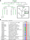
Distribution of SOD5-like SODs among fungi and oomycetes. A, phylogenetic representations of eukaryotes where one or more SOD5-like SODs can be identified. The distribution of SOD5-like SODs in the fungal kingdom (left) is indicated by green text. Within Saccharomycotina (true yeast), copper-only SODs appear to be segregated and are detected within members of the CTG but absent from WDG clade. The phylogenetic tree on right illustrates presence of SOD5-like SODs in distantly related fungi and oomycetes. B, the active site region spanning zinc binding ligands His-60 and Asp-63 of Saccharomyces cerevisiae Cu/Zn-SOD1 is aligned against the analogous region in SOD5-like SODs from select fungi and oomycetes (in the box), listed according to protein entry identifiers. The conserved Glu/Gln equivalent to SOD5 Glu-110 is highlighted in blue and green, respectively, whereas the invariant active site aspartate (corresponding to SOD5 Asp-113) is highlighted in red.
FIGURE 2.
FIGURE 2.
Structural comparison of the active sites of Cu/Zn-SOD1 versus C. albicans SOD5. A, active site of S. cerevisiae Cu/Zn-SOD1 (PDB code 1JCV). The copper and zinc metal cofactors are blue and gray spheres, respectively. The dynamic histidine His-63 that coordinates both Cu(II) and zinc is orange, and the ESL with conserved Asp-124 that H-bonds to His-46 is indicated in yellow. B, the active site of C. albicans wild type SOD5 (PDB code 4N3T). Copper is a blue sphere, Glu-110 and Asp-113 are pink, and the dynamic histidine His-93 residue is orange. The Asp-113 orientated water network is shown as small red and blue balls where blue represents the water molecule that interacts with the copper site in a fashion analogous to Asp-124 of the Cu/Zn-SOD1 ESL.
FIGURE 3.
FIGURE 3.
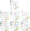
Structural comparisons of the active sites of wild type versus Glu-110 and Asp-113 mutants of SOD5. Active site Cu(I) structures of wild type (PDB code 4N3T), E110Q (PDB code 5KBK), E110A (PDB code 5KBC), and D113N SOD5 (PDB code 5KBM). The top two rows show individual structures, and the bottom row shows overlays of wild type and SOD5 mutants. Cu(I) is a blue sphere, and the dynamic His-93, Glu-110/Asp-113, and residues 154–159 are orange, pink, and yellow, respectively. Water molecules in wild type, E110Q, E110A, and D113N SOD5 are red, gray, light blue, and pink balls, respectively. Dashed lines indicate hydrogen bonds; superimposed structures show only hydrogen bonds of the mutants. Wild type SOD5 crystallized with a Tris molecule (blue, top panel).
FIGURE 4.
FIGURE 4.
SOD5 activity profiles as determined by pulse radiolysis. Open circles represent the second-order catalytic rate constants (kcat) as a function of pH for recombinant SOD5 and SOD5 Glu-110/Asp-113 mutants: wild type (WT) (black), E110A (red), E110Q (blue), and D113N (green). Values shown are the average of four or more independent measurements with range of values of ≤10%. The individual open squares represent the calculated k1 half-reaction (see Equation 1) for wild type and mutant SOD5 at the indicated pH. The reported k1 values are the average of two or more independent measurements with a range ≤10%.
FIGURE 5.
FIGURE 5.
Loss of SOD5 activity at alkaline pH reflects an inhibition in k2. A–C, time-dependent loss of superoxide substrate in the presence of wild type SOD5 as monitored by change in absorbance at 260 nm at pH 9.5. A, dose-dependent kinetic profiles for the decay of 3.6 μm superoxide with 3.2 μm Cu(II) SOD5. The first and second pulse with superoxide is shown in black and red, respectively. B, time-dependent decay of a 3.6 μm fixed superoxide substrate dose with 3.2 μm (black) and 7.9 μm (gray) Cu(II) SOD5. C, time-dependent decay of superoxide substrate as a function of pulse number employing 7.9 μm Cu(II) SOD5. Pulse sequence is indicated by color: pulse #1 (black), #2 (red), #3 (blue), #4 (green), #5 (magenta). D, SOD5 activity profile as a function of pH. Calculated second-order rate constants (kcat) are indicated as open circles. Calculated second-order rate constants for k1, as determined by either decay of superoxide anion at 260 nm under single turnover experiments or by loss of Cu(II) SOD5 d-d absorbance band at 720 nm, are shown as open and black squares, respectively. Reported kcat and k1 values are the averages of ≥4 and ≥2 independent measurements respectively, with a range of values obtained of ≤10%.
FIGURE 6.
FIGURE 6.
Reduction of Cu(II) SOD5 at elevated pH. Time-dependent loss of oxidized Cu(II) SOD5 as monitored by change in absorbance at 720 nm at elevated pH upon the substoichiometric pulse of superoxide substrate. The pseudo-first-order fit to the decay for each spectral time course is indicated by the solid red line.
FIGURE 7.
FIGURE 7.
Expression of wild type and Asp-113 mutant SOD5 in C. albicans cultures. C. albicans secreting either wild type or the indicated mutant alleles of SOD5 were cultured for 1 h at the designated pH. The growth medium containing freshly secreted SOD5 was concentrated and analyzed for: SOD5 activity by the native gel assay (no EndoH treatment) (top) and SOD5 protein levels by immunoblot in samples treated with EndoH with molecular weight makers indicated on the left (middle). Quantification of this immunoblot reveals expression levels relative to wild type SOD5 at pH 3.3 = 1.0; left, D113N pH 3.3 = 0.75 and wild type pH 7.5 = 0.76; D113N pH 7.5 = 1.2; right, D113A pH 3.3 = 0.65; wild type pH 7.5 = 0.79; D113N pH 7.5 = 0.54. Bottom, effects of pH on SOD5 glycosylation by immunoblot of samples not treated with EndoH with molecular weight makers indicated on the left. Glycosylated SOD5 exhibits more rapid mobility from cells cultured at pH 7.5, indicating that the change in mobility on the native gel (top) reflects alterations in SOD5 glycosylation. The very strong intensity of D113N, pH 7.5, without EndoH presumably reflects the impact of D113N SOD5 glycosylation on the immunoblot (e.g. antibody recognition), as it is not seen in the deglycosylated + EndoH sample.

References

    1. McCord J. M., and Fridovich I. (1969) Superoxide dismutase an enzymic function for erythrocuprein (hemocuprein). J. Biol. Chem. 244, 6049–6055 - PubMed
    1. Sheng Y., Abreu I. A., Cabelli D. E., Maroney M. J., Miller A. F., Teixeira M., and Valentine J. S. (2014) Superoxide dismutases and superoxide reductases. Chem. Rev. 114, 3854–3918 - PMC - PubMed
    1. Perry J. J., Shin D. S., Getzoff E. D., and Tainer J. A. (2010) The structural biochemistry of the superoxide dismutases. Biochim. Biophys. Acta 1804, 245–262 - PMC - PubMed
    1. Abreu I. A., and Cabelli D. E. (2010) Superoxide dismutases: a review of the metal-associated mechanistic variations. Biochim. Biophys. Acta 1804, 263–274 - PubMed
    1. Crapo J. D., Oury T., Rabouille C., Slot J. W., and Chang L. Y. (1992) copper, zinc superoxide-dismutase is primarily a cytosolic protein in human cells. Proc. Natl. Acad. Sci. U.S.A. 89, 10405–10409 - PMC - PubMed

MeSH terms

Associated data (V体育安卓版)

LinkOut - more resources