Skip to main page content
U.S. flag

An official website of the United States government

Dot gov

The VSports app下载. gov means it’s official. Federal government websites often end in . gov or . mil. Before sharing sensitive information, make sure you’re on a federal government site. .

Https

The site is secure. The https:// ensures that you are connecting to the official website and that any information you provide is encrypted and transmitted securely. V体育官网.

. 2016 Oct;15(10):2294-2301.
doi: 10.1158/1535-7163.MCT-16-0153. Epub 2016 Aug 2.

A Novel Small Molecule Activator of Nuclear Receptor SHP Inhibits HCC Cell Migration via Suppressing Ccl2

Affiliations

"VSports手机版" A Novel Small Molecule Activator of Nuclear Receptor SHP Inhibits HCC Cell Migration via Suppressing Ccl2

Zhihong Yang et al. Mol Cancer Ther. 2016 Oct.

VSports - Abstract

Small heterodimer partner (SHP, NR0B2) is a nuclear orphan receptor without endogenous ligands VSports手机版. Due to its crucial inhibitory role in liver cancer, it is of importance to identify small molecule agonists of SHP. As such, we initiated a probe discovery effort to identify compounds capable of modulating SHP function. First, we performed binding assays using small molecule microarrays (SMM) and discovered 5-(diethylsulfamoyl)-3-hydroxynaphthalene-2-carboxylic acid (DSHN) as a novel activator of SHP. DSHN transcriptionally activated Shp mRNA, but also stabilized the SHP protein by preventing its ubiquitination and degradation. Second, we identified Ccl2 as a new SHP target gene by RNA-seq. We showed that activation of SHP by DSHN repressed Ccl2 expression and secretion by inhibiting p65 activation of CCL2 promoter activity, as demonstrated in vivo in Shp-/- mice and in vitro in HCC cells with SHP overexpression and knockdown. Third, we elucidated a strong inhibitory effect of SHP and DSHN on HCC cell migration and invasion by antagonizing the effect of CCL2. Lastly, by interrogating a publicly available database to retrieve SHP expression profiles from multiple types of human cancers, we established a negative association of SHP expression with human cancer metastasis and patient survival. In summary, the discovery of a novel small molecule activator of SHP provides a therapeutic perspective for future translational and preclinical studies to inhibit HCC metastasis by blocking Ccl2 signaling. Mol Cancer Ther; 15(10); 2294-301. ©2016 AACR. .


Conflict of interest statement (V体育安卓版)

No conflict of interest to disclose for all authors.

Figures
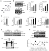
Figure 1
Figure 1. Identifying a small molecule that activates SHP gene transcription
(A) The structure of compound DSHN, which was identified by small molecule microarray (SMM). (B) Left: Semi-quantitative PCR of Shp mRNA in normal mouse hepatocyte Nmuli (upper panel) and hepatoma Hepa-1 (lower panel) cells treated with DSHN (20 μM). pos: positive control, cells transfected with a mouse Shp (mShp) expression plasmid; con, control, DMSO; neg: negative control, cells treated with an unrelated compound that does not induce Shp. Right: qPCR of Shp mRNA in mouse Hepa-1 (left) and human MHCC97H (right) cells treated with DSHN (20, 30 μM). Data is represented as mean ± SE (*p<0.01 vs. DMSO, grey bar). (C) Relative luciferase (luc) activity (RLU) of mShp promoter (pro.) without (first) or with co-transfected LRH-1 (second), HNF4α (third), and RXR/RARα (fourth) in the absence (-, DMSO) or presence of DSHN (50, 100 μm). Data is represented as mean ± SE of triplicate assays (§p<0.01 vs. white bar; *p<0.01 vs. gray bar). (D) Western blot of SHP protein in Hepa-1 cells treated with DSHN (20 μM) and protein synthesis inhibitor cycloheximide (CHX) to determine SHP protein half-life. The cells were transfected Flag-Shp plasmid (1 μg/well) for 24 hrs before harvested at indicated time point. (E) Ubiquitination analysis of SHP protein. HEK 293T cells were cotransfected with Flag-Shp (1 μg/well) and HA-Ub (2 μg/well) for 24 hrs before the treatment with Shp agonists (AHPN, AHPC), an unrelated compound (neg) and DSHN (20 μM). (F) Semi-quantitative PCR (upper) and qPCR (bottom) of Shp mRNA expression in mice treated with DSHN (upper: 5 mg/kg for i.v. injection or lower: 25 mg/25 kg for oral feeding). Data is represented as mean ± SE (*p<0.01 vs. solvent treated group). Mice of two month-old were used (n=5). ALL data are representative of two or more experiments.
Figure 2
Figure 2. SHP functions as a transcriptional suppressor of Ccl2
(A) RNA-seq of relative expression levels of Ccl family in sko vs wt liver. (B) Left: Semi-quantitative PCR of hepatic Ccl2, Ccr2 and Shp mRNA in wildtype (wt) and Shp−/− (sko). Middle and Right: ELISA of CCL2 levels in liver and serum of wt and sko. Data is represented as mean ± SE of triplicate assays (*p<0.01 vs. white bar). (C) Semi-quantitative PCR (left) and qPCR (middle and right) of CCL2 and SHP mRNA in Hep3B cells with Shp overexpression or knockdown (siSHP) using adenovirus (100 MOI). Lower band: mouse Shp (Ade-Shp); higher band: endogenous human SHP. Data is represented as mean ± SE of triplicate assays (*p<0.01 vs. white bar). (D) ELISA of CCL2 protein levels in culture medium of Hep3B cells infected with indicated adenovirus (Ad, 100 MOI). Data is represented as mean ± SE of triplicate assays (*p<0.01 vs. white bar; ¥p<0.01 vs. siSHP, black bar). (E) Promoter luciferase report assay. Hepa1 cells were cotransfected with the indicated plasmids for 48 hrs. Data is represented as mean ± SE of triplicate assays (*p<0.01 vs. white bar; ¥p<0.01 vs. 2nd bar). (F) Semi-quantitative PCR of SHP (left) and CCL2 (right) mRNA in Huh7 cells treated with DSHN for 24 hrs (10 μm). (G) Western blot of CCL2 protein in HCC cells treated with different doses of DSHN for 24 hr (MHCC97H, Huh7) or 48 hr (HepG2). Data are representative of two or more experiments.
Figure 3
Figure 3. Overexpression of SHP inhibits HCC cell invasion and migration
(A) Comparing the migration potential in Hep3B, Huh7, HMCC97L and MHCC97H cells. Migrated cells were visualized by the staining of crystal violet. (B) Left: The levels of secreted CCL2 protein in culture medium as examined by ELISA. Right: Semi-quantitative PCR of CCL2 mRNA in various HCC cells. (C) HCC cell migration assay. Huh7 (left) and Hepa1 (middle) cells were overexpressed Ade-SHP (adenovirus, 50 MOI) for 48 hrs. Data is represented as mean ± SE of triplicate assays (*p<0.01 vs. white bar). Right: Images of invaded cells stained with crystal violet. (D) HCC cell migration assay. MHCC97H cells were transduced with Ade-SHP (50 MOI) for 48 hrs and migrated cells were visualized by the staining of crystal violet. Data are representative of two or more experiments.
Figure 4
Figure 4. Activation of SHP inhibits CCl2-mediated HCC cell invasion and migration
(A) HCC invasion assay. MHCC97H cells were transduced with Ade-SHP for 48 hrs before subjected to invasion assay in the absence or presence of CCL2 (50 ng/ml). The invaded cells were stained with crystal violet and counted. Quantitative results on the right: *p<0.01 vs. GFP (-CCL2); ¥p<0.01 vs. GFP (+CCL2). (B) HCC migration assay. Huh7 cells were transduced with indicated adenovirus for 48 hrs before subjected to migration assay in the absence or presence of CCL2 (100 ng/ml). The migrated cells were stained with crystal violet and counted. *p<0.01 vs. GFP (-CCL2); ¥p<0.01 vs. GFP (+CCL2). (C) HCC migration assay. DSHN group: 50 μM for 48 hrs; CCL2 group: 50 ng/ml for 24 hrs. The migrated cells were stained with crystal violet and counted. Quantitative results on the right: *p<0.05 vs. DMSO (-CCL2); ¥p<0.01 vs. DMSO (+CCL2) and DSHN (-CCL2). (D) HCC migration assay in Huh7 cells treated with DSHN (50 μM). *p<0.01 vs. DMSO. Data are representative of two or more experiments.
Figure 5
Figure 5. The expression of SHP is negatively associated with cancer metastasis
(A) The expression of SHP in human HCC specimens. The data is derived from ONCOMINE (IMAGE: 418138). (B) The expression of SHP in human gastric cancer. The data is derived from Profile: GDS1210 / L76571_at / NR0B2. (C) Kaplan-Meier survival plots. The gastric cancer dataset was divided into two groups according to the expression levels of SHP from 305 patients. Grouped datasets were subjected to Kaplan-Meier survival analysis. (D) Kaplan-Meier survival plots. The breast cancer dataset was divided into two groups according to the expression levels of SHP from 3554 patients. Grouped datasets were subjected to Kaplan-Meier survival analysis.

References

    1. Conti I, Rollins BJ. CCL2 (monocyte chemoattractant protein-1) and cancer. Semin Cancer Biol. 2004;14:149–54. - "V体育2025版" PubMed
    1. Ueno T, Toi M, Saji H, Muta M, Bando H, Kuroi K, et al. Significance of macrophage chemoattractant protein-1 in macrophage recruitment, angiogenesis, and survival in human breast cancer. Clin Cancer Res. 2000;6:3282–9. - "VSports在线直播" PubMed
    1. Lee CC, Ho HC, Su YC, Lee MS, Hung SK, Lin CH. MCP1-Induced Epithelial-Mesenchymal Transition in Head and Neck Cancer by AKT Activation. Anticancer Res. 2015;35:3299–306. - PubMed
    1. Li X, Yao W, Yuan Y, Chen P, Li B, Li J, et al. Targeting of tumour-infiltrating macrophages via CCL2/CCR2 signalling as a therapeutic strategy against hepatocellular carcinoma. Gut. 2015 - "VSports最新版本" PubMed
    1. Chen TA, Wang JL, Hung SW, Chu CL, Cheng YC, Liang SM. Recombinant VP1, an Akt inhibitor, suppresses progression of hepatocellular carcinoma by inducing apoptosis and modulation of CCL2 production. PLoS One. 2011;6:e23317. - PMC - PubMed

Publication types

MeSH terms

V体育平台登录 - Substances