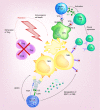
Anti-GD2 mAbs and next-generation mAb-based agents for cancer therapy
- PMID: 27485082
- PMCID: PMC5619016 (V体育2025版)
- DOI: 10.2217/imt-2016-0021
Anti-GD2 mAbs and next-generation mAb-based agents for cancer therapy
"V体育官网" Erratum in
-
Erratum.Immunotherapy. 2016 Nov;8(11):1349. doi: 10.2217/imt-2016-0021e1. Epub 2016 Oct 12. Immunotherapy. 2016. PMID: 27730843 Free PMC article. No abstract available.
"V体育平台登录" Abstract
Tumor-specific monoclonal antibodies (mAbs) have demonstrated efficacy in the clinic, becoming an important approach for cancer immunotherapy. Due to its limited expression on normal tissue, the GD2 disialogangloside expressed on neuroblastoma cells is an excellent candidate for mAb therapy VSports手机版. In 2015, dinutuximab (an anti-GD2 mAb) was approved by the US FDA and is currently used in a combination immunotherapeutic regimen for the treatment of children with high-risk neuroblastoma. Here, we review the extensive preclinical and clinical development of anti-GD2 mAbs and the different mechanisms by which they mediate tumor cell killing. In addition, we discuss different mAb-based strategies that capitalize on the targeting ability of anti-GD2 mAbs to potentially deliver, as monotherapy, or in combination with other treatments, improved antitumor efficacy. .
Keywords: cancer immunotherapy; monoclonal antibodies; neuroblastoma V体育安卓版. .
Conflict of interest statement
This research was supported by Hyundai Hope on Wheels Grant; Midwest Athletes Against Childhood; Stand Up 2 Cancer; The St. Baldrick’s Foundation; American Association of Cancer Research; University of Wisconsin-Madison Carbone Cancer Center; and supported in part by Public Health Service Grants CA21115, CA23318, CA66636, CA180820, CA180794, CA21076, CA180799, CA14958, CA180816, CA166105 and CA197078; from the National Cancer Institute. The Advanced Opportunity Fellowship through SciMed Graduate Research Scholars and the Molecular Bioscience Training Grant (MBTG) T32 GM07215 at University of Wisconsin – Madison, provided funding for ZP Horta and The Howard Hughes Medical Scholars Fellowship Program provided funding for JL Goldberg. The authors have no other relevant affiliations or financial involvement with any organization or entity with a financial interest in or financial conflict with the subject matter or materials discussed in the manuscript apart from those disclosed V体育ios版.
No writing assistance was utilized in the production of this manuscript.
Figures


References
-
- Siegel RL, Miller KD, Jemal A. Cancer statistics, 2015. CA Cancer J. Clin. 2015;65(1):5–29. - PubMed
-
- Mujoo K, Cheresh DA, Yang HM, Reisfeld RA. Disialoganglioside GD2 on human neuroblastoma cells: target antigen for monoclonal antibody-mediated cytolysis and suppression of tumor growth. Cancer Res. 1987;47(4):1098–1104. - PubMed
-
- US FDA. FDA approves first therapy for high-risk neuroblastoma [press release] 10 March (2015). www.fda.gov/newsevents/newsroom/pressannouncements/ucm437460.htm
-
- Cartron G, Dacheux L, Salles G, et al. Therapeutic activity of humanized anti-CD20 monoclonal antibody and polymorphism in IgG Fc receptor FcgammaRIIIa gene. Blood. 2002;99(3):754–758. - "V体育安卓版" PubMed
-
- Weng W-K, Levy R. Two immunoglobulin G fragment C receptor polymorphisms independently predict response to rituximab in patients with follicular lymphoma. J. Clin. Oncol. 2003;21(21):3940–3947. - PubMed
Publication types
MeSH terms
- Actions (V体育2025版)
- Actions (VSports最新版本)
- Actions (VSports app下载)
- Actions (VSports在线直播)
- "VSports注册入口" Actions
- "VSports最新版本" Actions
- "V体育安卓版" Actions
- V体育官网入口 - Actions
- V体育2025版 - Actions
Substances
- Actions (V体育平台登录)
- V体育官网 - Actions
Grants and funding
- R01 CA032685/CA/NCI NIH HHS/United States
- T32 GM007215/GM/NIGMS NIH HHS/United States
- R01 CA166105/CA/NCI NIH HHS/United States
- R35 CA197078/CA/NCI NIH HHS/United States
- U10 CA014958/CA/NCI NIH HHS/United States
- "V体育官网" U10 CA021115/CA/NCI NIH HHS/United States
- U10 CA021076/CA/NCI NIH HHS/United States (V体育官网入口)
- U10 CA066636/CA/NCI NIH HHS/United States (V体育ios版)
- U10 CA180820/CA/NCI NIH HHS/United States
- U10 CA023318/CA/NCI NIH HHS/United States
- "VSports" U10 CA180794/CA/NCI NIH HHS/United States
- U10 CA180799/CA/NCI NIH HHS/United States
- U10 CA180816/CA/NCI NIH HHS/United States
LinkOut - more resources
Full Text Sources
Other Literature Sources (VSports app下载)
Medical
