<style draggable="ByjYPSo"><noframes date-time="LU11O"><code dropzone="W970c"></code> Skip to main page content
U.S. flag

An official website of the United States government

Dot gov

The . gov means it’s official. Federal government websites often end in . gov or . mil. Before sharing sensitive information, make sure you’re on a federal government site. VSports app下载.

Https

The site is secure. The https:// ensures that you are connecting to the official website and that any information you provide is encrypted and transmitted securely. V体育官网.

. 2016 Jul 14;12(7):e1005761.
doi: 10.1371/journal.ppat.1005761. eCollection 2016 Jul.

"V体育官网" CD4+ T Cells Expressing PD-1, TIGIT and LAG-3 Contribute to HIV Persistence during ART

Affiliations

CD4+ T Cells Expressing PD-1, TIGIT and LAG-3 Contribute to HIV Persistence during ART

Rémi Fromentin et al. PLoS Pathog. .

"V体育安卓版" Abstract

HIV persists in a small pool of latently infected cells despite antiretroviral therapy (ART). Identifying cellular markers expressed at the surface of these cells may lead to novel therapeutic strategies to reduce the size of the HIV reservoir. We hypothesized that CD4+ T cells expressing immune checkpoint molecules would be enriched in HIV-infected cells in individuals receiving suppressive ART. Expression levels of 7 immune checkpoint molecules (PD-1, CTLA-4, LAG-3, TIGIT, TIM-3, CD160 and 2B4) as well as 4 markers of HIV persistence (integrated and total HIV DNA, 2-LTR circles and cell-associated unspliced HIV RNA) were measured in PBMCs from 48 virally suppressed individuals. Using negative binomial regression models, we identified PD-1, TIGIT and LAG-3 as immune checkpoint molecules positively associated with the frequency of CD4+ T cells harboring integrated HIV DNA. The frequency of CD4+ T cells co-expressing PD-1, TIGIT and LAG-3 independently predicted the frequency of cells harboring integrated HIV DNA. Quantification of HIV genomes in highly purified cell subsets from blood further revealed that expressions of PD-1, TIGIT and LAG-3 were associated with HIV-infected cells in distinct memory CD4+ T cell subsets. CD4+ T cells co-expressing the three markers were highly enriched for integrated viral genomes (median of 8. 2 fold compared to total CD4+ T cells). Importantly, most cells carrying inducible HIV genomes expressed at least one of these markers (median contribution of cells expressing LAG-3, PD-1 or TIGIT to the inducible reservoir = 76%). Our data provide evidence that CD4+ T cells expressing PD-1, TIGIT and LAG-3 alone or in combination are enriched for persistent HIV during ART and suggest that immune checkpoint blockers directed against these receptors may represent valuable tools to target latently infected cells in virally suppressed individuals VSports手机版. .


V体育官网 - Conflict of interest statement

The authors have declared that no competing interests exist.

Figures

Fig 1
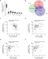
Fig 1. PD-1, TIGIT and LAG-3 are associated with virological markers of HIV persistence during ART.
(A) Expression of 7 ICs on CD4+ T cells in individuals receiving suppressive ART (n = 48). Data is represented as percentage of CD4+ T cells and horizontal bars indicate median values with interquartile ranges. (B) Size of the HIV reservoir measured by integrated HIV DNA, total HIV DNA, 2-LTR circles represented as copies per million CD4+ T cells and cell-associated US HIV RNA represented as copies per million copies of 18S. Horizontal bars indicate median values with interquartile ranges and open circles represent the limit of detection in the negative samples (based on cell input). (C), (D), (E) Associations between the frequency of CD4+ T cells harboring integrated HIV DNA and the frequency of CD4+ T cells expressing PD-1, TIGIT and LAG-3, respectively. P values were obtained from negative binomial regression analysis. Effect sizes for the associations are as follows: (C) A 2-fold increase in the percentage of PD-1+ CD4+ T cell was associated with 1.43-fold increase in the frequency of CD4+ T cell harboring integrated HIV DNA, (D) a 2-fold higher percentage of TIGIT+ CD4+ T cell was associated with 1.91-fold higher integrated HIV DNA and (E) a 2-fold higher percentage of LAG-3+ CD4+ T cell was associated with 1.62-fold higher integrated HIV DNA. Open circles represent the limit of detection in the negative samples (based on cell input).
Fig 2
Fig 2. Co-expression of PD-1, TIGIT and LAG-3 is a marker of HIV persistence during ART.
(A) Frequency of CD4+ T cells co-expressing PD-1 and/or TIGIT and/or LAG-3 (PD-1/TIGIT/LAG-3 triple–(P-T-L-), PD-1 single + (P+), TIGIT single + (T+), LAG-3 single + (L+), PD-1/TIGIT double + (P+T+), TIGIT/LAG-3 double + (T+L+), PD-1/LAG-3 double + (P+L+) and PD-1/TIGIT/LAG-3 triple + (P+T+L+)) determined by Boolean gating in cohort 1 (n = 48). Horizontal bars indicate median values with interquartile ranges. (B) Venn diagram showing the pattern of co-expression of PD-1, TIGIT and LAG-3. (C), (D), (E), (F) Associations between the frequency of CD4+ T cells harboring integrated HIV DNA and the frequency of CD4+ T cells expressing none of these markers (triple–), PD-1 and TIGIT (double +), TIGIT and LAG-3 (double +) and PD-1 and TIGIT and LAG-3 (triple +), respectively. P values were obtained from negative binomial regression analysis. Effect sizes for the associations are as follows: (C) 0.69-fold-change in integrated HIV DNA for 1 point increase in percentage of PD-1/TIGIT/LAG-3 triple—CD4+ T cells, (D) 1.18-fold-change in integrated HIV DNA for 1 point increase in percentage of PD-1/TIGIT double + CD4+ T cells, (E) 1.30-fold-change in integrated HIV DNA for 1 point increase in percentage of TIGIT/LAG-3 double + CD4+ T cells and (F) 1.94-fold-change in integrated HIV DNA for 1 point increase in percentage of PD-1/TIGIT/LAG-3 triple + CD4+ T cells. Open circles represent the limit of detection in the negative samples (based on cell input).
Fig 3
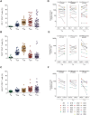
Fig 3. PD-1, TIGIT and LAG-3 identify HIV-infected cells in distinct memory CD4+ T-cell subsets during ART.
(A), (B), (C) Frequencies of memory CD4+ T-cell subsets (naïve (TN), central memory (TCM), transitional memory (TTM), effector memory (TEM) and terminally differentiated (TD)) expressing PD-1, TIGIT or LAG-3, respectively. Horizontal bars indicate median values with interquartile ranges. (D), (E), (F) Frequencies of cells harboring integrated
HIV DNA in TCM, TTM and TEM CD4+ T-cell subsets sorted based on their expression of PD-1 (n = 12), TIGIT (n = 9) or LAG-3 (n = 7), respectively. Results are expressed as the HIV copy number in million cells of a
given subset. P values were obtained from negative binomial regression analysis. Significant differences (p<0.05) are designated by a p value in bold. Open circles represent the limit of detection in the negative samples (based on cell input).
Fig 4
Fig 4. Co-expression of PD-1, TIGIT and LAG-3 identifies HIV-infected cells during ART.
(A) Pie chart representing the frequencies of memory CD4+ T cells co-expressing PD-1 and/or TIGIT and/or LAG-3 in cohort 1 (n = 48). Coloured bars on the right side designate categories of ICs expressing cells: Triple -: PD-1/TIGIT/LAG-3 triple—in blue; single +: PD-1 single +, TIGIT single +, LAG-3 single + in green; double +: PD-1/TIGIT double +, TIGIT/LAG-3 double +, PD-1/LAG-3 double + in orange and triple +: PD-1/TIGIT/LAG-3 triple + in red. (B) Frequency of memory CD4+ T cells harboring integrated HIV DNA represented as a fold change over frequency in total CD4+ T cells. Mean values and standard deviations from 5 independent donors are represented (n = 5). (C) Frequency of cells harboring inducible msRNA measured by TILDA in memory CD4+ T cells expressing any (i.e. at least one) versus none of PD-1, TIGIT or LAG-3 (mLPT+ and mLPT- respectively). P value and fold-difference were obtained from a maximum likelihood model.

References

    1. Perelson AS, Essunger P, Cao Y, Vesanen M, Hurley A, Saksela K, et al. Decay characteristics of HIV-1-infected compartments during combination therapy. Nature. 1997;387: 188–191. - PubMed
    1. Palella FJ, Delaney KM, Moorman AC, Loveless MO, Fuhrer J, Satten GA, et al. Declining morbidity and mortality among patients with advanced human immunodeficiency virus infection. HIV Outpatient Study Investigators. N Engl J Med. 1998;338: 853–860. - PubMed (V体育ios版)
    1. Davey RT, Bhat N, Yoder C, Chun TW, Metcalf JA, Dewar R, et al. HIV-1 and T cell dynamics after interruption of highly active antiretroviral therapy (HAART) in patients with a history of sustained viral suppression. Proc Natl Acad Sci U S A. 1999;96: 15109–15114. - PMC (V体育ios版) - PubMed
    1. Chun T-W, Justement JS, Murray D, Hallahan CW, Maenza J, Collier AC, et al. Rebound of plasma viremia following cessation of antiretroviral therapy despite profoundly low levels of HIV reservoir: implications for eradication. AIDS. 2010;24: 2803–2808. 10.1097/QAD.0b013e328340a239 - DOI (VSports) - PMC - PubMed
    1. International AIDS Society Scientific Working Group on HIV Cure, Deeks SG, Autran B, Berkhout B, Benkirane M, Cairns S, et al. Towards an HIV cure: a global scientific strategy. Nature Publishing Group. 2012;12: 607–614. - "VSports在线直播" PMC - PubMed

Publication types

V体育2025版 - MeSH terms