Skip to main page content (VSports在线直播)
U.S. flag

An official website of the United States government

Dot gov

The VSports app下载. gov means it’s official. Federal government websites often end in . gov or . mil. Before sharing sensitive information, make sure you’re on a federal government site. .

Https

The site is secure. The https:// ensures that you are connecting to the official website and that any information you provide is encrypted and transmitted securely V体育官网. .

. 2016 Jul 21;63(2):306-317.
doi: 10.1016/j.molcel.2016.05.041. Epub 2016 Jun 30.

Structural Basis for Cooperative Function of Mettl3 and Mettl14 Methyltransferases

Affiliations

VSports手机版 - Structural Basis for Cooperative Function of Mettl3 and Mettl14 Methyltransferases

VSports app下载 - Ping Wang et al. Mol Cell. .

Abstract

N(6)-methyladenosine (m(6)A) is a prevalent, reversible chemical modification of functional RNAs and is important for central events in biology. The core m(6)A writers are Mettl3 and Mettl14, which both contain methyltransferase domains. How Mettl3 and Mettl14 cooperate to catalyze methylation of adenosines has remained elusive. We present crystal structures of the complex of Mettl3/Mettl14 methyltransferase domains in apo form as well as with bound S-adenosylmethionine (SAM) or S-adenosylhomocysteine (SAH) in the catalytic site. We determine that the heterodimeric complex of methyltransferase domains, combined with CCCH motifs, constitutes the minimally required regions for creating m(6)A modifications in vitro. We also show that Mettl3 is the catalytically active subunit, while Mettl14 plays a structural role critical for substrate recognition. Our model provides a molecular explanation for why certain mutations of Mettl3 and Mettl14 lead to impaired function of the methyltransferase complex. VSports手机版.


Figures

Figure 1
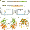
Figure 1. Structure of the Mettl3/Mettl14 methyltransferase domain complex
(A) Domain architecture of human Mettl3 and Mettl14. Crystallization constructs are indicated with color-coded bars underneath, with corresponding residue numbers. The terminal extensions of MTD14 are colored in yellow. CCCH indicates a Cys-Cys-Cys-His motif. (B) Size-exclusion chromatography with Multi-Angle Light Scattering (SEC-MALS) profile for the full-length Mettl3/Mettl14 complex. The left axis represents the absorbance at 280 nm (blue peak) and the right axis represents the measured molecular weight from the scattering (orange line) at each elution volume. The theoretical molecular weight for 1:1 Mettl3/Mettl14 complex is 118 KDa and the average measured molecular weight from MALS is 122 KDa. (C) SEC-MALS profile for the MTD3/MTD14 complex. Similar representation as (B). The theoretical molecular weight for 1:1 MTD3/MTD14 complex is 65KDa and the average measured molecular weight from MALS is 68KDa. (D) Cartoon representation of the overall structure of the MTD3/MTD14 complex. MTD3 is shown in green and MTD14 is colored orange. The N-terminal (N) and C-terminal (C) termini are as indicated, and the terminal extensions of MTD14 are colored yellow. (E) Superimposition of MTD3 and MTD14 in cartoon representation. See also Figure S1.
Figure 2
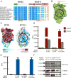
Figure 2. Mettl3 is the active subunit, and Mettl14 catalytic motif is dispensable
(A) Sequence alignment of the region containing the catalytic motifs DPPW (Mettl3) and EPPL (Mettl14). The secondary structures of the segments are indicated above the sequences. (B) Superimposition of MTD3 (green surface representation) and MTD14 (orange cartoon and sticks representation). The proposed catalytic cavity (black circle) is more occluded in MTD14. (C) Sequence conservation shown on surface representation of MTD3 and MTD14. Both MTD3 and MTD14 are colored according to the conservation score. The proposed active site is marked with a black circle. (D) In vitro methyltransferase activity of the full-length Mettl3/Mettl14 complexes expressed in E. coli, with indicated point mutations for each polypeptide. Bars correspond to amounts of tritium incorporated into methylated RNA substrates shown as disintegrations per minute (DPM) with cognate (GGACU=red) or mutant (GGAUU=gray) sequence. Data shown as mean +/- SD from three replicates. (E) In vitro methyltransferase activity of the full-length Mettl3/Mettl14 complex with indicated point mutations purified from HEK293 cells (blue bars). Data shown as mean +/- SD from three replicates. (F) SDS-PAGE analysis (visualized by Stain-Free dye) followed by western blot of the proteins expressed in HEK293 cells used in the in vitro methylation assay in Figure 2E. See also Figures S2 and S3.
Figure 3
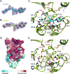
Figure 3. The SAM and SAH binding pockets of MTD3
(A) Composite omit map contoured at 1.0 σ shows clear electron density (blue mesh) for SAM, shown in stick representation with backbone colored in cyan. (B) Composite omit map contoured at 1.0 σ shows clear electron density (blue mesh) for SAH, shown in stick representation with backbone colored in yellow. (C) Detailed interactions of SAM (cyan sticks) and the active site of MTD3 (green cartoon representation). Specific residues making contacts with SAM are shown in green stick representation and labeled with black text. The catalytic motif DPPW is labeled with red text. Important water molecules involved in SAM binding are shown as red spheres. Hydrogen bonds are indicated with black dashed lines. The distance between the methyl group of SAM and the proposed catalytic residue D395 is marked with a double-headed arrow (3.8Å). (D) Evolutionary sequence conservation projected on the surface of MTD3 shows high conservation near the SAM binding site. SAM is shown in stick representation, backbone colored in cyan. (E) Detailed interactions of SAH (yellow sticks) and the active site of MTD3 (green cartoon representation). Specific residues making contacts with SAH are shown in green stick representation and labeled with black text. The active site DPPW motif is labeled with red text. Important water molecules involved in SAM binding are shown as red spheres. Hydrogen bonds are marked as black dashed lines. See also Figure S4.
Figure 4
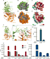
Figure 4. Mettl14 stabilizes and facilitates Mettl3 catalytic activity
(A) The overall structure of the SAM-bound MTD3/MTD14 complex shown in cartoon representation. SAM is shown in stick representation with backbone colored in cyan. Residues identified by Ligplot to be interdomain contacts are shown with sticks for side chains (green for MTD3 and orange for MTD14). (B) Green surface representation of MTD3 with residues that contact MTD14 shown in gray. (C) MTD3 in the same orientation as in (B), but colored by sequence conservation. (D) Orange surface representation of MTD14 with residues that contact MTD3 shown in gray. (E) MTD14 in the same orientation as in (D), but colored by sequence conservation. (F) The overall structure of the SAM-bound MTD3/MTD14 complex shown in cartoon representation. SAM is shown as cyan sticks. The dashed rectangle provides a close-up view of the MTD3/MTD14 binding interface. Critical residues for heterodimer formation are shown as green sticks and labeled with green text (MTD3) or orange sticks and labeled with red text (MTD14). Water molecules mediating hydrogen bonds are shown as small red spheres and hydrogen bonds are shown as black dashed lines. (G) In vitro methyltransferase activity of the full-length Mettl3/Mettl14 complex or Mettl3 alone expressed in HEK293 cells. Data shown as mean +/- SD from three replicates. (H) In vitro methyltransferase activity of the full-length Mettl3/Mettl14 complexes expressed in E. coli with indicated point mutations or truncation (MTD). Data shown as mean +/- SD from three replicates. (I) In vitro methyltransferase activity of the full length Mettl3/Mettl14 complexes expressed in HEK293 cells with indicated point mutations. Data shown as mean +/- SD from three replicates. See also Figure S5.
Figure 5
Figure 5. Proposed RNA substrate interactions with the Mettl3/Mettl14 complex
(A) In vitro methyltransferase activity of the full-length Mettl3/Mettl14 complexes expressed in E. coli with indicated point mutations or truncations (MTD). (B) In vitro methyltransferase activity of the full length Mettl3/Mettl14 complexes expressed in HEK293 cells with indicated point mutations. Data shown as mean +/- SD from three replicates. (C) Modeling of the RNA binding site by superimposition of the mDNMT1-DNA complex structure (PDB: 4DA4) onto the MTD3/MTD14 complex. The modeled DNA substrate of the complex structure of mDNMT1-DNA is shown in purple, cartoon representation and MTD of mDNMT1 is omitted for simplicity. Close-up view (dashed rectangle) shows the Y406 residue 6.3Å from the methyl group of SAM (cyan sticks). (D) In vitro methyltransferase activity of the full-length Mettl3/Mettl14 complexes expressed in E. coli with indicated point mutations. Data shown as mean +/- SD from three replicates. (E) In vitro methyltransferase activity of the full length Mettl3/Mettl14 complexes expressed in HEK293 cells with indicated point mutations. Data shown as mean +/- SD from three replicates. (F) Surface representation colored by vacuum electrostatic potential of the MTD3/MTD14 complex in the same orientation as in (C). The basic patch close to the modeled DNA is indicated by a green dashed box. SAM is shown in stick representation, colored in cyan. See also Figure S6.
Figure 6
Figure 6. Annotated model of the MTD3/MTD14 complex structure
The MTD3 (green cartoon) and MTD14 (orange cartoon, terminal extensions in yellow) complex structure is shown. The terminal extensions of MTD14 reach over to MTD3 (yellow ribbon). SAM (cyan sticks) binds one end of a catalytic cavity, which extends to the interdomain interface (light purple sphere). Concave space between the MTDs is surrounded by three “fence” loops (purple). Specific residues mutated in this study and shown to dramatically reduce enzymatic activity are shown with dark blue sphere representation, and the weak mutation of MTD14 catalytic site is shown with gray spheres. All mutated residues are labeled (MTD3: green, MTD14: orange).

V体育平台登录 - Comment in

References

    1. Adams PD, Afonine PV, Bunkóczi G, Chen VB, Davis IW, Echols N, Headd JJ, Hung LW, Kapral GJ, Grosse-Kunstleve RW, et al. PHENIX: a comprehensive Python-based system for macromolecular structure solution. Acta Crystallogr D Biol Crystallogr. 2010;66:213–221. - PMC - PubMed
    1. Alarcón CR, Lee H, Goodarzi H, Halberg N, Tavazoie SF. N6-methyladenosine marks primary microRNAs for processing. Nature. 2015;519:482–485. - PMC - PubMed
    1. Bell DW. Novel genetic targets in endometrial cancer. Expert Opinion on Therapeutic Targets. 2014;18:725–730. - PMC - PubMed
    1. Ben-Haim M, Moshitch-Moshkovitz S, Rechavi G. FTO: linking m6A demethylation to adipogenesis. Cell Research. 2014;25:3–4. - PMC - PubMed
    1. Bokar JA, Shambaugh ME, Polayes D, Matera AG, Rottman FM. Purification and cDNA cloning of the AdoMet-binding subunit of the human mRNA (N6-adenosine)-methyltransferase. RNA. 1997;3:1233–1247. - "VSports在线直播" PMC - PubMed

MeSH terms

"V体育官网入口" Grants and funding