V体育安卓版 - Skip to main page content
U.S. flag

An official website of the United States government

Dot gov

The . gov means it’s official. Federal government websites often end in . gov or . mil VSports app下载. Before sharing sensitive information, make sure you’re on a federal government site. .

Https

The site is secure. The https:// ensures that you are connecting to the official website and that any information you provide is encrypted and transmitted securely. V体育官网.

. 2017 Oct;66(10):1748-1760.
doi: 10.1136/gutjnl-2015-310847. Epub 2016 Jul 1.

Hemidesmosome integrity protects the colon against colitis and colorectal cancer (V体育ios版)

Affiliations

"VSports手机版" Hemidesmosome integrity protects the colon against colitis and colorectal cancer

Adèle De Arcangelis et al. Gut. 2017 Oct.

Abstract

Objective: Epidemiological and clinical data indicate that patients suffering from IBD with long-standing colitis display a higher risk to develop colorectal high-grade dysplasia VSports手机版. Whereas carcinoma invasion and metastasis rely on basement membrane (BM) disruption, experimental evidence is lacking regarding the potential contribution of epithelial cell/BM anchorage on inflammation onset and subsequent neoplastic transformation of inflammatory lesions. Herein, we analyse the role of the α6β4 integrin receptor found in hemidesmosomes that attach intestinal epithelial cells (IECs) to the laminin-containing BM. .

Design: We developed new mouse models inducing IEC-specific ablation of α6 integrin either during development (α6ΔIEC) or in adults (α6ΔIEC-TAM) V体育安卓版. .

Results: Strikingly, all α6ΔIEC mutant mice spontaneously developed long-standing colitis, which degenerated overtime into infiltrating adenocarcinoma. The sequence of events leading to disease onset entails hemidesmosome disruption, BM detachment, IL-18 overproduction by IECs, hyperplasia and enhanced intestinal permeability V体育ios版. Likewise, IEC-specific ablation of α6 integrin induced in adult mice (α6ΔIEC-TAM) resulted in fully penetrant colitis and tumour progression. Whereas broad-spectrum antibiotic treatment lowered tissue pathology and IL-1β secretion from infiltrating myeloid cells, it failed to reduce Th1 and Th17 response. Interestingly, while the initial intestinal inflammation occurred independently of the adaptive immune system, tumourigenesis required B and T lymphocyte activation. .

Conclusions: We provide for the first time evidence that loss of IECs/BM interactions triggered by hemidesmosome disruption initiates the development of inflammatory lesions that progress into high-grade dysplasia and carcinoma. Colorectal neoplasia in our mouse models resemble that seen in patients with IBD, making them highly attractive for discovering more efficient therapies VSports最新版本. .

Keywords: CELL MATRIX INTERACTION; COLORECTAL CANCER; IBD; INTEGRINS; INTESTINAL BARRIER FUNCTION. V体育平台登录.


Conflict of interest statement

Competing interests: None declared.

"V体育平台登录" Figures

Figure 1
Figure 1
Efficient deletion of Itga6 in α6ΔIEC mice results in compromised hemidesmosomes and epithelial fragility. (A) Strategy to generate an intestinal epithelium-specific Itga6 knockout (for details, see online supplementary figure S1A). The Itga6 floxed allele (α6fl) was obtained after insertion of two loxP cassettes (green triangles) at the Itga6 3′ end including the TM and the cytoplasmic A and B (α6A; α6B) exons. Crossing of the α6fl/fl mice with the transgenic Villin-Cre line results in a truncated Itga6 copy, denoted α6ΔIEC. (B) Morphology of the colorectal region in 15-week-old WT and α6ΔIEC mice. White stars indicate stools. Scale bars, 5 mm. (C–H) Immunodetection of hemidesmosome markers in the colon of E16.5 embryos (C and D) and in intestinal segments of mice aged 9–16 weeks (E–H); (E) rectum; (F and G) jejunum; (H) colon; 4',6-diamidino-2-phenylindole (DAPI) marks nuclei (blue). (C and E) α6-integrin chain and (D) β4-integrin chain (green) with the mucin Muc2 (red). The remaining signal in (C and E) corresponds to α6 integrin in blood vessels, confirming the specificity of the deletion in the epithelium. (F) Plectin and (G) K8/K18 intermediate filaments (green), with collagen IV (red). (H) Laminin-γ2 chain (green). White arrows, epithelium/lamina propria interface; yellow arrowheads, hemidesmosome patches; stars, areas of epithelial detachment in mutants. Scale bars, 50 µm. (I) Histological analysis of the colon from 3-week-old mice; bracket, detached cells from the surface epithelium. Scale bar, 100 µm. (J) Scattered dot plots showing the protein concentration of epithelial cell lysates obtained from small intestinal tissue of pups aged 2 (P2) and 14 (P14) days submitted to a detachment assay; error bars, SD; *p<0.05, **p<0.01. (K) Scattered dot plots showing the plasma concentration of FITC-dextran (FD4) as a measurement of intestinal permeability in 6-week-old animals fed with FD4; error bars, SD; ****p<10−4. A, anus; C, colon; e, epithelium; lp, lamina propria; lu, lumen; m, muscle layer; mes, mesenchyme; P, prolapse; R, rectum; TM, transmembrane; w, weeks; WT, wildtype.
Figure 2
Figure 2
α6ΔIEC Mice develop colitis soon after weaning. (A) Representative images of WT and α6ΔIEC adult colons observed by mini-endoscopy. Black arrow, lesion affecting the mutant mucosa; arrowheads, presence of abundant mucinous material in the lumen of the mutant colon. (B) Longitudinal H&E stained sections of the recto-anal region from animals aged 15 weeks. The bracket in mutants highlights the hyperplasia in mucosa and thickened muscle layers (arrowheads, ulcerated surface). Scale bars, 500 µm. (C) Histological scores displayed as scattered dot plots (error bars, SD) measured in the proximal and distal colons of WT and α6ΔIEC mice from 2 to 9 weeks (w) of age. (D) Transverse H&E stained sections of the distal colons of WT and α6ΔIEC mice at 4 weeks. Double arrows, crypt height. Scale bar, 50 µm. (E) Immunohistochemical BrdU detection (brown nuclei) on colon sections of 3-week-old animals; dashed lines and double arrows, area of expanded proliferation in the mutant colon. Scale bar, 50 µm. (F and G) Proliferation and crypt height measurement in colons of WT and α6ΔIEC mice aged 2 and 3 weeks, and adult stages displayed as scattered dot plots (error bars, SD). (F) Quantification of crypt height. (G) Quantification of BrdU-positive cells. *p<0.05, **p<0.01. e, epithelium; lu, lumen; ns, not significant; w, weeks; WT, wildtype.
Figure 3
Figure 3
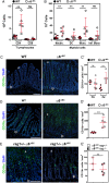
Inflammation onset in α6ΔIEC mice is mediated by myeloid cells recruitment, independently of the adaptive immune system. (A and B) FACS analysis and quantification of immune cell subpopulations present in colonic lamina propria extracts from animals raised in conventional conditions. LPMCs originate from WT and α6ΔIEC animals aged 9–15 weeks. Results are displayed as scattered dot plots (error bars, SD); each dot represents the number of positive cells present in the extract for each animal. (A) CD4+ and CD8+ T lymphocytes; (B) Cells of myeloid origin defined as: neutrophils, Ly-6G+ CD11b+; DC, Ly-6G CD11c+ MHC class IIintermediate; monocytes, Ly-6G Ly-6C+ MHC class II; pro-inflammatory monocytes, Ly-6G Ly-6C+ CD11clow MHC class II+ CD64+. (C–E) Immunodetection of CD11b+ cells (green) in the colon of WT and α6ΔIEC mice aged 4 (C) and 9 (D) weeks, and of combined rag1+/− α6ΔIEC and rag1−/− α6ΔIEC animals (E). Scale bars, 100 µm. (C′–E′) Quantification of the number of CD11b+ infiltrating cells per mm2 of colon from images illustrated in (C–E), displayed as scattered dot plots (error bars, SD). (C′) WT and α6ΔIEC mice aged 3 and 4 weeks; (D′) 9-week-old WT and α6ΔIEC mice; (E′) combined rag1+/− α6ΔIEC and rag1−/− α6ΔIEC; rag1 α6ΔIEC mice were raised in SPF conditions. **p<0.01. DC, dendritic cells; e, epithelium; FACS, fluorescence activated cell sorting; Infl. Mono, pro-inflammatory monocytes; lp, lamina propria; LPMCs, lamina propria mononuclear cells; MHC, major histocompatibility complex; Mono., monocytes; Neutro., neutrophils; ns, not significant; SPF, specific pathogen-free; w, weeks.
Figure 4
Figure 4
Colitis initiation in α6ΔIEC mice is triggered by strong epithelial IL-18 secretion and colitis worsening by IL-1β hypersecretion. (A and B) Scattered dot plot quantification of IL-1β and IL-18 levels measured by ELISA starting from colon explants of WT and α6ΔIEC mice at 9 weeks (A) and at 2, 3 or 4 weeks of age (B) (error bars, SD; ns, not significant; *p<0.05, **p<0.01, ***p<0.001). (C) Western blot analysis performed on protein extracts obtained from an enriched epithelial fraction (enriched IECs) or from whole colon segments (total colon lysates) of 2-week-old WT and α6ΔIEC mice. Representative blots of three independent experiments performed on at least five animals per group are illustrated. Western blot with GAPDH and β-Tubulin are presented as loading controls. Casp1, caspase-1; enriched IECs, enriched intestinal epithelial cells; GAPDH, glyceraldehyde 3-phosphate dehydrogenase; w, weeks.
Figure 5
Figure 5
Colitis in α6ΔIEC mice is partially improved by antibiotic treatment. (A–C) Scattered dot plot representations showing the quantification of cytokine levels measured by ELISA starting from colon explants (A), of the CD11b+ and CD4+ cells infiltrating the colonic mucosa (B), and of histological scores (C), in 9-week-old WT and α6ΔIEC mice treated with antibiotics (+ATB) or not. In each scattered dot plot, error bars represent SD; *p<0.05, **p<0.01, ***p<0.001. (A) ELISA quantification of IL-1β and IL-18 levels. (B) Quantification of CD11b+ and CD4+ infiltrating cells per surface of colon. (C) Colitis histological scores established in the proximal and distal colons. +ATB, with antibiotics; ns, not significant.
Figure 6
Figure 6
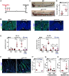
Colorectal inflammation occurs quickly after α6 integrin ablation in adult IECs. (A) Experimental procedure of TAM treatment (red arrows indicate the days of TAM administration) and subsequent analysis (black arrow) of WT and α6ΔIEC-TAM mice, illustrated for the time point 15 days post-TAM administration. Animal age is indicated in weeks. (A–D and F–H) Eight-week-old WT and α6ΔIEC-TAM mice were treated with TAM and analysed 15 days after the first TAM gavage. (B) Immunodetection of α6-integrin and β4-integrin chains (green) on colon sections of WT and α6ΔIEC-TAM. DAPI marks nuclei (blue). Scale bars, 50 µm. (C) Morphology of the colorectal region of WT and α6ΔIEC-TAM mice. Scale bars, 5 mm. (D) Scattered dot plots showing the plasma concentration of FITC-dextran (FD4) in WT and α6ΔIEC-TAM mice fed with FD4; error bars, SD. (E) ELISA quantification of the levels of IL-1β and IL-18 secreted by colon explants cultured for 24 hours. Samples were analysed 6, 10 and 15 days after the first TAM gavage in WT and α6ΔIEC-TAM mice treated with antibiotics or not. Results are displayed as scattered dot plots; error bars, SD; ns, not significant. (F and F′) Immunodetection (F) and quantification (F′) of CD11b+ cells (green) in the colon of 10-week-old TAM treated WT and α6ΔIEC-TAM mice, 15 days post TAM gavage. Scale bar, 100 µm. (G and H) Quantification by FACS analysis of the immune cell subpopulations present in LPMCs of 10-week-old TAM treated WT and α6ΔIEC-TAM mice, 15 days post TAM gavage. (G) CD4+ and CD8+ T lymphocytes; (H) Cells of myeloid origin defined as Ly-6G+ CD11b+. Results are displayed as scattered dot plots (error bars, SD); each dot represents the number of positive cells present in the extract for each animal. *p<0.05; **p<0.01; ***p<0.001. A, anus; Asterisks (*), stools; +ATB, with antibiotics; C, colon; Ca, caecum; d, day; e, epithelium; FACS, fluorescence activated cell sorting; IECs, intestinal epithelial cells; LPMCs, lamina propria mononuclear cells; lp, lamina propria; lu, lumen; ns, not significant; R, rectum; TAM, tamoxifen; w, week.
Figure 7
Figure 7
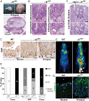
All α6ΔIEC mice spontaneously develop colitis-associated adenocarcinomas. (A–G) Tumour features in ≥1-year-old α6ΔIEC mutant mice. (A) Wide-field view (top) and histological section (bottom) of a large prolapse with an infiltrating mucinous adenocarcinoma invading the submucosa and muscle layers (m). Dotted line, region comprising tumour lesions. Scale bars: 5 mm (top); 500 µm (bottom). (B) Views of prolapse-associated tumours: (left) intraepithelial adenocarcinoma and (right) highly infiltrating adenocarcinoma (dotted lines, arrows). Higher magnifications are shown in the lower panels. Scale bars: 100 µm. (C) Ki67 immunostaining (brown nuclei) on rectal sections from 1-year-old WT and α6ΔIEC mice. Ki67+ cells are located at the base of the crypts (bar), and in the invading glands of the prolapse associated-adenocarcinoma (right panel, arrows). Scale bars: 100 µm. (D) Schematic representation showing the repartition of 1-year-old affected mice according to their genotype and the type of intestinal lesions they displayed; numbers mentioned in histogram columns and percentage (y-axis) of affected mice as well as the lesion types are indicated. The genotype of mice and the housing conditions (conventional (conv.) vs SPF) are indicated under the chart. (E) FDG-PET images of WT and α6ΔIEC mice showing a significant FDG uptake in the mutant large intestine and the rectal prolapse area (arrows). (F) Section through the recto-anal region of a combined rag1−/− α6ΔIEC mutant showing a low grade dysplasia (dotted line, arrows). Lower panel: high magnification of the dysplasia. Scale bars: 100 µm. (G) Immunodetection of laminin γ2 chain (green) in WT rectum and α6ΔIEC prolapse. DAPI (in blue) marks nuclei. Arrowheads, basement membrane underlying the surface epithelium. Arrows, increased deposits of laminin γ2 in the basement membrane and around tumour areas in α6ΔIEC prolapse. Asterisks (*), invasive gland infiltrated into the sm. Scale bar: 100 µm. ADK, adenocarcinoma; Dys, low grade dysplasia; e, epithelium; FDG, fluorodeoxyglucose; Inf, inflammation; lp, lamina propria; lu, lumen; m, muscle layer; sm, submucosa; SPF, specific pathogen-free; PET, positron emission tomography.
Figure 8
Figure 8
Model of the sequence of events leading to colorectal inflammation and carcinogenesis. Schematic drawing illustrating two IECs of the large intestine attached through α6β4 integrin to the BM via the HDs. By compelling the data obtained from α6ΔIEC and α6ΔIEC-TAM models, we suggest that the following sequence of events occurs. First, loss of the integrin impairs epithelial integrity causing epithelium fragility and detachment from the BM. The absence of tight anchorage to the BM results in disruption of the IFs network and impaired cell architecture. Second, epithelial damage triggers activation of caspase-1 in IECs followed by a drastic activation and secretion of IL-18. Intestinal permeability increases, and hyperplasia occurs in the epithelium (not illustrated). Third, presumably as a secondary consequence (defective cytoskeleton, changes in mucosal pH, cytokine release), the integrity and composition of the mucin layer become altered, favouring bacterial penetration and translocation. Concomitantly, altered epithelial barrier function triggers an inflammatory response, induced in part by exposure of the immune system to bacteria. Inflammation relies on engagement of the innate immune system, mostly independently of the lymphocyte-mediated immunity. It is characterised by enhanced secretion of epithelial IL-18 and immune-mediated IL-1β, and infiltration of CD11b+ myeloid cells into the colonic mucosa. In parallel, lymphoid cells secrete IL-17, IL-22 and IFNγ and sustain chronic inflammation over a long period. Lastly, perpetuation of long-standing colitis associated with alterations of the microbiota invariably induces the spontaneous development of adenocarcinomas in all α6ΔIEC mutants, tumour progression being dependent on the adaptive immune system. BM, basement membrane; HDs, hemidesmosomes; IECs, intestinal epithelial cells; IFs, intermediate filaments.

VSports - Comment in

VSports注册入口 - References

    1. McGuckin MA, Eri R, Simms LA, et al. . Intestinal barrier dysfunction in inflammatory bowel diseases. Inflamm Bowel Dis 2009;15:100–13. 10.1002/ibd.20539 - "V体育2025版" DOI - PubMed
    1. Okamoto R, Watanabe M. Role of epithelial cells in the pathogenesis and treatment of inflammatory bowel disease. J Gastroenterol 2016;51:11–21. 10.1007/s00535-015-1098-4 - DOI - PubMed
    1. Salim SY, Söderholm JD. Importance of disrupted intestinal barrier in inflammatory bowel diseases. Inflamm Bowel Dis 2010;17:362–81. 10.1002/ibd.21403 - "V体育ios版" DOI - PubMed
    1. Abraham C, Cho JH. Inflammatory bowel disease. N Engl J Med 2009;361:2066–78. 10.1056/NEJMra0804647 - DOI - PMC - PubMed
    1. Grivennikov SI. Inflammation and colorectal cancer: colitis-associated neoplasia. Semin Immunopathol 2013;35:229–44. 10.1007/s00281-012-0352-6 - DOI - PMC - PubMed

Publication types

MeSH terms