Skip to main page content
U.S. flag

An official website of the United States government

Dot gov

The . gov means it’s official. Federal government websites often end in . gov or VSports app下载. mil. Before sharing sensitive information, make sure you’re on a federal government site. .

Https

The site is secure. The https:// ensures that you are connecting to the official website and that any information you provide is encrypted and transmitted securely V体育官网. .

. 2016 May 27:6:26298.
doi: 10.1038/srep26298.

PGE2 maintains self-renewal of human adult stem cells via EP2-mediated autocrine signaling and its production is regulated by cell-to-cell contact

Affiliations

VSports注册入口 - PGE2 maintains self-renewal of human adult stem cells via EP2-mediated autocrine signaling and its production is regulated by cell-to-cell contact

Byung-Chul Lee et al. Sci Rep. .

"V体育官网入口" Abstract

Mesenchymal stem cells (MSCs) possess unique immunomodulatory abilities. Many studies have elucidated the clinical efficacy and underlying mechanisms of MSCs in immune disorders. Although immunoregulatory factors, such as Prostaglandin E2 (PGE2), and their mechanisms of action on immune cells have been revealed, their effects on MSCs and regulation of their production by the culture environment are less clear. Therefore, we investigated the autocrine effect of PGE2 on human adult stem cells from cord blood or adipose tissue, and the regulation of its production by cell-to-cell contact, followed by the determination of its immunomodulatory properties. MSCs were treated with specific inhibitors to suppress PGE2 secretion, and proliferation was assessed. PGE2 exerted an autocrine regulatory function in MSCs by triggering E-Prostanoid (EP) 2 receptor. Inhibiting PGE2 production led to growth arrest, whereas addition of MSC-derived PGE2 restored proliferation. The level of PGE2 production from an equivalent number of MSCs was down-regulated via gap junctional intercellular communication. This cell contact-mediated decrease in PGE2 secretion down-regulated the suppressive effect of MSCs on immune cells. In conclusion, PGE2 produced by MSCs contributes to maintenance of self-renewal capacity through EP2 in an autocrine manner, and PGE2 secretion is down-regulated by cell-to-cell contact, attenuating its immunomodulatory potency VSports手机版. .


"VSports" Figures

Figure 1
Figure 1. Inhibition of COX-2/mPGES-1 reduces the cellular growth of hUCB-MSCs and hAD-MSCs through G1 cell cycle arrest.
hUCB-MSCs and hAD-MSCs were treated with celecoxib (selective inhibitor for COX-2) or cay10526 (selective inhibitor for mPGES-1) at indicated concentrations. (a) COX-2 and mPGES-1 protein levels in hUCB-MSCs were examined by Western blot analysis. Cell proliferation was determined by (b) bromodeoxyuridine (BrdU) assay and (c) CPDL. (d) Phase-contrast images, bar = 500 μm. Upper panel: hUCB-MSCs. lower panel: hAD-MSCs. ▼; flattened or spread out cell bodies. (e) FACS analysis of cell cycle and (f) apoptosis. Gel electrophoresis was conducted under the same experimental conditions, and images of blots were cropped. Uncropped blot images are shown in Suppl. Fig. S5. Results show a representative experiment. *P < 0.05, ***P < 0.001. Results are shown as the mean ± SEM.
Figure 2
Figure 2. PGE2 produced by hMSCs restores the COX-2-mediated inhibition of cell proliferation.
(a) COX-2 inhibited cells were co-cultured with naive cells for 24 hours, and the proliferation was determined by BrdU assay. After siRNA transfection, (b) sustained expression levels of COX-2 on day 1 and 3 were detected by Western blot analysis. (c) siCOX-2 transfected cells were treated with PGE2 or co-cultured with naive and COX-2 suppressed cells, and proliferation was measured by direct cell counts and BrdU assay. Gel electrophoresis was conducted under the same experimental conditions, and images of blots were cropped. *P < 0.05, ***P < 0.001. Results are shown as the mean ± SEM.
Figure 3
Figure 3. EP2 receptor has a crucial role in hMSC self-renewal.
(a) EP receptor expression in hUCB-MSCs and hAD-MSCs was assessed by Western blot analysis. HMO6, a human microglia cell line, was used as a positive control. In the presence of selective blockers for EPs, (b) cell growth rates were examined by BrdU assay and (c) results of repeated experiments are presented. Selective blockers for EP1: SC-51089, EP2: AH-6809, EP3: L-798106, and EP4: L-161,982. (d) After celecoxib treatment, cells were treated with selective agonists for EP2 or EP3, and proliferation was measured with a BrdU assay kit. Gel electrophoresis was conducted under the same experimental conditions, and images of blots were cropped. Selective agonist for EP2: Butaprost, EP3: Sulprostone. *P < 0.05, **P < 0.01. Results are shown as the mean ± SEM.
Figure 4
Figure 4. Cell contact regulates PGE2 secretion and expression of EP receptor in hMSCs.
Identical numbers of cells were seeded on culture plates of different widths for 24 hours to achieve different cellular confluencies. (a,b) COX-2 and mPGES-1 protein levels were examined by (a) Western blot analysis and (b) immunocytochemistry. Bar = 500 μm. PGE2, IL-6, IL-8, TGF-β1, NO and IDO levels were determined from culture supernatant. (c–g) ELISA, (h) Western blotting of the IFNγ treatment group was used as a positive control. (i) Protein level of EP receptors was measured by Western blot analysis. Gel electrophoresis was conducted under the same experimental conditions, and images of blots were cropped. Uncropped blot images are shown in Suppl. Fig. S5. *P < 0.05, **P < 0.01, ***P < 0.001. Results are shown as the mean ± SEM.
Figure 5
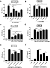
Figure 5. Gap junction intercellular communication (GJIC) is responsible for cellcontact-mediated PGE2 suppression in hMSCs.
hMSCs under cell contact were treated with 100 μM carbenoxolone (CBX), a gap junction decoupler, for 24 hours. Protein expression levels of (a) COX-2 and (b) mPGES-1 were examined by Western blot analysis. (c) PGE2 concentrations were measured from the cultured media by ELISA. Gel electrophoresis was conducted under the same experimental conditions, and images of blots were cropped. Uncropped blot images are shown in Suppl. Fig. S5. *P < 0.05, ***P < 0.001. Results are shown as the mean ± SEM.
Figure 6
Figure 6. Cell contact-mediated decrease in PGE2 secretion is followed by the attenuation of the immunosuppressive effects of hMSCs.
(a–e) Proliferation levels of mitogen-activated hMNCs were determined by BrdU, referred to as the MLR (mixed lymphocyte reaction) assay. (a) hMNCs were co-cultured with hUCB-MSCs that were pre-treated with selective inhibitors for various factors (Direct-MLR), or (b) cultured in the presence of conditioned media harvested from hUCB-MSCs (CM-MLR). Celecoxib: selective COX-2 inhibitor, 1-MT: selective IDO inhibitor, L-NAME: selective NOS inhibitor, aIL-10: neutralized with anti-IL-10 antibody. (c) hMNCs were co-culture with hUCB-MSCs treated with various doses of celecoxib, or (d) cultured in the presence of CM from hUCB-MSCs after treatment of various doses of celecoxib. (e) hMNCs were cultured in the presence of CM from the same number of hUCB-MSCs cultured under non-contact or contact condition. (f) IL-10 production in hMNCs cultured with CM from non-contact or contact hUCB-MSCs was measured by ELISA. *P < 0.05, **P < 0.01, ***P < 0.001. Results are shown as the mean ± SEM.

References (VSports)

    1. Uccelli A., Moretta L. & Pistoia V. Mesenchymal stem cells in health and disease. Nat Rev Immunol 8, 726–736, 10.1038/nri2395 (2008). - DOI - PubMed
    1. Bianco P. et al. The meaning, the sense and the significance: translating the science of mesenchymal stem cells into medicine. Nat Med 19, 35–42, 10.1038/nm.3028 (2013). - V体育官网 - DOI - PMC - PubMed
    1. Bernstein I. D. & Delaney C. Engineering stem cell expansion. Cell Stem Cell 10, 113–114, 10.1016/j.stem.2012.01.012 (2012). - DOI - PubMed
    1. Cho Y. H. et al. Enhancement of MSC adhesion and therapeutic efficiency in ischemic heart using lentivirus delivery with periostin. Biomaterials 33, 1376–1385, 10.1016/j.biomaterials.2011.10.078 (2012). - "VSports注册入口" DOI - PubMed
    1. Hahn J. Y. et al. Pre-treatment of mesenchymal stem cells with a combination of growth factors enhances gap junction formation, cytoprotective effect on cardiomyocytes, and therapeutic efficacy for myocardial infarction. J Am Coll Cardiol 51, 933–943, 10.1016/j.jacc.2007.11.040 (2008). - DOI - PubMed

"V体育官网" Publication types

MeSH terms

"VSports在线直播" LinkOut - more resources