V体育平台登录 - Skip to main page content
U.S. flag

An official website of the United States government

Dot gov

The . gov means it’s official. Federal government websites often end in VSports app下载. gov or . mil. Before sharing sensitive information, make sure you’re on a federal government site. .

Https

The site is secure V体育官网. The https:// ensures that you are connecting to the official website and that any information you provide is encrypted and transmitted securely. .

. 2016 Jul;17(7):851-860.
doi: 10.1038/ni.3441. Epub 2016 May 9.

BACH2 regulates CD8(+) T cell differentiation by controlling access of AP-1 factors to enhancers

Affiliations

VSports - BACH2 regulates CD8(+) T cell differentiation by controlling access of AP-1 factors to enhancers

Rahul Roychoudhuri et al. Nat Immunol. 2016 Jul.

V体育2025版 - Abstract

T cell antigen receptor (TCR) signaling drives distinct responses depending on the differentiation state and context of CD8(+) T cells. We hypothesized that access of signal-dependent transcription factors (TFs) to enhancers is dynamically regulated to shape transcriptional responses to TCR signaling. We found that the TF BACH2 restrains terminal differentiation to enable generation of long-lived memory cells and protective immunity after viral infection. BACH2 was recruited to enhancers, where it limited expression of TCR-driven genes by attenuating the availability of activator protein-1 (AP-1) sites to Jun family signal-dependent TFs. In naive cells, this prevented TCR-driven induction of genes associated with terminal differentiation. Upon effector differentiation, reduced expression of BACH2 and its phosphorylation enabled unrestrained induction of TCR-driven effector programs VSports手机版. .


Conflict of interest statement (VSports手机版)

The authors declare no competing financial interests.

Figures (VSports手机版)
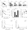
Figure 1
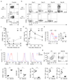
Figure 1. BACH2 is required for maintenance of primary and secondary CD8+ T cell responses in vivo and establishment of protective immunity.
a, Pre-transfer flow cytometry of WT and KO naïve OT-I cells mixed at ~1:1 ratio. b-c, Kinetic analysis of cells in a following transfer into recipient mice and infection with VV-OVA. Representative flow cytometry (b) and replicate measurements (c) are shown. d, Ratio of WT and KO CD8+ T cells in indicated organs at day 21 following infection normalized to ratios found in the spleen. e, Bacterial titers following LM-OVA infection of animals which were previously administered individual transfers of naïve WT or KO OT-I cells and infected with VV-OVA. Primary infection with VV-OVA occurred 60 days prior to secondary infection with LM-OVA. f-g, Secondary recall responses mediated by WT and KO central memory (f) and effector memory (g) cells isolated at day 14 following primary infection as in c upon ~1:1 mixed transfer into infection-naïve recipients subsequently infected with VV-OVA. Numbers in gates indicate percentages. *P<0.05; **P<0.01; ***P<0.005; ****P<0.001. Data (a-g) are representative of two independent experiments with 4-6 mice per genotype and timepoint (c-g; mean and s.e.m.).
Figure 2
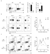
Figure 2. BACH2 prevents terminal differentiation and apoptosis of CD8+ T cells following viral infection.
a-d, Surface phenotype of cells before (a) or at indicated timepoints (b) after mixed transfer of naïve WT and KO OT-I CD8+ T cells into recipient mice and infection with VV-OVA. Representative flow cytometry (a-b) and replicate measurements of CD62L (c) and KLRG1 (d) on the surface of transferred cells are shown. Too few KO cells were detected at day 90 for phenotypic analysis. e, Intracellular cytokine staining of IFN-γ expression by WT and KO CD8+ T cells from spleens of animals 5 days following infection. f, Overexpression of BACH2 prevents terminal effector differentiation following secondary responses to infection in vivo. Expression of CD62L, CD44 and KLRG1 on the surface of CD8+ T cells 5 days following transfer of GFP+ (transduced) OT-I CD8+ T into animals subsequently infected with VV-OVA. g, Expression of IFN-γ and TNF by transferred cells following brief restimulation ex vivo. Numbers in gates indicate percentages. *P<0.05; **P<0.01; ***P<0.005; ****P<0.001. Data (a-g) are representative of two independent experiments with 4-6 mice per genotype and timepoint (c-g; graphs represent mean and s.e.m.).
Figure 3
Figure 3. BACH2 suppresses differentiation of short-lived effector cells and apoptosis during acute CD8+ T cell responses to viral infection.
a, Expression of CD127 and KLRG1 on the surface of OT-I cells at day 7 following mixed transfer of naïve FACS-sorted WT and KO OT-I CD8+ T cells into recipient mice and infection with VV-OVA. b, BrdU incorporation by WT and Bach2 KO CD8+ T cells at the indicated timepoints following infection. Mice were injected intraperitoneally with BrdU 16 h preceding harvest at each timepoint. *P<0.05, **P<0.005. c, Annexin V and propidium iodide staining of cells at indicated timepoints following infection. Numbers in gates indicate percentages. *P<0.01, **P<0.001. Data are representative of two independent experiments with 4-6 mice per genotype and timepoint (a-c; graphs represent mean and s.e.m.).
Figure 4
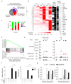
Figure 4. Enrichment of BACH2 at AP-1 sites within CD8+ T cell genomes.
a, Relationship of genome-wide BACH2 binding sites in d5 in vitro activated CD8+ T cells to annotated genes. b, Consensus motif enriched within BACH2 binding sites in CD8+ T cells (left). AP-1 consensus motif (JASPAR; right). c, Genome-wide co-localization analysis of BACH2 with indicated enhancer associated post-transcriptional histone modifications or p300 within d5 in vitro activated CD8+ T cells. d-f, Co-localization of JunB (d), c-Jun (e) and JunD (f) at genome-wide BACH2 binding sites in CD8+ T cells. Histograms indicate normalized average tag density at indicated distances from BACH2 peak summits. Pie charts indicate proportions of Jun binding sites that directly overlap with BACH2 binding sites (red indicates proportion of Jun sites exhibiting co-localization with BACH2). Binomial tests were used to determine peak significance within ChIP-Seq data and a threshold of p<1 × 10⁻⁵ was used for peak calling.
Figure 5
Figure 5. BACH2 represses TCR-driven transcriptional programs in CD8+ T cells.
Naïve WT and KO CD8+ T cells isolated ex vivo by flow cytometric sorting were stimulated briefly with platebound anti-CD3 and anti-CD28. a, Pie chart showing genes upregulated in KO compared with WT cells at 0, 6 or 18h (p<0.05, log2 FC>1). Of these, 192 TCR-induced genes (upregulated by stimulation in WT or KO cells at 6 or 18 h) were identified. b, Hierarchical cluster analysis of TCR-induced genes upregulated in KO cells. FPKM values are normalized to row maxima. TCR-inducibility in WT and KO cells, and BACH2 binding are indicated to right of heatmap. c, Fraction of genes in b differentially expressed in KO compared with WT cells at 0, 6 and 18h (p<0.05, log2 FC>1). d, Enrichment of genes in b with transcriptional differences between terminally differentiated KLRG-1+ OT-I cells and naïve cells. Normalized enrichment score (NES) = 1.9; Q<0.0001. e, Alignments showing mRNA expression in WT and KO naïve CD8+ T cells (top) and mapped BACH2, JunD and H3K4me1 binding sites (bottom). f, Naïve WT and KO CD8+ T cells pretreated for 1 h with JNKi (12.5µM) or vehicle at 37°C were stimulated with platebound anti-CD3 and anti-CD28 and mRNA expression was measured relative to Actb. *P<0.01; **P<0.001. g, mRNA expression at indicated timepoints following anti-CD3 restimulation of GFP+ (transduced) cells infected with indicated retroviruses during primary stimulation. Average FPKM values (b) and analyses of two replicate measurements per condition (a, c-e) are shown. Data (f-g; mean and s.e.m.) are representative of two independent experiments.
Figure 6
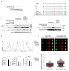
Figure 6. BACH2 restricts access of Jun family TFs to regulatory elements of TCR-induced genes.
a, Histogram of JunD enrichment centered around genome-wide BACH2 binding sites in WT and Bach2–/– d5 in vitro activated CD8+ T cells. Western blot shows abundance of indicated total proteins in these cells. b, Average ATAC-Seq signal around BACH2 binding sites in WT and Bach2–/– d5 in vitro activated CD8+ T cells. c, Scatterplot comparing differences in average JunD binding at BACH2 binding sites (x-axis) with differences in mRNA expression of associated genes (y-axis). TCR-induced genes (induced by 2 h anti-CD3 stimulation of d5 in vitro activated CD8+ T cells) and non-induced genes are shown separately. Statistical significance was evaluated using two-sample Kolmogorov-Smirnov test. d, Representative alignments of ChIP-, ATAC- and RNA-Seq measurements in d5 in vitro activated CD8+ T cells at selected loci identified in c. Arrows indicate increased JunD binding at BACH2 binding sites in Bach2–/– cells. e, mRNA expression at specific timepoints following restimulation of transduced (GFP+) d5 in vitro activated CD8+ T cells infected with indicated retroviruses during primary stimulation. f, mRNA expression in WT and KO CD8+ T stimulated in vitro for 5 days in the presence of JNKi (12.5µM) or vehicle. *P<0.001. g, Congenically distinct GFP+ (transduced) WT and KO OT-I cells infected with indicated retroviruses during primary stimulation were co-transferred at ~1:1 ratio into recipient mice. KO/WT ratios within spleens of recipient animals were measured 5 days after infection with VV-OVA. *P<0.01. Data and analyses are based upon one (a), four (b) and two (c) measurements or represent two repeated experiments (e-g; mean and s.e.m.).
Figure 7
Figure 7. Dynamic regulation of BACH2 expression enables stage-specific control of TCR-driven gene expression.
a-b, Genomic alignments (a) and quantification (b) of H3K4me3 in naïve, memory and effector CD8+ T cells at the Bach2 gene (UCSC). c, mRNA expression in indicated subsets of OT-I cells sorted 30 days after infection with VV-OVA. *P<0.05, **P<0.01. d, mRNA expression normalized to Actb (left), and protein expression (right) in naïve and CD62L KLRG1 effector OT-I cells isolated 7 days after infection with VV-OVA (d7 Eff). *P<0.001. e, Comparison of TCR-induced gene expression in KO and WT naïve and d7 Eff cells. Sorted naïve and d7 Eff cells were subjected to brief (6h) stimulation ex vivo with platebound anti-CD3 and anti-CD28. TCR-induced genes (induced by 6 h stimulation of either naïve WT (blue arc) or KO cells (red arc); p<0.05, log2 FC>1) significantly upregulated in KO compared with WT cells at either 0 or 6 h (p<0.05, log2 FC>1) were selected for further analysis. f, Comparison of expression of genes in e in Naïve and d7 Eff cells at 0 or 6 h after stimulation. Color scale of heatmap normalized to row maxima. g, Fraction of genes in f that are significantly differentially expressed between KO and WT cells at either 0 or 6h following stimulation (p<0.05, log2 FC>1). Data representative of two independent experiments (c-d; mean and s.e.m.), or average FPKM values (f) and analyses (e,g) of two replicate measurements per genotype and timepoint.
Figure 8
Figure 8. Phosphorylation of BACH2 at Ser520 results in functional inactivation.
a, d5 in vitro activated CD8+ T cells were restimulated for 30 minutes with crosslinked anti-CD3 and cellular lysates were treated with or without lambda phosphatase (LP). BACH2 migration was resolved by immunostaining. b, Alignment of BACH2 amino acid sequences in indicated vertebrates. S520A substitution mutation is shown. c, CD8+ T cells were transduced with indicated retroviruses during primary stimulation and restimulated with crosslinked anti-CD3 4 days after transduction, preincubating with indicated pharmacological inhibitors for 1 h. α-Flag-immunoprecipitated proteins or total lysates were subjected to immunostaining. Phospho-BACH2 was detected using an RXXpS phospho-Akt substrate motif-specific antibody. d, Purified Akt1 was combined with ATP and immunoprecipitated Flag-tagged WT or BACH2(S520A). Phospho-BACH2 was detected using an RXXpS phospho-Akt substrate motif-specific antibody. e, CD8+ T cells were transduced with indicated retroviruses during primary stimulation in rhIL-2 and IL-12 prior to restimulation on anti-CD3-coated plates in the presence of Brefeldin A for intracellular cytokine staining. *P<0.05, **P<0.01, ***P<0.005. f-g, CD8+ T cells were transduced with indicated retroviruses and cultured as in e. Cells were briefly restimulated with platebound α-CD3 (2h) and stained for the retroviral transduction marker (Thy-1.1) during the last 30 mins of culture. Fixed cells were stained with DAPI and localization of BACH2-GFP was resolved by imaging flow cytometry. Representative cells (f) and the ratio of nuclear/cytoplasmic mean pixel intensity (MPI) in replicate cells (g) are shown. ***P<0.005. Data representative of two (e, g; mean and s.e.m.) and three (a, c-d) independently repeated experiments.

Comment in

References

    1. Kaech SM, Cui W. Transcriptional control of effector and memory CD8+ T cell differentiation. Nat Rev Immunol. 2012;12:749–761. - PMC - PubMed
    1. Belz GT, Kallies A. Effector and memory CD8+ T cell differentiation: toward a molecular understanding of fate determination. Curr Opin Immunol. 2010;22:279–285. - "VSports" PubMed
    1. D’Cruz LM, Rubinstein MP, Goldrath AW. Surviving the crash: transitioning from effector to memory CD8+ T cell. Semin Immunol. 2009;21:92–98. - PMC - PubMed
    1. Williams MA, Bevan MJ. Effector and memory CTL differentiation. Annu Rev Immunol. 2007;25:171–192. - PubMed
    1. Restifo NP, Gattinoni L. Lineage relationship of effector and memory T cells. Curr Opin Immunol. 2013;25:556–563. - PMC - PubMed

Publication types

"V体育ios版" MeSH terms

Substances