Skip to main page content
U.S. flag

An official website of the United States government

Dot gov

The VSports app下载. gov means it’s official. Federal government websites often end in . gov or . mil. Before sharing sensitive information, make sure you’re on a federal government site. .

Https

The site is secure. The https:// ensures that you are connecting to the official website and that any information you provide is encrypted and transmitted securely V体育官网. .

. 2016 Jun;49(3):362-72.
doi: 10.1111/cpr.12254. Epub 2016 Apr 29.

Inflammatory cytokines induce caveolin-1/β-catenin signalling in rat nucleus pulposus cell apoptosis through the p38 MAPK pathway

Affiliations

Inflammatory cytokines induce caveolin-1/β-catenin signalling in rat nucleus pulposus cell apoptosis through the p38 MAPK pathway

Jianxi Wang et al. Cell Prolif. 2016 Jun.

Abstract

Objectives: Apoptosis of nucleus pulposus (NP) cells is a major cause of intervertebral disc degeneration. To elucidate relationships between caveolin-1 and cytokine-induced apoptosis, we investigated the role of caveolin-1 in cytokine-induced apoptosis in rat NP cells and the related signalling pathway. VSports手机版.

Materials and methods: Rat NP cells were treated with interleukin (IL)-1β or tumour necrosis factor alpha (TNF-α), and knockdown of caveolin-1 and β-catenin was achieved using specific siRNAs. Then, apoptotic level of rat NP cells and expression and activation of caveolin-1/β-catenin signalling were assessed by flow cytometric analysis, qRT-PCR, western blotting and luciferase assays V体育安卓版. The relationship between the mitogen-activated protein kinase (MAPK) pathway and caveolin-1 promoter activity was also determined by luciferase assays. .

Results: IL-1β and TNF-α induced apoptosis, upregulated caveolin-1 expression and activated Wnt/β-catenin signalling in rat NP cells, while the induction effect of cytokines was reversed by caveolin-1 siRNA and β-catenin siRNA. Promotion of rat NP cell apoptosis and nuclear translocation of β-catenin induced by caveolin-1 overexpression were abolished by β-catenin siRNA V体育ios版. Furthermore, pretreatment with a p38 MAPK inhibitor or dominant negative-p38, blocked cytokine-dependent induction of caveolin-1/β-catenin expression and activity. .

Conclusions: The results revealed the role of p38/caveolin-1/β-catenin in inflammatory cytokine-induced apoptosis in rat NP cells. Thus, controlling p38/caveolin-1/β-catenin activity seemed to regulate IL-1β- and TNF-α-induced apoptosis in the NP during intervertebral disc degeneration. VSports最新版本.


"VSports app下载" Figures

Figure 1
Figure 1
Expression of caveolin‐1 in rat NP cells treated with inflammatory cytokines. After treatment with IL‐1β (10 ng/ml) or TNF‐α (50 ng/ml), the expression of caveolin‐1, at both the mRNA level (by real‐time PCR; (a) and (b), respectively) and the protein level (by Western blot analysis; (c) and (d), respectively), was analysed at the designated time points (0, 1, 6, 12, 24 and 48 h) in rat NP cells. Each sample was analysed in triplicate (**P < 0.01). Error bars represent SD.
Figure 2
Figure 2
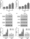
Activation of Wnt/β‐catenin signalling after inflammatory cytokine treatment in rat NP cells. After treatment with IL‐1β (10 ng/ml) or TNF‐α (50 ng/ml), the expression of β‐catenin, at both the mRNA level (by qRTPCR; (a) and (b), respectively) and the protein level (by Western blot analysis; (c) and (d), respectively; in both the cytosol and nucleus), was analysed in rat NP cells. The activation of Wnt/β‐catenin signalling in rat NP cells was also assessed by a dual luciferase assay (TOPflash/FOPflash), after treatment with IL‐1β (e) or TNF‐α (f). Each sample was analysed in triplicate. Error bars represent SD. **P < 0.01; ns, not significant.
Figure 3
Figure 3
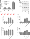
Role of caveolin‐1/β‐catenin signalling in rat NP cell apoptosis. After treatment with inflammatory cytokines or siRNA specific for Cav‐1 or β‐catenin, the expression of caveolin‐1 and β‐catenin was determined in rat NP cells by Western blotting and the apoptotic rate was determined by an FITC‐annexin V/PI FCM assay. (a) Protein expression levels of caveolin‐1 (a) and β‐catenin (b) were significantly downregulated by siRNA. (c) Scattergram of FITC‐annexin V/PI FCM of NP cells. The graphs are representative of several experiments. (d) Cytokine‐induced apoptosis was significantly reduced by Cav‐1 and β‐catenin siRNA. Values represent the mean of three experiments. Results are shown as mean ± SD. * versus Ctr; # versus IL‐1β or TNF‐α; *,#P < 0.05, **P < 0.01. Ctr, control; Cav‐1, Caveolin‐1; β‐cat, β‐catenin.
Figure 4
Figure 4
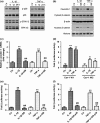
Effect of caveolin‐1 si RNA on the Wnt/β‐catenin signalling pathway and rat NP cell apoptosis. (a) Western blot analysis of the expression of nuclear and cytosolic β‐catenin and histones in NP cells treated with IL‐1β or TNF‐α, with or without siRNA against caveolin‐1 (Cav‐1). Results shown that the expression of nuclear β‐catenin was blocked by caveolin‐1 siRNA in cytokine‐treated rat NP cells. (b–d) Caspase‐3, caspase‐8 and caspase‐9 activity was determined by a caspase activity assay. Caspase‐3 and caspase‐9 were completely inhibited by Cav‐1 siRNA or β‐catenin siRNA in rat NP cells treated with IL‐1β or TNF‐α. Each sample was analysed in triplicate. Error bars represent SD. * versus Ctr; # versus IL‐1β or TNF‐α; *, #P < 0.05, **, ##P < 0.01. Ctr, control; Cav‐1, Caveolin‐1; β‐cat, β‐catenin.
Figure 5
Figure 5
Effect of caveolin‐1 overexpression on the Wnt/β‐catenin signalling pathway and rat NP cell apoptosis. (a) Caveolin‐1 overexpression was performed in rat NP cells. Caveolin‐1 expression at both the protein and mRNA level was significantly upregulated. Activation of Wnt/β‐catenin signalling and apoptosis of rat NP cells were tested by Western blotting and FITC‐annexin V/PI FCM, respectively. Caspase‐3, caspase‐8 and caspase‐9 activity was also measured. (b) Caveolin‐1 overexpression promoted nuclear translocation of β‐catenin. (c) Contour diagram of FITC‐annexin V/PI FCM of rat NP cells. Caveolin‐1 overexpression induced rat NP cell apoptosis, but Cav‐1‐induced apoptosis was inhibited by β‐catenin siRNA. (d–f) Increased caspase‐3, caspase‐8 and caspase‐9 activity in Cav‐1 overexpressing cells was reduced by β‐catenin siRNA. Each sample was analysed in triplicate. Error bars represent SD. * versus Ctr; # versus Cav‐1; *, #P < 0.05, **, ##P < 0.01. Ctr, control; Vec, Vector; Cav‐1, Caveolin‐1; β‐cat, β‐catenin.
Figure 6
Figure 6
Effect of MAPK signalling on the caveolin‐1/β‐catenin signalling pathway in cytokine‐treated rat NP cells. (a) Expression levels of p‐p38 and p‐ERK1/2 MAPK were determined by Western blotting in rat NP cells after IL‐1β and TNF‐α stimulation. (b) Pretreatment with the specific p38 MAPK inhibitor SB203580 blocked the protein expression of Cav‐1 and nuclear translocation of β‐catenin induced by IL‐1β and TNF‐α in rat NP cells. (c) The increase in the caveolin‐1 mRNA level induced by IL‐1β and TNF‐α was also decreased by SB203580. (d) To investigate the mechanism of cytokine action on caveolin‐1 expression, we measured the activity of two promoter fragments Cav‐1‐W1 (−1296/−1) and Cav‐1‐W2 (−222/−1). The activity of the Cav‐1‐W1 reporter was significantly induced by the cytokines (~7.2‐fold), whereas Cav‐1‐W2 showed only a ~2.8‐fold induction upon cytokine treatment. To elucidate the role of p38 MAPK in controlling promoter activity in rat NP cells, we measured Cav‐W1 activity following IL‐1β and TNF‐α treatment with or without p38 MAPK inhibitor SB203580 or dominant negative (DN)‐p38 transfection. Cytokine‐mediated induction of Cav‐W1 activity was completely blocked by the p38 MAPK pathway inhibitor (e) and DN‐p38 (f). Results are shown as the mean ± SD from three independent experiments. * versus Ctr; # versus IL‐1β or TNF‐α; *P < 0.05, **, ##P < 0.01. Ctr, control; SB, SB203580.

References

    1. Katz JN (2006) Lumbar disc disorders and low‐back pain: socioeconomic factors and consequences. J. Bone Joint Surg. Am. 88(Suppl 2), 21–24. - PubMed
    1. Hadjipavlou AG, Tzermiadianos MN, Bogduk N, Zindrick MR (2008) The pathophysiology of disc degeneration: a critical review. J. Bone Joint Surg. Br. 90, 1261–1270. - PubMed
    1. Ding F, Shao ZW, Xiong LM (2013) Cell death in intervertebral disc degeneration. Apoptosis 18, 777–785. - "V体育官网入口" PubMed
    1. Liu G, Cao P, Chen H, Yuan W, Wang J, Tang X (2013) MiR‐27a regulates apoptosis in nucleus pulposus cells by targeting PI3K. PLoS ONE 8, e75251. - PMC - PubMed
    1. Aguiar DJ, Johnson SL, Oegema TR (1999) Notochordal cells interact with nucleus pulposus cells: regulation of proteoglycan synthesis. Exp. Cell Res. 246, 129–137. - PubMed

MeSH terms (VSports在线直播)

Substances

"VSports在线直播" Associated data