Skip to main page content
U.S. flag

An official website of the United States government

Dot gov

The . gov means it’s official. Federal government websites often end in . gov or . mil. Before sharing sensitive information, make sure you’re on a federal government site. VSports app下载.

Https

The site is secure V体育官网. The https:// ensures that you are connecting to the official website and that any information you provide is encrypted and transmitted securely. .

. 2016 Apr 26;55(16):2411-21.
doi: 10.1021/acs.biochem.6b00093. Epub 2016 Apr 13.

Effect of the Spiroiminodihydantoin Lesion on Nucleosome Stability and Positioning

Affiliations

VSports - Effect of the Spiroiminodihydantoin Lesion on Nucleosome Stability and Positioning

Erika M Norabuena et al. Biochemistry. .

Abstract

DNA is constantly under attack by oxidants, generating a variety of potentially mutagenic covalently modified species, including oxidized guanine base products. One such product is spiroiminodihydantoin (Sp), a chiral, propeller-shaped lesion that strongly destabilizes the DNA helix in its vicinity VSports手机版. Despite its unusual shape and thermodynamic effect on double-stranded DNA structure, DNA duplexes containing the Sp lesion form stable nucleosomes upon being incubated with histone octamers. Indeed, among six different combinations of lesion location and stereochemistry, only two duplexes display a diminished ability to form nucleosomes, and these only by ∼25%; the other four are statistically indistinguishable from the control. Nonetheless, kinetic factors also play a role: when the histone proteins have less time during assembly of the core particle to sample both lesion-containing and normal DNA strands, they are more likely to bind the Sp lesion DNA than during slower assembly processes that better approximate thermodynamic equilibrium. Using DNase I footprinting and molecular modeling, we discovered that the Sp lesion causes only a small perturbation (±1-2 bp) on the translational position of the DNA within the nucleosome. Each diastereomeric pair of lesions has the same effect on nucleosome positioning, but lesions placed at different locations behave differently, illustrating that the location of the lesion and not its shape serves as the primary determinant of the most stable DNA orientation. .


Figures

Figure 1:
Figure 1:. Sequence and Starting Model of the Nucleosome Core Particle.
(a) Sequenceof the 146-bp DNA duplex containing an Sp lesion at one of three defined locations, called here Lesion 7 (green), Lesion 9 (orange), and Lesion 12 (purple). (b) Location of the Sp lesions highlighted on the unmodified nucleosome model of Luger and colleagues (PDB ID 1AOI, reference 42), assuming that the DNA position on the protein core is not changed by the presence of the lesion. All three lesions were placed near the dyad axis of the particle. The histone H3-H4 tetramer is colored in dark grey, while the H2A/H2B dimers are colored in light grey. (c) Proposed location of the Sp lesions shown from the edge of the particle down along the dyad axis. (d, e) Proposed location of the Sp lesions, highlighting their proximity to the histone protein core and (in)accessibility from the solution. Note that the actual DNA bases shown are the control DNA (G, A, and C) and not Sp. Two slightly different conformations, ~1 bp apart, are possible because the model structure is pseudopalindromic.
Figure 2:
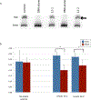
Figure 2:. Stability of Nucleosome Formation Depends upon the Rate of Assembly.
(a) Gel shift showing the results of nucleosome exchange from unlabeled random-sequence DNA to 146-mer duplex under “fast” single dialysis conditions. Nucleosomes were assembled using the control DNA or DNA containing the Sp lesion. DNA duplexes containing the two Sp diastereomers at location 12 were evaluated independently (12-1 and 12-2). The bottom band (“free”) contains protein-free DNA and the top bands (“nuc”) contain mononucleosomes. Double arrows highlight the double band illustrating the presence of at least two nucleosome conformations. (b) Percent of DNA that forms nucleosomes under “fast” single dialysis or “slow” double dialysis conditions. The undamaged control forms nucleosomes equally well under either condition, although the variability in the data is diminished under the slower assembly conditions (F-test, >95% confidence). The DNA containing the Sp lesion at location 12 forms fewer nucleosomes when allowed equilibrate more gradually (student’s t test, >95% confidence). Error bars show the standard deviation of the mean (n=5 for all samples, except control double dialysis n=10).
Figure 3:
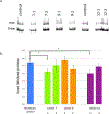
Figure 3:. Stable Lesion-Containing Nucleosomes Are Formed under Slow, Double-Dialysis Conditions.
(a) Gel shift showing the results of “slow” nucleosome exchange from unlabeled random-sequence DNA to 146-mer duplex. All seven possible duplexes are shown, including the control parent DNA and DNA containing both diastereomers of the Sp lesion at locations 7, 9, and 12. Lesion DNA was radiolabeled on the 5’ end of the lesion strand; control DNA was 5’ end labeled on strand 1 (lane 1) or strand 2 (lane 6). The bottom band (“free”) contains protein-free DNA and the top band (“nuc”) contains mononucleosomes. (b) DNA containing the Sp lesion forms nucleosomes almost as well as the control. Error bars show the standard error of the mean (control n=10, lesion n=5). Only two duplexes, Lesion 7-1 and Lesion 12-1, bind significantly more weakly than the control (student’s t-test, > 95% confidence).
Figure 4:
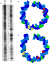
Figure 4:. DNase I Footprinting of Control DNA in Nucleosomes.
(a) DNase I cleavage of bare and nucleosomal control DNA duplexes. The + symbol indicates a location of relatively increased cleavage in the nucleosomal DNA, while * indicates areas of relatively diminished cleavage. Note that the cleavage in the nucleosomal DNA is globally lower than in the bare DNA, both due to shielding by histones and to the presence of unlabeled chicken DNA; both nuclease concentration and visual contrast have been increased in the nucleosomal sample to compensate. (b,c) Changes in DNase I cleavage mapped to the model nucleosome upon which this 146 base pair sequence is loosely based (PDB ID 1AOI, reference 42). The histone octamer has been removed for clarity. Regions of strong cleavage are colored green, weak cleavage is colored yellow, and absent or drastically reduced cleavage is red. No data are available where the DNA is colored blue. The most robustly cleaved regions correlate with locations where the minor groove is open to the nuclease and the backbone points outward; regions of diminished or absent cleavage point inward toward the octamer.
Figure 5:
Figure 5:. Computationally Predicted Regions of DNase I Accessibility and Cleavage.
In order to determine where the DNase I would be expected to cleave the model nucleosome structure, the energetic stability of DNAse I-nucleosome complexes was evaluated at every position of the endonuclease along (a) strand 1 and (b) strand 2 of the undamaged 146-base pair DNA duplex. In order to display the large range of interaction energies, y-values represent the log10 of the absolute value of the interaction energies (in kcal/mol). Signs were retained so that negative energies correspond to energetically favorable complexes, while positive energies reflect steric clashes between the histones and the DNase I. The x axes are calibrated to the base pair nearest the active site histidines. In addition to reiterating the experimentally well-known 10 bp periodicity characteristic of nucleosomes, this model illustrates the relatively small “bite size” of the enzyme, since only ~4 bp are accessible for cleavage on each strand as it passes the outside of the nucleosome core particle. At the bottom of each graph are the positions of experimentally-observed DNase I cleavage on our 146 bp sequence. As in figure 4, regions of strong cleavage are colored green, weak cleavage is colored yellow, and absent or drastically reduced cleavage is red. Generally the experimentally-determined DNase I cleavage lines up well with the modeled negative interaction energies, and the small discrepancies do not show a consistent 5’ or 3’ trend that would indicate a different rotational or translational position from the model.
Figure 6:
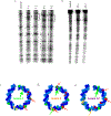
Figure 6:. DNase I Footprinting of Nuclesomes Containing Site-Specifically Incorporated Sp Lesions.
(a) Footprinting analysis of Lesions 7 and 9 and control DNA under double dialysis conditions. Globally, the five samples are quite similar, but some small differences are seen between the two different lesion positions, although not between diastereomers placed in the same location. Bands that are missing relative to the control are marked with asterisks (*); bands augmented relative to the control are indicated with plus signs (+). Location of the lesions are highlighted with the letter L. The brackets have been added to highlight how the pattern of cleavage is shifted by ~3-4 base pairs between the two lesion positions. (b) Footprinting analysis of Lesion 12 and control DNA under double dialysis conditions. All three samples are essentially identical except for the strong band around the lesion (marked L and *). (c-e) Summary of DNase I footprinting results for DNA containing Sp lesions at locations 7, 9, and 12 superimposed on the structure of the nucleosome core particle DNA with the proteins removed for clarity. Since each pair of diastereomers behaved identically with respect to footprinting, only one picture is shown for each lesion location. Regions of strong cleavage are colored green, weak cleavage is colored yellow, and absent or drastically reduced cleavage is colored red. Since most of the cleaved bases are the same as in the control (Fig. 4bc), red and green arrows highlight locations where the pattern of cleavage is different than the control. Large green arrows correlate with the location of the lesions in each case. For lesion 12, changes in footprints distal from the lesion were observed only under single dialysis conditions.

References

    1. Friedberg EC, Walker GC, Siede W, Wood RD, Schultz RA, and Ellenberger T (2006) DNA Repair and Mutagenesis 2nd ed. ASM Press, Washington, D.C.
    1. Dedon PC (2011) Oxidation and deamination of DNA by endogenous sources In Chemical Carcinogenesis: Current Cancer Research (Penning TM, Ed.) pp. 209–225, Springer Science and Business Media.
    1. Evans MD, Dizdaroglu M, and Cooke MS (2004) Oxidative DNA damage and disease: induction, repair and significance. Mutat. Res. 567, 1–61. - PubMed
    1. Steenken S, and Jovanovic S (1997) How easily oxidizable is DNA? One-electron reduction potentials of adenosine and guanosine radicals in aqueous solution. J. Am. Chem. Soc. 119, 617–618.
    1. Helbock H, Beckman K, Shigenaga M, Walter P, Woodall A, Yeo H, and Ames B (1998) DNA oxidation matters: the HPLC-electochemical detection assay of 8-oxo-deoxyguanosine and 8-oxo-guanine. Proc. Natl. Acad. Sci. USA 95, 288–293. - PMC - PubMed

Publication types

LinkOut - more resources