Skip to main page content
U.S. flag

An official website of the United States government

Dot gov

The . gov means it’s official. Federal government websites often end in VSports app下载. gov or . mil. Before sharing sensitive information, make sure you’re on a federal government site. .

Https

The site is secure. The https:// ensures that you are connecting to the official website and that any information you provide is encrypted and transmitted securely. V体育官网.

. 2016 Jul;100(1):233-40.
doi: 10.1189/jlb.4TA0815-391RR. Epub 2016 Mar 31.

Human MAIT-cell responses to Escherichia coli: activation, cytokine production, proliferation, and cytotoxicity

Affiliations

"V体育安卓版" Human MAIT-cell responses to Escherichia coli: activation, cytokine production, proliferation, and cytotoxicity

VSports最新版本 - Joana Dias et al. J Leukoc Biol. 2016 Jul.

Abstract

Mucosa-associated invariant T cells are a large and relatively recently described innate-like antimicrobial T-cell subset in humans. These cells recognize riboflavin metabolites from a range of microbes presented by evolutionarily conserved major histocompatibility complex, class I-related molecules. Given the innate-like characteristics of mucosa-associated invariant T cells and the novel type of antigens they recognize, new methodology must be developed and existing methods refined to allow comprehensive studies of their role in human immune defense against microbial infection. In this study, we established protocols to examine a range of mucosa-associated invariant T-cell functions as they respond to antigen produced by Escherichia coli These improved and dose- and time-optimized experimental protocols allow detailed studies of MR1-dependent mucosa-associated invariant T-cell responses to Escherichia coli pulsed antigen-presenting cells, as assessed by expression of activation markers and cytokines, by proliferation, and by induction of apoptosis and death in major histocompatibility complex, class I-related-expressing target cells. The novel and optimized protocols establish a framework of methods and open new possibilities to study mucosa-associated invariant T-cell immunobiology, using Escherichia coli as a model antigen VSports手机版. Furthermore, we propose that these robust experimental systems can also be adapted to study mucosa-associated invariant T-cell responses to other microbes and types of antigen-presenting cells. .

Keywords: MR1; antimicrobial activity; functional evaluation; innate-like T cells V体育安卓版. .


Figures

Figure 1.
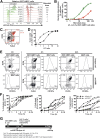
Figure 1.. Optimization of an E. coli mediated MAIT-cell activation assay.
(A) A representative example from 11 donors of the MAIT-cell population and CD69 and IFNγ expression by resting and activated MAIT cells. (B–G) Unless otherwise indicated, MAIT cells were stimulated with 3 min-fixed E. coli (100 CFU/monocyte) for 24 h, in the presence of 1.25 μg/ml anti-CD28 and at a Vα7.2+ cell:monocyte ratio of 2:1. (B, D–G) Frequency of CD69+IFNγ+ MAIT cells. Lines represent individual donors. (B) E. coli was fixed for 1–60 min or heat killed at 95°C for 10 min. Data were normalized to 1 min-fixed E. coli. (C) Monocytes were fed live or 3 min-fixed E. coli. FACS plots are representative of 7 donors. E. coli CFU:monocyte ratio (D) and anti-CD28 concentration (E) ranged from 0.01 to 300 and 0 to 5 μg/ml, respectively. (F) The Vα7.2+ cell:monocyte ratio ranged from 0.25:1 to 64:1. (G) MAIT cells were harvested every 2 h between 6 and 24 h of activation. (H) Timeline of the MAIT-cell activation assay.
Figure 2.
Figure 2.. Assessment of E. coli mediated MR1-dependent MAIT-cell activation by expression of CD25.
(A) Expression of activation markers on MAIT cells in resting condition (solid gray) or upon 24 h activation by E. coli fed monocytes, in the presence of anti-MR1 mAb (gray traces) or isotype ctrl (black traces). Representative examples of CD25 and CD69 (B) and CD69 and IFNγ (D) expression by MAIT cells and frequency of CD25+CD69+ (C) and CD69+IFNγ+ (E) MAIT cells, upon 24 h activation by E. coli fed monocytes and in the presence of 20 μg/ml anti-MR1 mAb or isotype ctrl. Before the Vα7.2+ cells were added, the medium was washed away, and fresh complete medium was added, followed by anti-MR1 mAb or isotype ctrl (A–E). Data are from 4 (A–C) or 6 (D–E) individual donors.
Figure 3.
Figure 3.. Optimization of an assay for proliferation of MAIT cells in response to E. coli.
Representative example of CTV dilution (A) and expansion index (B) of the MAIT-cell population, after 3, 5, or 7 d culture of Vα7.2+ cells with monocytes fed with different doses of 3 min-fixed E. coli, and in the presence of anti-CD28 and IL-2, as well as of MR1-blocking mAb (red) or isotype ctrl (green). Data for unstimulated MAIT cells (solid gray) are also shown (n = 3 donors). (C) Summarized timeline of the optimized MAIT-cell proliferation assay.
Figure 4.
Figure 4.. Optimization of a MAIT-cell cytotoxicity assay.
Representative example of pHrodo Red staining of 293T-hMR1 cells (A) and frequency of pHrodo Red+ 293T-hMR1 cells (B) after 3 h incubation with pHrodo Red-labeled E. coli at different doses and in the presence (red) or absence (green) of cytochalasin D. Data in (B) are technical triplicates from 1 of 4 independent experiments. (C) Representative example of FLICA and DCM staining of 293T-hMR1 cells left alone (black) or after 24 h culture with MAIT cells (red) (E:T = 10). (D) E:T curve of dead E. coli pulsed 293T-hMR1 cells (defined as FLICA+DCM+). The lines represent individual donors from 1 of 5 independent experiments. (E) Representative FACS plots of (F) the kinetics of apoptotic (FLICA+) and dead (FLICA+ DCM+) E. coli pulsed target 293T-hMR1 cells, and MAIT-cell degranulation (CD107a+) in the presence of MR1-blocking mAb or isotype ctrl (n = 3 donors). (G) Summarized layout of the optimized MAIT-cell-mediated cytotoxicity assay.

V体育官网入口 - References

    1. Le Bourhis L., Mburu Y. K., Lantz O. (2013) MAIT cells, surveyors of a new class of antigen: development and functions. Curr. Opin. Immunol. 25, 174–180. - PubMed
    1. Treiner E., Duban L., Bahram S., Radosavljevic M., Wanner V., Tilloy F., Affaticati P., Gilfillan S., Lantz O. (2003) Selection of evolutionarily conserved mucosal-associated invariant T cells by MR1. Nature 422, 164–169. - PubMed
    1. Huang S., Martin E., Kim S., Yu L., Soudais C., Fremont D. H., Lantz O., Hansen T. H. (2009) MR1 antigen presentation to mucosal-associated invariant T cells was highly conserved in evolution. Proc. Natl. Acad. Sci. USA 106, 8290–8295. - PMC - PubMed
    1. Leeansyah E., Ganesh A., Quigley M. F., Sönnerborg A., Andersson J., Hunt P. W., Somsouk M., Deeks S. G., Martin J. N., Moll M., Shacklett B. L., Sandberg J. K. (2013) Activation, exhaustion, and persistent decline of the antimicrobial MR1-restricted MAIT-cell population in chronic HIV-1 infection. Blood 121, 1124–1135. - PMC - PubMed
    1. Walker L. J., Kang Y. H., Smith M. O., Tharmalingham H., Ramamurthy N., Fleming V. M., Sahgal N., Leslie A., Oo Y., Geremia A., Scriba T. J., Hanekom W. A., Lauer G. M., Lantz O., Adams D. H., Powrie F., Barnes E., Klenerman P. (2012) Human MAIT and CD8αα cells develop from a pool of type-17 precommitted CD8+ T cells. Blood 119, 422–433. - PMC - PubMed

MeSH terms