Skip to main page content
U.S. flag

An official website of the United States government

Dot gov

The . gov means it’s official. Federal government websites often end in VSports app下载. gov or . mil. Before sharing sensitive information, make sure you’re on a federal government site. .

Https

The site is secure. The https:// ensures that you are connecting to the official website and that any information you provide is encrypted and transmitted securely V体育官网. .

. 2016 Apr 12;7(15):19327-40.
doi: 10.18632/oncotarget.8451.

Capsules of virulent pneumococcal serotypes enhance formation of neutrophil extracellular traps during in vivo pathogenesis of pneumonia

Affiliations

Capsules of virulent pneumococcal serotypes enhance formation of neutrophil extracellular traps during in vivo pathogenesis of pneumonia

Anandi Narayana Moorthy (V体育官网) et al. Oncotarget. .

Abstract

Neutrophil extracellular traps (NETs) are released by activated neutrophils to ensnare and kill microorganisms. NETs have been implicated in tissue injury since they carry cytotoxic components of the activated neutrophils. We have previously demonstrated the generation of NETs in infected murine lungs during both primary pneumococcal pneumonia and secondary pneumococcal pneumonia after primary influenza VSports手机版. In this study, we assessed the correlation of pneumococcal capsule size with pulmonary NETs formation and disease severity. We compared NETs formation in the lungs of mice infected with three pneumococcal strains of varying virulence namely serotypes 3, 4 and 19F, as well as a capsule-deficient mutant of serotype 4. In primary pneumonia, NETs generation was strongly associated with the pneumococcal capsule thickness, and was proportional to the disease severity. Interestingly, during secondary pneumonia after primary influenza infection, intense pulmonary NETs generation together with elevated myeloperoxidase activity and cytokine dysregulation determined the disease severity. These findings highlight the crucial role played by the size of pneumococcal capsule in determining the extent of innate immune responses such as NETs formation that may contribute to the severity of pneumonia. .

Keywords: Immune response; Immunity; Immunology and Microbiology Section; Streptococcus pneumoniae; capsule; neutrophil extracellular traps; secondary pneumonia; serotype V体育安卓版. .


Conflict of interest statement (VSports手机版)

CONFLICTS OF INTEREST

The authors declare no conflicts of interest.

Figures
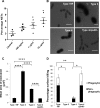
Figure 1
Figure 1. Capsule polysaccharide induces NETs and protects pneumococci from neutrophil-mediated surface killing
A. Bone marrow-derived neutrophils were stimulated for 2 h with various concentrations of purified capsule polysaccharide from serotype 4 (x-axis). Percentage NETs data (y-axis) are presented as means ± SE (n = 3 independent experiments per group). The purified capsule polysaccharide induced NETs in a concentration-dependent manner. *indicates P value < 0.05. B. FITC-dextran (2000 kDa) exclusion assay was performed to estimate the capsule size of pneumococcal strains. Representative images from FITC-dextran exclusion assay. Scale bars = 2 μm. C. Serotype 3 had the largest capsule (∼8-fold > 19F) followed by serotypes 4 and 19F. Serotype 4 capsule mutant had significantly smaller (∼3-fold) capsule than its wild-type. n = 100 bacterial cells per strain. D. Serotype 3 was completely resistant to neutrophil-mediated killing, whereas serotype 19F was easily killed (almost 70%). Serotype 4 was killed to a lesser extent (∼10%), while its capsule mutant (4cps4D- lacking the cps4D gene) was highly susceptible to killing (∼80%). For all strains, non-phagocytic killing predominated over phagocytic killing. Data are presented as means ± SE (n = 3 per strain). *indicates P < 0.05, **P < 0.01, ****P < 0.0001.
Figure 2
Figure 2. Capsule thickness correlates with pneumococcal virulence during primary lung infection in mice
BALB/c mice were intratracheally infected with 107 CFU of serotypes 19F, 3 and 4 as well as the capsule mutant of type 4 (4cps4D-). Lungs were harvested on 1, 2 and 3 days post-infection (dpi). A. Infection with both serotypes 3 and 4 caused significant body weight loss until 3 dpi, whereas serotype 19F recovered by 3 dpi, while 4cps4D- failed to cause any clinical symptoms. B. Both serotypes 3 and 4 induced 100% morbidity, with type 3 causing higher mortality. Type 19F and 4cps4D- did not cause any morbidity. C. Primary infection of mice with serotype 3 led to the highest bacterial load in the lungs. Serotype 4 (wild-type) exhibited bacterial load, whereas its capsule mutant was absent in the lungs. D. Hematoxylin and eosin staining of lung sections revealed considerable neutrophil infiltration (arrows) in the alveolar spaces (AV) arising from infection with types 3, 4 and 19F. Mock control and 4cps4D- displayed the least neutrophil presence in lungs. Magnification and scale bars: top panels = 100× & 500 μm; lower panels = 1000× & 25 μm. E. Histopathologic scoring revealed the worst lung pathology caused by serotype 3, while types 19F, 4 and 4cps4D- showed less infiltration by 3 dpi. Data are presented as means ± SE (n = 8 per group). *indicates P < 0.05, **P < 0.01, ***P < 0.001.
Figure 3
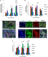
Figure 3. NETs score, but not H2O2 or MPO activity, correlates with overall virulence after primary pneumococcal lung infection
A. Serotype 4 induced the highest H2O2 concentration in the lungs of infected mice. B. MPO activity partially corresponded with H2O2 concentration, and was the lowest after serotype 4 mutant infection. C. NETs were identified and scored in the lung sections by triple immunolabeling (DAPI = blue, histone H2B = green, MPO = red). Arrows indicate single NET strands, whereas asterisks represent clusters. Scale bars = 50 μm. D. NETs score correlated with capsule thickness and clinical severity. Serotype 3 induced the highest NETs at 2 and 3 dpi. Infection with 4cps4D- was significantly lower than its wild-type for all parameters. Data are presented as means ± SE (n = 8 per group). *indicates P < 0.05, **P < 0.01, ***P < 0.001.
Figure 4
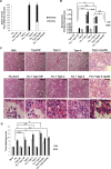
Figure 4. Capsule thickness alone does not determine the severity of secondary pneumococcal pneumonia after primary influenza infection
BALB/c mice were first intratracheally infected with 5 PFU of PR8 influenza virus (sub-lethal dose), followed by 100 CFU of S. pneumoniae after 7 days. Mice were monitored daily until they lost 30% of initial body weight or were euthanized at specific time-points. Lungs were harvested at 24 and 48 h after secondary infection. A. Secondary infections with both serotypes 3 and 4 led to 100% morbidity by day 10, while mice in other groups survived until the experimental end-point. Kaplan-Meier analyses revealed that survival rates of secondary infections with types 3 and 4 were statistically lower than influenza alone, and secondary infections with types 19F and 4cps4D-. n = 3 per group. *represents P < 0.05. B. All the serotypes replicated efficiently in the lungs during secondary infection (after influenza) compared to bacteria infection alone. Secondary infections with serotype 3 followed by serotype 4 led to the highest pulmonary bacterial loads. However, the capsule mutant was not recovered from the lungs. C. Hematoxylin and eosin staining of lung sections revealed intense neutrophil infiltration (arrows) in the alveolar spaces of secondary infected groups of serotypes 3, 4 and 19F. Notably, secondary infection with 4cps4D- showed strong inflammatory cell infiltration, comprising mostly lymphocytes (asterisks). Magnification and scale bars: 100× & 500 μm (top two panels); 1000× & 25 μm (bottom panel). D. Histopathologic scoring revealed that secondary infections with serotype 4 (followed by serotype 3) suffered from the worst lung pathology. Data are presented as means ± SE (n = 6 per group). *denotes P < 0.05, **P < 0.01, ***P < 0.001.
Figure 5
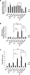
Figure 5. NETs and neutrophil activity in lungs generally correspond to disease severity after secondary pneumococcal infection
A. Lung H2O2 levels were generally elevated after secondary infection especially with serotype 3. B. Secondary infections with serotype 4 (followed by serotype 3) exhibited the greatest MPO activity in lungs. C. Secondary infections with serotype 4 (followed by serotype 3) also induced the highest pulmonary NETs score. Secondary infection with 4cps4D- resulted in significantly lower NETs and MPO activity than its wild-type. Data are presented as means ± SE (n = 6 per group). *P < 0.05, **P < 0.01.
Figure 6
Figure 6. Secondary infection with virulent pneumococci induces augmented levels of pro-inflammatory cytokines in lungs
Secondary infections with serotype 4 (followed by serotype 3) elicited the highest cytokine levels. IL-17 was not detectable in the lung homogenates. Data are presented as means ± SE (n = 4 per group). *indicates P < 0.05, **P < 0.01, ***P < 0.001.

References

    1. Geno KA, Gilbert GL, Song JY, Skovsted IC, Klugman KP, Jones C, Konradsen HB, Nahm MH. Pneumococcal capsules and their types: past, present, and future. Clin Microbiol Rev. 2015;28:871–899. - PMC - PubMed
    1. Hathaway LJ, Brugger SD, Morand B, Bangert M, Rotzetter JU, Hauser C, Graber WA, Gore S, Kadioglu A, Mühlemann K. Capsule type of Streptococcus pneumoniae determines growth phenotype. PLoS Pathog. 2012;8:e1002574. - PMC - PubMed
    1. Weinberger DM, Trzciński K, Lu YJ, Bogaert D, Brandes A, Galagan J, Anderson PW, Malley R, Lipsitch M. Pneumococcal capsular polysaccharide structure predicts serotype prevalence. PLoS Pathog. 2009;5:e1000476. - PMC - PubMed
    1. Briles DE, Crain MJ, Gray BM, Forman C, Yother J. Strong association between capsular type and virulence for mice among human isolates of Streptococcus pneumoniae. Infect Immun. 1992;60:111–116. - PMC - PubMed
    1. Morona JK, Miller DC, Morona R, Paton JC. The effect that mutations in the conserved capsular polysaccharide biosynthesis genes cpsA, cpsB and cpsD have on virulence of Streptococcus pneumoniae. J Infect Dis. 2004;189:1905–1913. - PubMed

MeSH terms

Substances (VSports)