Skip to main page content
U.S. flag

An official website of the United States government

Dot gov

The . gov means it’s official. Federal government websites often end in . gov or . mil VSports app下载. Before sharing sensitive information, make sure you’re on a federal government site. .

Https

The site is secure. The https:// ensures that you are connecting to the official website and that any information you provide is encrypted and transmitted securely. V体育官网.

. 2016 May 13;118(10):1525-39.
doi: 10.1161/CIRCRESAHA.116.308501. Epub 2016 Mar 22.

Caspase-1 Inflammasome Activation Mediates Homocysteine-Induced Pyrop-Apoptosis in Endothelial Cells (V体育2025版)

Affiliations

V体育官网 - Caspase-1 Inflammasome Activation Mediates Homocysteine-Induced Pyrop-Apoptosis in Endothelial Cells

Hang Xi et al. Circ Res. .

Abstract

Rationale: Endothelial injury is an initial mechanism mediating cardiovascular disease VSports手机版. .

Objective: Here, we investigated the effect of hyperhomocysteinemia on programed cell death in endothelial cells (EC) V体育安卓版. .

Methods and results: We established a novel flow-cytometric gating method to define pyrotosis (Annexin V(-)/Propidium iodide(+)) V体育ios版. In cultured human EC, we found that: (1) homocysteine and lipopolysaccharide individually and synergistically induced inflammatory pyroptotic and noninflammatory apoptotic cell death; (2) homocysteine/lipopolysaccharide induced caspase-1 activation before caspase-8, caspase-9, and caspase-3 activations; (3) caspase-1/caspase-3 inhibitors rescued homocysteine/lipopolysaccharide-induced pyroptosis/apoptosis, but caspase-8/caspase-9 inhibitors had differential rescue effect; (4) homocysteine/lipopolysaccharide-induced nucleotide-binding oligomerization domain, and leucine-rich repeat and pyrin domain containing protein 3 (NLRP3) protein caused NLRP3-containing inflammasome assembly, caspase-1 activation, and interleukin (IL)-1β cleavage/activation; (5) homocysteine/lipopolysaccharide elevated intracellular reactive oxygen species, (6) intracellular oxidative gradient determined cell death destiny as intermediate intracellular reactive oxygen species levels are associated with pyroptosis, whereas high reactive oxygen species corresponded to apoptosis; (7) homocysteine/lipopolysaccharide induced mitochondrial membrane potential collapse and cytochrome-c release, and increased B-cell lymphoma 2-associated X protein/B-cell lymphoma 2 ratio which were attenuated by antioxidants and caspase-1 inhibitor; and (8) antioxidants extracellular superoxide dismutase and catalase prevented homocysteine/lipopolysaccharide -induced caspase-1 activation, mitochondrial dysfunction, and pyroptosis/apoptosis. In cystathionine β-synthase-deficient (Cbs(-/-)) mice, severe hyperhomocysteinemia-induced caspase-1 activation in isolated lung EC and caspase-1 expression in aortic endothelium, and elevated aortic caspase-1, caspase-9 protein/activity and B-cell lymphoma 2-associated X protein/B-cell lymphoma 2 ratio in Cbs(-/-) aorta and human umbilical vein endothelial cells. Finally, homocysteine-induced DNA fragmentation was reversed in caspase-1(-/-) EC. Hyperhomocysteinemia-induced aortic endothelial dysfunction was rescued in caspase-1(-/-) and NLRP3(-/-) mice. .

Conclusions: Hyperhomocysteinemia preferentially induces EC pyroptosis via caspase-1-dependent inflammasome activation leading to endothelial dysfunction VSports最新版本. We termed caspase-1 responsive pyroptosis and apoptosis as pyrop-apoptosis. .

Keywords: apoptosis; caspase-1; endothelial cells; homocysteine; pyroptosis V体育平台登录. .


Figures (VSports手机版)

Figure 1
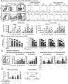
Figure 1. Hcy suppresses cell viability, induces pyroptosis/apoptosis in ECs. HUVECs were cultured to 80% confluence, synchronized (serum free, 6hr), and then switched to 0.5%FBS medium containing L-Hcy, L-Cys, LPS, and H2O2 (500µM) for 24hr as indicated
A, Pyroptosis and apoptosis gating (FCM, AV/7AAD/Casp staining). Cells were treated and stained by AnnexinV-Pacific Blue and 7AAD, and Casp1/9 activity kit for FCM analysis (dot plot shown in online Figure II). Necrotic cells were excluded from intact cells by their content of nuclear debris. Casp9 activity was used as a marker of apoptosis. Q2+Q3 cells were define as apoptosis. Q4 was defined as pyroptosis. B, Cell viability. Cells in 96-well plate were fixed, stained by crystal violet for cell viability. C, Hcy/LPS induced Pyroptosis/apoptosis (dot plot shown in online Figure IV). Cell death forms were identified by FCM using Annexin V-FITC/PI staining (dot plot shown in online Figure II). D, DNA fragmentation. DNA fragmentation was determined by TUNEL staining (images shown in online Figure V). Data are representative of 4 separated experiments and presented as Mean±SEM. Value on the top of bars are normalized by the mean of the control or Q1. *, P<0.05 vs control; #, P<0.05 vs LPS alone in same group; $, P<0.05 vs same dose of Hcy alone; Syn is synergy effect. £, p<0.05. §, p<0.05 vs Q4. FCM, flow cytometry.
Figure 2
Figure 2. Hcy activates endothelial casp1 prior to casp8,9,3 activations in the presence/absence of LPS
HUVECs were cultured and treated with L-Hcy (500µM) and/or LPS (10 µg/mL) as described in Fig. 1. Casp1,8,9,3 activities were measured by using a manufacture’s kit and Western Blotting respectively. Caspase activated cells were FAM positive and labeled by green fluorescence (images shown in online Figure VI). A, Casps Activity (24hr). Casp1,8,9,3 activities were detected by fluorescence spectrometry through a 0~24hr time-course. See supplement Table 1 for values and statistical analysis. B, Casp1 activity (FCM, 2 & 24Hr). Casp1 activity was examined after 2hr and 24hr treatment by FCM. C, Casp8,9,3 activities (FCM, 24 Hr). Casp8,9,3 activities were detected by FCM. D, Casp1 protein and activity. A 20kDa cleavage fragment indicated activated Casp1 and detected by WB, after 2hr and 24hr L-Hcy incubation, with or without LPS presence. E, Schematic sketch show casp1 activation and detection by WB, Fluorometer or FCM. *, p<0.05 vs control; £, p<0.05 vs L-Hcy treatment alone. In A, ‡, p<0.05 vs Casp1 activity at 0hr; ‖, p<0.05 vs Casp8 activity at 0hr; ╫, p<0.05 vs Casp9 activity at 0hr; §, p<0.05 vs Casp3 activity at 0hr. Values are Mean±SEM; n=4.
Figure 3
Figure 3. Caspase-1 mediates Hcy induced pyroptosis/apoptosis and casp9/3 activities in presence/absence of LPS
HUVEC was pretreated by indicated caspase inhibitors for 30m, and then treated with L-Hcy (500µM) and/or LPS (10 µg/mL) for additional 24hr as described in Fig. 1. Cell death forms and caspase activities were determined as described in Fig. 1/2. A, Pyroptosis/apoptosis population (caspase inhibition test). Representative dot plots of Annexing V-FITC/PI staining. R1 was considered as pyroptosis, and R2 as apoptosis. B, Quantification. C, Rescue efficacy on Hcy/LPS-induced pyroptosis/apoptosis (RE=A/B × 100%). Rescue efficacy (RE) for each inhibitor was calculated for their inhibitory capacity to pyrotosis/apoptosis. D, Caspase activities (Casp1 inhibitor). Cells were pretreated with casp1 inhibitor and assayed for casp1,8,9,3 activities by FCM. Numbers above each bar is the percentage normalized by the mean of control. #, P<0.05 vs vehicle with same treatment. Arrows indicate the direction of significant changes. Values are Mean±SEM; n=4. RE, rescue efficacy.
Figure 4
Figure 4. Hcy induces inflammasome assembly, leading to casp1 activation, and IL-1β cleavage in the presence/absence of LPS
Celle were treated with L-Hcy (500µM)/LPS (10 µg/mL) as described in Fig. 1. Precursor and substrate of casp1, component of inflammasome and IL-1β were detected by IP and WB, respectively. A, Schematic sketch for Inflammasome complex assembly and cleavage. B, Inflammasome complex analysis. After Hcy/LPS treatment, cell lysate was used for IP and WB. C, IL-1β expression and activation. Cells were pretreated with Casp1 inhibitor for 30m prior to Hcy/LPS treatment. Protein levels were normalized by β-actin density. Numbers above on each bar is the percentage normalized by the mean of control. Arrows indicate the direction of significant changes. *, p < 0.05 vs control in same group; £, p < 0.05 vs Hcy treatment in same group; #, p < 0.05 vs same treatment in vehicle. LRRs: leucine-rich repeat; NACHT, acronym standing for NAIP (neuronal apoptosis inhibitor protein); PYD, pyrin domain; CARD, caspase activation and recruitment domain; P20, protein 20; P10, protein 10; Act-IL-1β, activated interleukin-1β. Values are Mean±SEM; n=4.
Figure 5
Figure 5. Hcy derived ROS trigger endothelial casp1 activation
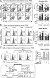
HUVECs were cultured in 6cm dishes as in Fig.1. A&B, Cells were transduced by adenoviral ec-SOD (1 MOI) for 48hr, treated with PEG-catalase (25mg/mL) for 30m with refreshed medium, and then with L-Hcy (500µM) and/or LPS (10 µg/mL) for additional 24hr before subjected for cell death and casp1 activity analysis. A, Pyroptosis/apoptosis (anti-oxidants rescue, FCM). B, Casp1 activity (anti-oxidants rescue, FCM). C, H2O2 on Casp1 activity (casp1 inhibition rescue, FCM). Cells were pretreated with casp1 inhibitor or inhibitor of cathepsin B (a lysosomal cysteine protease) for 30m, and then treated with H2O2 (500 µM) for additional 24 hours before subjected for casp1 activity analysis. Numbers above on each bar is the percentage normalized by the mean of control. Data are representative of 4 separated experiments and presented as Mean±SEM. #, P<0.05 vs vehicle in A, B; *, P<0.05 vs non-treatment control; †, P<0.05 vs H2O2 treatment alone in C. Arrows indicate the direction of significant changes. Values are Mean±SEM; n=4.
Figure 6
Figure 6. Hcy/LPS-induced intracellular ROS levels determines cell destiny (pyroptosis/apoptosis/viable cell) in ECs
HUVECs were cultured and treated with L-Hcy (500µM), LPS (10 µg/mL) or H2O2 (500µM) for 24hr as described in Fig. 1. Triple staining was applied to detect intracellular ROS (DHE) and apoptosis/pyroptosis simultaneously (Annexin-V-FITC and 7-AAD). A, ROS+/apoptosis and ROS+/pyroptosis cells (FCM). The ROS+/apoptosis and ROS+/pyroptosis cells were defined and quantified (dot blot and histogram shown in online Figure VIII). B, ROS level quantification in viable, pyroptotic, apoptotic cell population. ROS level was quantified by mean fluorescence intensity (MFI) of DHE in apoptotic (Annexin V+), pyroptotic (7-AAD+/Annexin V), and viable (7-AAD/Annexin V) cells. C, ROS gradient and apoptosis/pyroptosis. Four regions of ROS level (low, intermediate, high, and extreme high) were defined by their DHE fluorescence intensities. Apoptosis/pyroptosis was quantified in each region. Numbers above each bar is the percentage normalized by the mean of control. *, p < 0.05 vs control in same group or same ROS gradient; #, p < 0.05 vs Hcy in same group or same ROS gradient; †, p < 0.05 vs same treatment in pyroptosis; $, p < 0.05 vs same treatment in viable cells; ‖, p < 0.05 vs same treatment in G1; ‡, p < 0.05 vs same treatment in G2; £, p < 0.05 vs same treatment in G3.) Values are Mean±SEM; n=4.
Figure 7
Figure 7. Hcy induces Δψm collapse and cytochrome-c release, and increase Bax/Bcl-2 Ratio via oxidative stress, Casp1 activation in ECs
HUVECs were cultured in 6cm dish and treated with L-Hcy (500µM) and/or LPS (10 µg/mL) as described in Fig. 1. Cells were pretreated with antioxidants-adenoviral ec-SOD (1MOI) (WB shown in online Figure IX) for 48hr and PEG-catalase (25mg/mL) for 30min, or Casp1, 9 inhibitors for 30min, prior to Hcy/LPS treatment. Mitochondrial function was accessed by JC-1 staining to determine Δψm. Apoptosis signaling was examined by WB for Bax (pro-apoptotic protein) to Bcl-2(anti-apoptotic) ratio. A, Δψm quantification (JC-1 staining, FCM). Δψm collapse cells were quantitated as percentage in gate i. Δψm detection by JC-I staining by fluorescent microscope images shown in online Figure X. B, Cytochrome-c (WB). Cell homogenates were separated to cytosolic and mitochondrial fraction for cytochrome-c protein content by WB. Cytochrome-c ratio (cytosolic fraction/mitochondrial fraction content) was calculated to reflect its leakage from mitochondrial to cytosol. Coomassie blue staining and VADC-1 (mitochondrial protein) were used as loading controls. C, Bax/Bcl-2 (WB). Bax/Bcl-2 ratio was examined by WB. Bax/Bcl-2 ratio is used as apoptosis/pyroptosis index. Arrows indicate the direction of significant changes. Values are Mean±SEM; n=4. Numbers above each bar is the percentage normalized by the mean of control. *, P<0.05 vs control in same group; #, P<0.05 vs same treatment in vehicle. Δψm, mitochondrial potential.
Figure 8
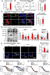
Figure 8. HHcy-induced Casp1 activation mediates EC death in MAEC and impaired endothelial-dependent vessel relaxation in mouse aorta
Plasma Hcy level, Casp1/9 activity, and Bax and Bcl-2 protein expression were measured in Cbs−/− and control mice. Vascular reactivity were determined in aorta from Casp1−/−, Cbs−/−, Casp1−/−/Cbs−/− and NLRP3−/− mice. A, Plasma Hcy levels in mice. Hcy levels were measured in plasma by LC-ESI-MS/MS. B, Casp1 activation in mouse MLECs. Lung ECs (CD31+, MLECs) were isolated from Cbs−/− mice and assayed for Casp1 activity by FCM. C, Casp1 protein levels (aorta). Mouse aortas were isolated. Aortic cross sections were double-stained with antibodies against Casp1 (Red) and endothelial maker-CD31 (Green). D, Casp1,9 activities and Bax/Bcl-2 ratio (mouse aorta). Whole aortae were isolated from hCBS/Cbs−/− and control mice (n=5), homogenized for WB, and quantified. Casp1/9 activities are expressed as Act/Pro, apoptosis index as Bax/Bcl-2 ratio. E, DNA fragmentation (MAEC, TUNEL). Aortic EC were isolated from Casp1−/− mice, cultured till 80% confluence and then treated with L-Hcy (500µM) for 24hr as described in Fig. 1. Cell death was examined by TUNEL staining. F, Endothelial-dependent vessel relaxation. Aortic rings were pre-constricted with PE (1uM) and tested for endothelial-dependent relaxation to cumulative addition of Ach. Vessels from NLRP3−/− mice were incubated with Hcy (500µM, 48Hr) prior to relaxation assessment. G. Endothelial-independent vessel relaxation. Aortic rings were pre-constricted (PE) and tested for endothelial-independent relaxation to cumulative addition of SNP. Numbers above each bar are the percentage normalized by the mean of control. *, P<0.05 vs control mice; #, P<0.05 vs. mouse with Casp1−/− or NLRP3−/− mice; †, P<0.05 vs Cbs+/+ MAEC control; ‡, P<0.05 vs Cbs+/+ MAEC treated with Hcy. Arrows indicate the direction of significant changes. Values are Mean±SEM; n=4. Ach, acetylcholine. SNP, sodium nitroprusside; PE, phenylephrine; MLEC, mouse lung endothelial cells.

References (VSports最新版本)

    1. Eikelboom JW, Lonn E, Genest J, Jr, Hankey G, Yusuf S. Homocyst(e)ine and cardiovascular disease: A critical review of the epidemiologic evidence. Annals of internal medicine. 1999;131:363–375. - PubMed
    1. van den Berg M, Stehouwer CD, Bierdrager E, Rauwerda JA. Plasma homocysteine and severity of atherosclerosis in young patients with lower-limb atherosclerotic disease. Arteriosclerosis, thrombosis, and vascular biology. 1996;16:165–171. - PubMed (V体育ios版)
    1. McCully KS. Homocysteine and vascular disease. Nature medicine. 1996;2:386–389. - PubMed
    1. Wang H, Jiang X, Yang F, Gaubatz JW, Ma L, Magera MJ, Yang X, Berger PB, Durante W, Pownall HJ, Schafer AI. Hyperhomocysteinemia accelerates atherosclerosis in cystathionine beta-synthase and apolipoprotein e double knock-out mice with and without dietary perturbation. Blood. 2003;101:3901–3907. - PubMed
    1. Wang H, Yoshizumi M, Lai K, Tsai JC, Perrella MA, Haber E, Lee ME. Inhibition of growth and p21ras methylation in vascular endothelial cells by homocysteine but not cysteine. The Journal of biological chemistry. 1997;272:25380–25385. - PubMed

"V体育2025版" Publication types

MeSH terms

Substances (V体育安卓版)