Skip to main page content
U.S. flag

An official website of the United States government

Dot gov

The . gov means it’s official. Federal government websites often end in . gov or VSports app下载. mil. Before sharing sensitive information, make sure you’re on a federal government site. .

Https

The site is secure. The https:// ensures that you are connecting to the official website and that any information you provide is encrypted and transmitted securely. V体育官网.

. 2016 Mar 10;11(3):e0151335.
doi: 10.1371/journal.pone.0151335. eCollection 2016.

Activation of Myenteric Glia during Acute Inflammation In Vitro and In Vivo

Affiliations

Activation of Myenteric Glia during Acute Inflammation In Vitro and In Vivo

Corinna Rosenbaum et al. PLoS One. .

Abstract

Background: Enteric glial cells (EGCs) are the main constituent of the enteric nervous system and share similarities with astrocytes from the central nervous system including their reactivity to an inflammatory microenvironment. Previous studies on EGC pathophysiology have specifically focused on mucosal glia activation and its contribution to mucosal inflammatory processes observed in the gut of inflammatory bowel disease (IBD) patients VSports手机版. In contrast knowledge is scarce on intestinal inflammation not locally restricted to the mucosa but systemically affecting the intestine and its effect on the overall EGC network. .

Methods and results: In this study, we analyzed the biological effects of a systemic LPS-induced hyperinflammatory insult on overall EGCs in a rat model in vivo, mimicking the clinical situation of systemic inflammation response syndrome (SIRS). Tissues from small and large intestine were removed 4 hours after systemic LPS-injection and analyzed on transcript and protein level. Laser capture microdissection was performed to study plexus-specific gene expression alterations V体育安卓版. Upon systemic LPS-injection in vivo we observed a rapid and dramatic activation of Glial Fibrillary Acidic Protein (GFAP)-expressing glia on mRNA level, locally restricted to the myenteric plexus. To study the specific role of the GFAP subpopulation, we established flow cytometry-purified primary glial cell cultures from GFAP promotor-driven EGFP reporter mice. After LPS stimulation, we analyzed cytokine secretion and global gene expression profiles, which were finally implemented in a bioinformatic comparative transcriptome analysis. Enriched GFAP+ glial cells cultured as gliospheres secreted increased levels of prominent inflammatory cytokines upon LPS stimulation. Additionally, a shift in myenteric glial gene expression profile was induced that predominantly affected genes associated with immune response. .

Conclusion and significance: Our findings identify the myenteric GFAP-expressing glial subpopulation as particularly susceptible and responsive to acute systemic inflammation of the gut wall and complement knowledge on glial involvement in mucosal inflammation of the intestine. V体育ios版.


Conflict of interest statement

Competing Interests: The authors have declared that no competing interests exist.

Figures

Fig 1
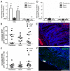
Fig 1. Gfap gene expression upregulation in hyperinflamed intestinal rat tissue.
(A) Quantitative PCR analysis of glial marker expression in whole intestinal wall preparations from duodenal, ileal and colonic segments shows that Gfap gene expression but not S100b expression is upregulated in hyperinflamed gut tissue. We found increases in Gfap expression in duodenal segments by 6.8-fold and in colonic segments by 5.5-fold with a concurrent decrease of S100b expression to the 0.4-fold. In ileal samples no significant changes were observed. Absolute normalized gene expression of Gfap (B) and S100b (C) normalized to the reference genes (ΔΔCq). Means of LPS-treated samples (filled symbols) are indicated with lines, means of sham-treated samples (empty symbols) are indicated with dashed lines. (D) Quantification of immunohistochemical stainings for intramuscular GFAP (E, representative duodenal staining) and the glial transcription factor Sox10 (F, representative duodenal staining). 4 h after LPS injection we did not observe any significant alterations on protein level. Counts of Sox10-positive nuclei in the myenteric plexus show a tendency to be reduced upon LPS treatment in the duodenum, on average we counted 3.6 Sox10-positive nuclei per image in LPS-injected animals and 5.8 nuclei in sham-treated controls. Results (mean ± standard deviation) are given as fold change relative to sham group set to 1, as indicated by the horizontal line, n = 5, # p < 0.10, ** p < 0.01, scale = 100 μm.
Fig 2
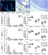
Fig 2. LPS-induced Gfap and S100b gene expression alterations are confined to the myenteric plexus.
(A) Immunohistochemical staining of p75 neurotrophin receptor (p75NTR) indicating the position of myenteric and submucosal plexus structures within the tissue. (B-C) Cresylviolet-stained tissue section showing exemplary myenteric, submucosal and mucosal tissue segments cut and catapulted for mRNA transcript level analysis (B) and sequential excision of segments, here shown for the myenteric region (C). (D, F, H) Quantitative PCR analysis of laser-dissected myenteric, submucosal and mucosal tissue segments of duodenal (D), ileal (F) and colonic (H) samples. In all intestinal regions the significant upregulation of Gfap up to the 17.2±5.2 fold change in the duodenum and downregulation of S100b gene expression is confined to the myenteric plexus. Results (mean ± standard deviation) are given as fold change relative to sham group set to 1. (E, G, I) Absolute normalized gene expression of Gfap (sqares) and S100b (circles) in duodenal (E), ileal (G) and colonic (I) segments, plotted on a logarithmic scale. Means of LPS-treated samples (filled symbols) are indicated with lines, means of sham-treated samples (empty symbols) are indicated with dashed lines. n = 3, *p<0.05, **p<0.01, scale = 100 μm.
Fig 3
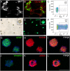
Fig 3. GFAP-expressing glia sorted for EGFP reporter via flow cytometry and expanded in culture.
(A) Endogenous GFP fluorescence and (B) overlay with GFAP antibody staining in LMMP strips from transgenic postnatal day 7 mice visualizes that only cells within the plexus structures express GFP in varying intensities. (C) Flow-cytometry analysis of the muscle tissue digest; the gated GFAP-GFP-positive population was sorted and cultured in vitro to propagate gliospheres. (D) Brightfield and (E) fluorescence microscopy of gliospheres cultured for 2 weeks in vitro shows the formation of gliospheres with a majority of cells expressing GFP. (F) Flow-cytometry analysis of glial culture 2 weeks after initial cell sorting reveals a strong GFP-positivity of about 60% of the total cell population. Immunohistochemical stainings show that the major proportion within gliospheres consists of glial cells positive for GFAP (G) and S100B (H) but spheres also contain cells expressing alpha smooth muscle actin (αSMA, Fig 3K) but are devoid of strongly vimentin-positive cells (J). Scale = 100 μm.
Fig 4
Fig 4. Signaling molecules secreted by GFAP-expressing glia.
(A) Secretion of selected pro-inflammatory signaling molecules by untreated (-) and LPS-challenged (+) gliospheres. Each data point represents an independent experiment, n = 3, ***p<0.001. (B) Tabular summary of all signaling molecules measured.
Fig 5
Fig 5. Significantly upregulated glial genes upon LPS stimulation are enriched in immunologic processes.
(A-C) Bar charts displaying significantly enriched annotations of differentially expressed genes referring to cellular compartments (A), molecular functions (B) and biological processes (C). The level of significance is given as logarithm (log10) of the p-value calculated using the Benjamini correction. Enrichment is given as the count of genes associated to the respective term in the dataset relative to the expected count (Count/ExpCount). (D) Voronoi diagram displaying each significantly differentially expressed gene. Genes are plotted based on their absolute expression level and the relative expression change in response to LPS treatment given as fold change. The plotting area is segmented and a field is assigned to each gene, its size represents the unique character of the expression parameters. Yellow coloring indicates association of a gene product to the GO-term “immune response”, which is enriched with the highest significance. Genes associated to this term include some of those with the highest fold-change increase upon LPS-stimulation. Increased labeling font size indicates validation of expression with the nCounter technology. (E) Spearman correlation of 60 genes analyzed with the Illumina microarray and the Nanostring nCounter technology. The Spearman coefficient of 0.74 shows highly significant correlation (p<0.0001) of the results obtained with both technologies. Data points for the glial marker genes Gfap and S100b are highlighted in red.
Fig 6
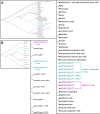
Fig 6. Comparative meta-analyses of multiple GSE datasets reveal genetic similarities among different tissue.
(A) and (B) display complete dendrograms, the length along the line connecting two datasets indicates the degree of similarity; the shorter the distance of connecting lines, the more similar these datasets are. For better legibility the list of datasets are enlarged right of the dendrogram overview. (A) Comparative analysis of gene expression profiles of GFAP-expressing glia and profiles of different tissues/cell types reveals a high similarity of GFAP-glia in vivo to CNS and PNS nerve cell types and a much lower similarity to GFAP-glia expanded in vitro. Gene expression profiles of the latter show similarities to mesenchymal cell types. (B) Hierarchical clustering of glial-specific GSE datasets to show similarities of enteric GFAP-glia to CNS and PNS glia including LPS-challenged cells. Comparative analysis reveals high similarity of GFAP-glia in vivo to astrocytes in vivo. If cultured in vitro, the GFAP-glia genetic profile has higher similarity to PNS sciatic nerve cells and CNS microglia. LPS stimulation leads to severe alterations in gene expression profiles of CNS astrocytes and microglia, which seems not the case for EGCs.

"V体育平台登录" References

    1. Neunlist M, Aubert P, Bonnaud S, van Landeghem L, Coron E, Wedel T et al. (2007) Enteric glia inhibit intestinal epithelial cell proliferation partly through a TGF-beta1-dependent pathway. Am. J. Physiol. Gastrointest. Liver Physiol. 292 (1): G231–41. - PubMed
    1. Bohórquez DV, Samsa LA, Roholt A, Medicetty S, Chandra R, Liddle RA et al. (2014) An Enteroendocrine Cell—Enteric Glia Connection Revealed by 3D Electron Microscopy. PLoS ONE 9 (2): e89881 10.1371/journal.pone.0089881 - "V体育ios版" DOI - PMC - PubMed
    1. Hanani M, Reichenbach A (1994) Morphology of horseradish peroxidase (HRP)-injected glial cells in the myenteric plexus of the guinea-pig. Cell Tissue Res 278 (1): 153–160. - PubMed
    1. Rühl A (2005) Glial cells in the gut. Neurogastroenterology & Motility 17 (6): 777–790. - PubMed
    1. Boesmans W, Lasrado R, Vanden Berghe P, Pachnis V (2015) Heterogeneity and phenotypic plasticity of glial cells in the mammalian enteric nervous system. Glia 63 (2): 229–241. 10.1002/glia.22746 - DOI - PubMed

Publication types

MeSH terms

Substances