Skip to main page content
U.S. flag

An official website of the United States government

Dot gov

The . gov means it’s official. Federal government websites often end in VSports app下载. gov or . mil. Before sharing sensitive information, make sure you’re on a federal government site. .

Https

The site is secure V体育官网. The https:// ensures that you are connecting to the official website and that any information you provide is encrypted and transmitted securely. .

. 2016 Apr;34(4):430-4.
doi: 10.1038/nbt.3461. Epub 2016 Feb 22.

Inclusion of Strep-tag II in design of antigen receptors for T-cell immunotherapy

Affiliations

Inclusion of Strep-tag II in design of antigen receptors for T-cell immunotherapy

Lingfeng Liu et al. Nat Biotechnol. 2016 Apr.

Abstract

Adoptive immunotherapy with genetically engineered T cells has the potential to treat cancer and other diseases. The introduction of Strep-tag II sequences into specific sites in synthetic chimeric antigen receptors or natural T-cell receptors of diverse specificities provides engineered T cells with a marker for identification and rapid purification, a method for tailoring spacer length of chimeric receptors for optimal function, and a functional element for selective antibody-coated, microbead-driven, large-scale expansion. These receptor designs facilitate cGMP manufacturing of pure populations of engineered T cells for adoptive T-cell therapies and enable in vivo tracking and retrieval of transferred cells for downstream research applications. VSports手机版.


Conflict of interest statement (VSports注册入口)

Competing Financial Interests Statement

L. L. and S. R V体育安卓版. R. are co-inventors of a patent “Tagged chimeric effector molecules and receptors thereof” filed by Fred Hutchinson Cancer Research Center (PCT/US2014/072007), and licensed to Juno Therapeutics.

S. R. R V体育ios版. holds equity stake in, and is a cofounder of, Juno Therapeutics and is on the advisory board for and consults for Cell Medica.

No potential conflicts of interest were disclosed by the other authors.

Figures
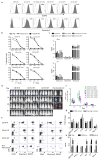
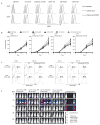
Figure 1
Figure 1. Expression and function of CD19 CARs that contain Strep-tag II
(a) Analysis of CD19 CAR expression. Primary human CD8+ T cells were transduced with epHIV7 lentiviral vectors encoding a CD19-Hi CAR or CD19 CARs with Strep-tag in various extracellular locations. Each CAR contained a 4-1BB/CD3ζ intracellular signaling domain and EGFRt downstream of a T2A element. Transduced cells were sorted for EGFRt+ cells by fluorescence-activated cell sorting (FACS), and purity confirmed by staining with anti-EGFR (grey – top panels). Cell surface expression of the Strep-tag CARs was evaluated by staining with anti-Strep-tag II antibodies (grey - bottom panels). Non-transduced cells (white) served as controls for staining. (b) Cytolytic activity of CD19-Hi and Strep-tag 4-1BBζ CAR-T cells. After sorting for EGFRt expression, CD8+ T cells transduced with each of the CARs were tested for lysis of CD19+ Raji lymphoma and K562 leukemia transduced with CD19 (K562/CD19) or ROR1 (K562/ROR1) at various effector/target (E:T) ratios. (c) IFN-γ and IL2 production by CD19-Hi and Strep-tag 4-1BBζ CAR-T cells 24h after stimulation with K562/CD19 and K562/ROR1. PMA/Ionomycin treated T cells were used as a positive control. The data in a–c is representative of three experiments with CD8+ T cells from different donors. (d) Cohorts of NSG mice were inoculated with 0.5×10⁶ firefly luciferase expressing CD19+ Raji lymphoma cells (Raji-ffluc) via tail vein injection and treated 7 days after tumor inoculation with 2.5×10⁶ CD19 4-1BBζ CAR-T cells with Hi, 1ST, and 3ST spacers, respectively. CAR-T cells were formulated in a CD8:CD4 ratio of 1:1. Tumor progression and distribution were evaluated by serial bioluminescence imaging. (e) Tracking CAR-expressing T cells in vivo by staining with anti-Strep-tag II mAb. Blood obtained from mice 8 days after the T cell infusions was stained with anti-human CD45, CD8, CD4, anti-Strep-tag II and anti-EGFR mAbs, and analyzed by flow cytometry. Expression of Strep-tag II and EGFRt on CD45+ CD8+ and CD45+ CD4+ T cells is shown. (f) Kinetics of expansion and contraction of CD19 CAR-T cells in the blood after adoptive transfer to NSG mice bearing Raji tumors. The mean frequency of CD45+CD8+ EGFRt+ and CD45+CD4+ EGFRt+ human T cells in blood of the mice (n=5) of each group at various times after the T cell infusion is shown. The data in d–f are representative of three experiments. (g) Fold-change in expression of selected cytokine genes in CD19-1ST/4-1BBζ CAR-T cells after infusion to Raji tumor bearing and non-tumor bearing NSG mice. CAR-T cells were sorted 2 days after infusion from blood, bone marrow and spleen after staining with anti-EGFR or anti-Strep-tag mAb. Gene expression was analyzed using a human common cytokine PCR array. The mean fold change values of cytokine genes in the sorted CAR-T cells from NSG/Raji vs sorted CAR-T cells from non-tumor bearing NSG mice were calculated. Samples were run in triplicate and data are presented as the mean fold increase ± SD. (h) Cytokine production by CD8+ T cells expressing CD19-1ST/4-1BBζ CARs 2 days after stimulation with Raji cells in vitro. Supernatants of CAR-T cells co-cultured with CD19+ Raji cells (intra-assay triplicates) for 48 h were pooled together and analyzed using the Luminex Multiplex platform to validate the production of cytokines that were upregulated in CAR-T cells in NSG mice bearing Raji tumors. PMA/Ionomycin treated and non-treated T cells were used as positive and negative control, respectively. The results are representative of biological replicates.
Figure 2
Figure 2. Activation, proliferation and function of Strep-tag CAR-T cells after stimulation with anti-Strep tag II mAb
(a) CD4+ and CD8+ T cells expressing each of the CD19 CARs were sorted for EGFRt expression and stimulated with anti-Strep-tag II or anti-Strep-tag II/CD28 mAb–coated microbeads. After 48 h of stimulation, expression of the CD25 activation marker was determined by flow cytometry. Unstimulated cells (medium) were used as controls. (b) Growth curves of Strep-tag CAR-T cells. FACS sorted EGFRt+ CD19 CAR-T cells (CD8+ and CD4+) were cultured with anti-Strep-tag II or anti-Strep-tag II/CD28 mAb coated microbeads in CTL media containing IL-2 (30–50 U/ml) and IL-15 (2 ng/ml) for 9 days. Aliquots of T cells were removed from the cultures for counting on days 3, 6, and 9 and the fold-increase in cell number determined. The data show the mean fold expansion obtained in three experiments with T cells from different donors. (c) Stimulation of Strep-tag CAR-T cells with anti-Strep-tag II/CD28 beads induces selective outgrowth of transduced cells. CD8+ and CD4+ T cells were transduced with CD19 1ST/4-1BBζ and 1ST/CD28ζ CARs and 10 days later stimulated with anti-Strep-tag II/CD28 microbeads plus IL-2 or cultured with IL-2 alone for 9 additional days. The percentage of Strep-tag II positive cells at day 0 (before stimulation) and day 9 (after stimulation) were measured by flow cytometry. The results are representative of three experiments. (d) Anti-tumor activity of CD19 1ST/4-1BBζ or 1ST/CD28ζ CAR-T cells in Raji-ffluc-bearing NSG mice. 2.5×10⁶ CD19 CAR-T cells expanded with anti-CD3/CD28 beads or with anti-Strep-tag II/CD28 microbeads, and control non-transduced T cells were formulated in a CD8:CD4 ratio of 1:1 and infused into cohorts of NSG mice 7 days after inoculation with 0.5×x106 Raji/ffluc tumor cells. Tumor progression and distribution were evaluated by serial bioluminescence imaging.
Figure 3
Figure 3. Strep-tag CAR-T cells can be enriched and exhibit potent anti-tumor activity in vivo
(a) Enrichment of Strep-tag CAR-T cells containing 1, 2, or 3 Strep-tag II sequences in the spacer region using StrepTactin coated beads on the automated T-CATCH device. Flow cytometric analysis of the frequency of Strep-tag CAR-T cells before and after enrichment using anti-Strep-tag II staining. Data is representative of six experiments using T cells from 3 donors. (b) Yield of Strep-tag CAR-T cells after T-CATCH enrichment. Yield was determined by the absolute numbers of Strep-tag CAR-T cells in the enriched fraction divided by the absolute numbers of Strep-tag CAR-T cells in the starting population. Data is derived from four experiments and expressed as means ± SD. Statistical analysis was performed using the Student’s t test. *P<0.05. (c) Experimental scheme for adoptive transfer of Strep-tag CAR-T cells enriched by StrepTactin selection or using EGFR mAb. CD8+ and CD4+ T cells were stimulated in independent cultures with anti-CD3/CD28 microbeads and transduced with the CD19-3ST/41BBζ CAR. Cultures were established at different times so that T cell administration into tumor bearing mice occurred simultaneously. Anti-CD3/CD28 beads were removed at day 5 in all groups and CAR-T cells were prepared for inoculation into mice either by selection on the T-CATCH at day 8, FACS sorting for EGFRt+ cells on day 10, or FACS sorting of EGFRt+ cells followed by 8 days of culture on irradiated CD19+ LCL cells with IL2 to remove residual bound anti EGFR mAb. (d) Flow cytometric analysis of CD19 CAR expression using Strep-tag II staining of CD8+ and CD4+ CAR-T cells before enrichment, after T-CATCH purification, after EGFR mAb sorting, and after EGFR mAb sorting followed by culture. (e) NSG mice engrafted 7 days earlier with 0.5×10⁶ Raji/ffluc were treated with a total dose of 2.5×10⁶ CAR-T cells selected by T-CATCH, EGFR sorting or EGFR sorting followed by culture and formulated in a CD4:CD8 ratio of 1:1. Tumor progression and distribution were evaluated by serial bioluminescence imaging after injection of luciferin substrate. (f) Persistence of CD19 CAR-T cells in each cohort of NSG/Raji mice. Flow cytometric analysis of CD4+ and CD8+ CAR T cells in the peripheral blood of each group of mice after staining with CD45, CD8, CD4 and EGFR Ab at different time points after T cell infusion. The frequency of CAR-T cells is presented as percentage of live peripheral blood cells. (g) Survival of mice treated with different CAR-T cell products or with non-transduced T cells depicted as Kaplan-Meier curves. The data in d-g are representative of two independent experiments.

References

    1. Brentjens RJ, et al. CD19-targeted T cells rapidly induce molecular remissions in adults with chemotherapy-refractory acute lymphoblastic leukemia. Science translational medicine. 2013;5:177ra138. doi: 10.1126/scitranslmed.3005930. - DOI - PMC - PubMed
    1. Kochenderfer JN, et al. Chemotherapy-refractory diffuse large B-cell lymphoma and indolent B-cell malignancies can be effectively treated with autologous T cells expressing an anti-CD19 chimeric antigen receptor. Journal of clinical oncology : official journal of the American Society of Clinical Oncology. 2015;33:540–549. doi: 10.1200/jco.2014.56.2025. - DOI - PMC - PubMed
    1. Robbins PF, et al. A pilot trial using lymphocytes genetically engineered with an NY-ESO-1-reactive T-cell receptor: long-term follow-up and correlates with response. Clinical cancer research : an official journal of the American Association for Cancer Research. 2015;21:1019–1027. doi: 10.1158/1078-0432.ccr-14-2708. - DOI - PMC - PubMed
    1. Morgan RA, et al. Cancer regression in patients after transfer of genetically engineered lymphocytes. Science (New York, NY) 2006;314:126–129. doi: 10.1126/science.1129003. - DOI - PMC - PubMed
    1. Maude SL, et al. Chimeric antigen receptor T cells for sustained remissions in leukemia. The New England journal of medicine. 2014;371:1507–1517. doi: 10.1056/NEJMoa1407222. - DOI - PMC - PubMed

Publication types

MeSH terms

Substances