Skip to main page content
U.S. flag

An official website of the United States government

Dot gov

The . gov means it’s official. Federal government websites often end in . gov or VSports app下载. mil. Before sharing sensitive information, make sure you’re on a federal government site. .

Https

The site is secure V体育官网. The https:// ensures that you are connecting to the official website and that any information you provide is encrypted and transmitted securely. .

. 2016 Feb 25;164(5):1015-30.
doi: 10.1016/j.cell.2016.01.009. Epub 2016 Feb 18.

"VSports注册入口" TGF-β Tumor Suppression through a Lethal EMT

Affiliations

"V体育平台登录" TGF-β Tumor Suppression through a Lethal EMT

Charles J David et al. Cell. .

Abstract

TGF-β signaling can be pro-tumorigenic or tumor suppressive VSports手机版. We investigated this duality in pancreatic ductal adenocarcinoma (PDA), which, with other gastrointestinal cancers, exhibits frequent inactivation of the TGF-β mediator Smad4. We show that TGF-β induces an epithelial-mesenchymal transition (EMT), generally considered a pro-tumorigenic event. However, in TGF-β-sensitive PDA cells, EMT becomes lethal by converting TGF-β-induced Sox4 from an enforcer of tumorigenesis into a promoter of apoptosis. This is the result of an EMT-linked remodeling of the cellular transcription factor landscape, including the repression of the gastrointestinal lineage-master regulator Klf5. Klf5 cooperates with Sox4 in oncogenesis and prevents Sox4-induced apoptosis. Smad4 is required for EMT but dispensable for Sox4 induction by TGF-β. TGF-β-induced Sox4 is thus geared to bolster progenitor identity, whereas simultaneous Smad4-dependent EMT strips Sox4 of an essential partner in oncogenesis. Our work demonstrates that TGF-β tumor suppression functions through an EMT-mediated disruption of a lineage-specific transcriptional network. .


Conflict of interest statement

The authors declare to have no financial interests in connection with this work.

Figures
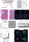
Figure 1
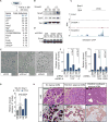
Figure 1. TGF-β induces EMT followed by apoptosis in the premalignant pancreas
(A) Experimental scheme to model TGF-β-mediated tumor suppression by re-expression of Smad4 in Smad4-mutant PDA. (B) Immunohistochemistry (IHC) for cleaved Caspase 3 (CC3) in KC or KSC pancreata collected two days after caerulein treatment, quantification at right. Scale bars 100 μm. (C) H&E staining of a representative KSIC autochthonous tumor (top) and orthotopic tumor (bottom). Scale bars 200 μm. (D) Representative H&E staining and GFP/E-cadherin immunofluorescence after orthotopic injections of the indicated cells, collected 3.5 days post-injection. Apoptotic cells are indicated with arrows (see high magnification inset, Smad4+ H&E section.) Scale bars 50 μm. (E) Cells grown in monolayer in high serum medium containing 2.5 μM MK2206 were treated with TGF-β (100 pM) or SB505124 (SB; 2.5 μM) and assayed for cleaved Caspase 3/7 activity at the indicated times. (F) Time lapse microscopy of Smad4+ cells at the indicated times following TGF-β treatment (see also Supplementary Video 1). (G) Immunoblot of EMT marker E-cadherin (E-cad) and apoptosis marker cleaved Caspase 3 (CC3) after TGF-β treatment. (H) Pancreatic organoids cultured from KIC or KSIC mice were treated with MK2206 and TGF-β or SB505124, and were assayed for cleaved Caspase 3/7 activity at the indicated times. (I) 3-dimensional images of E-cadherin and CC3 immunofluorescence performed in KIC or KSIC PDA organoids treated as indicated for 24 h. Scale bars 10 μm. Values in panels E and H are results of 3 biological replicates. Bar graphs represent mean +/− SD.
Figure 2
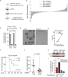
Figure 2. Snail and Zeb1 promote EMT and apoptosis
(A). List of most strongly induced genes after 90 min of TGFβ treatment in Smad4+ cells. (B) Immunoblot for Snail and Zeb1 in Smad4-mutant and Smad4+ cells after 24 h of TGFβ. (C) Smad2/3 binding to the Snai1 locus after 1 h of TGFβ treatment in Smad4-mutant or Smad4+ cells. (D) Immunoblot for E-cadherin in Smad4+ cells expressing the indicated shRNAs 24 h post-TGFβ/SB treatment. (E) Images of Smad4+ cells treated with TGFβ for 72h expressing control shRNAs, or shRNAs targeting Snail or Zeb1. (F) Smad4+ cells expressing Snai1-targeting (left panel) or Zeb1-targeting shRNAs (right panel) were treated with TGFβ or SB for 36 h then assayed for Caspase 3/7 activity. (G) Smad4+ cells transduced with Tet-On Snail were treated with or without dox for 12 h followed by TGF-β or SB, then assayed for caspase activity after 18 h. (H) H&E and CK19 IHC for representative ADM or atypical ADM lesions in KC or KSnSnC mice. Scale bars 100 μm. Values in panels F and G are the average of 3 biological replicates. Bar graphs represent mean +/− SD.
Figure 3
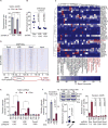
Figure 3. Sox4 is required for TGF-β-driven apoptosis
(A) Experimental scheme to screen for factors required for TGF-β-induced cell death. (B) Plot depicting shRNA enrichment/depletion in TGF-β vs SB treated cells. Positive control shRNAs targeting Tgfbr1 and Tgfbr2 and negative control shRNAs targeting Renilla luciferase are indicated. See also Table S2. (C) Indicated TetOn Sox4 shRNAs were transduced into Smad4+ cells, cells were pre-treated with or without dox for 24 h, then treated as indicated. Caspase activity was assayed after 36 h. (D) Images of control or Sox4 KD cells treated with TGF-β for 37 h (see also Supplementary Video 2). (E) Immunoblot for the indicated factors after Sox4 depletion and 24 h of SB/TGFβ treatment. (F) Sox4 immunoblot after 24 h of TGFβ treatment in cells bearing control shRNAs or shRNAs targeting the Snai1 or Zeb1 transcripts. Samples and load control same as Figure 2D. (G) Publicly available RNA-seq expression data from The Cancer Genome Atlas (TCGA) derived from 179 PDA tumors was used to plot SOX4 and CDH1 levels. Spearman and Pearson correlation coefficients are indicated. (H) TetOn Sox4 Smad4+ PDA cells were treated as indicated and assayed for apoptosis after 18 h. Values in panels C and H are averages of 2 and 3 biological replicates, respectively. Bar graphs represent mean +/− SD.
Figure 4
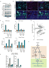
Figure 4. Sox4 is part of a Smad4-independent group of TGF-β-induced genes
(A) Sox4 mRNA levels in the indicated cells after 24 h of the indicated treatment. Values are the mean and range of four technical replicates, and representative of at least 3 separate experiments. (B) Immunblot for Sox4 after 24 h of the indicated treatment in mPDA (top) and BxPC3 cells (bottom). (C) Immunohistochemistry for Sox4 performed 72 h after Smad4(−) or Smad4(+) PDA cells were implanted in the pancreata of syngeneic mice. Inset shows higher magnification image of Sox4+ EMT cancer cell. (D) Smad4 dependence of 90-minute TGF-β genes. Fold induction in Smad4-mutant and Smad4+ cells is plotted on the x- and y-axes respectively. Select Smad4-independent genes were assayed by qRT-PCR after 12 h induction and plotted (red-shaded region). (E) Smad2/3 binding to the Sox4 locus as determined by ChIP-seq performed in Smad4-mutant and Smad4(+) cells. (F) Summary diagram showing cooperation between Smad4-dependent and Smad4-independent branches to induce apoptosis.
Figure 5
Figure 5. Sox4 plays a pro-tumorigenic role in PDA cells
(A) Experimental scheme for identification of tumorigenic transcription factors in PDA. (B) Waterfall plot depicting results of an shRNA screen of the 93 most highly expressed TFs in PDA cells. Cells expressing the library were grown as oncospheres for 18 days with one passage. Data presented is the merged results of two separate pools. See also Table S3. (C) PDA cells expressing the indicated shRNAs were plated as oncospheres and counted after one week. (D) Bright-field images of cells expressing a control or Sox4 shRNA grown as oncospheres for 2 weeks. Scale bar 200 μm. (E) PDA cells transduced with a TetOn Sox4 shRNA were cultured in full serum medium in 2 dimensional culture, and were assayed for cell viability at the indicated times (in h). (F) Tumor-free survival after KSIC mPDA cells tranduced with TetOn Sox4 shRNAs were injected subcutaneously into mice maintained on or off dox diet. Plot reflects results of pooled experiments using Sox4.965 and Sox4.2509 shRNAs. For each condition (on or off dox), n = 10 mice, survival compared by log-rank test. (G) Luciferase labeled KSIC cells transduced with the indicated shRNAs were injected orthotopically in mice and imaged after four weeks. Luminescence was normalized to day 0 values. Statistal significance was calculated by a two-tailed unpaired t test. (H) Sox4 protein expression after SB/TGF-β treatment of cells grown in adherent conditions (right) or after three days in oncosphere conditions (left). (I) Secondary oncosphere formation of Smad4-mutant and Smad4+ PDA cells. Primary oncospheres were cultured for two weeks in the presence or absence of 20 pM TGF-β. Spheres were dissociated by trypsinization and replated for one week prior to counting. Values in panels C and I are the average of 3 biological replicates. Bar graphs represent mean +/− SD.
Figure 6
Figure 6. Klf5 is a lineage survival gene that determines Sox4 function
(A) KSIC mPDA cells transduced with TetOn Klf5 shRNAs were grown as oncospheres for two weeks as indicated then quantified. (B) KSIC cells were transduced with the indicated shRNAs and implanted orthotopically in mice maintained on doxycycline feed. Mice were imaged after four weeks. Control group same as Figure 5G. (C) Heatmap showing highly expressed TFs in cancer exhibiting lineage specificity. TCGA RNA-seq data was used to rank TFs by average expression levels in the indicated tumor types. To identify lineage specific TFs, we included any TF with expression in the top 0.5% in at least one tumor type, while excluding TFs that exhibited expression in the top 10% in more than half of the tumor types analyzed. The latter step excluded 22 TFs. Tumor types exhibiting >10% of loss of TGF-β signaling are indicated in red. (D) Heatmap showing overlap between Sox4, Klf5, and Med1 ChIP-seq peaks. Below is a venn diagram depicting overlap between Sox4 and Klf5 ChIP-seq peaks (E) Smad4-mutant PDA cells transduced with the indicated TetOn shRNAs were treated with dox for 48 h to deplete the target protein, followed by treatment with SB/TGF-β. Cells were assayed for Caspase 3/7 activity after 36 h SB/TGF-β treatment. (F) A TetOn Klf5 construct was introduced into constitutive Smad4(+) cells. Klf5 was induced with doxycycline for 12 hours prior to the addition of MK2206. Cells were then treated with SB/TGF-β for 36 h, and assayed for caspase 3/7 activity. (G) TetOn Klf5 cells were treated as in (F), then immunoblotted after 24 h of the indicated treatment. (H) 3000 Smad4(+) TetOn Klf5 cells were injected orthotopically in caerulein-treated mouse pancreata. Mice were sacrificed after 2 weeks, and pancreata subjected to serial sectioning and quantification of maximal tumor surface area, plotted as indicated (On dox n=14, Off dox n=10; unpaired two-tailed t test). (I) Klf5.1343 shRNA was cloned into a tandem TetOn shRNA vector together with shRNAs targeting Renilla luciferase or Sox4. Cells were incubated with dox for 48 h, then treated as indicated and assayed for Caspase 3/7 activity after 36 h. Values in panels A, E, F, and I are the average of 3 biological replicates. SD and p values were calculated by a two-tailed t test.
Figure 7
Figure 7. EMT-linked Klf5 repression promotes an apoptotic Sox4 program
(A) Immunoblot for highly expressed endoderm-associated TFs after 24 h of the indicated treatment. (B) The indicated cells were injected as in Figure 1D. Klf5 and GFP were then detected by immunofluorescence. Scale bars 50 μm. (C) Smad2/3 ChIP-qRT-PCR performed at the indicated loci in the presence of control, or Snail/Zeb1 shRNAs. Error bars represent the mean and range of 4 technical replicates, representative of two separate experiments. Cells were treated with SB/TGF-β for 12 h prior to crosslinking. (D) Sox4-dependent gene expression in TGF-β-treated Smad4+ cells. Cells bearing TetOn Sox4 shRNAs were cultured +/− dox for 24 h prior to treatment with TGF-β. After another 24 h, RNA was extracted and RNA-seq was performed. Sox4-dependent differentially expressed genes were then plotted. (E) qRT-PCR validation of Bim and Bmf as Sox4-dependent gene responses in TGF-β-treated Smad4+ cells. Values are the result of 4 technical replicates. (F) Diagram summarizing effects of TGF-β signaling in naïve PDA cells.

Comment in

"VSports手机版" References

    1. Aguirre AJ, Bardeesy N, Sinha M, Lopez L, Tuveson DA, Horner J, Redston MS, DePinho RA. Activated Kras and Ink4a/Arf deficiency cooperate to produce metastatic pancreatic ductal adenocarcinoma. Genes & development. 2003;17:3112–3126. - PMC - PubMed
    1. Bardeesy N, Cheng KH, Berger JH, Chu GC, Pahler J, Olson P, Hezel AF, Horner J, Lauwers GY, Hanahan D, et al. Smad4 is dispensable for normal pancreas development yet critical in progression and tumor biology of pancreas cancer. Genes & development. 2006;20:3130–3146. - PMC - PubMed
    1. Bialkowska AB, Liu Y, Nandan MO, Yang VW. A colon cancer-derived mutant of Kruppel-like factor 5 (KLF5) is resistant to degradation by glycogen synthase kinase 3beta (GSK3beta) and the E3 ubiquitin ligase F-box and WD repeat domain-containing 7alpha (FBW7alpha) The Journal of biological chemistry. 2014;289:5997–6005. - PMC (VSports注册入口) - PubMed
    1. Boj SF, Hwang CI, Baker LA, Chio II, Engle DD, Corbo V, Jager M, Ponz-Sarvise M, Tiriac H, Spector MS, et al. Organoid models of human and mouse ductal pancreatic cancer. Cell. 2015;160:324–338. - PMC - PubMed
    1. Burk U, Schubert J, Wellner U, Schmalhofer O, Vincan E, Spaderna S, Brabletz T. A reciprocal repression between ZEB1 and members of the miR-200 family promotes EMT and invasion in cancer cells. EMBO reports. 2008;9:582–589. - PMC - PubMed

Publication types

MeSH terms