Skip to main page content
U.S. flag

An official website of the United States government

Dot gov

The . gov means it’s official VSports app下载. Federal government websites often end in . gov or . mil. Before sharing sensitive information, make sure you’re on a federal government site. .

Https

The site is secure. The https:// ensures that you are connecting to the official website and that any information you provide is encrypted and transmitted securely V体育官网. .

. 2016 Feb 9;11(2):e0148452.
doi: 10.1371/journal.pone.0148452. eCollection 2016.

IL-15 Superagonist Expands mCD8+ T, NK and NKT Cells after Burn Injury but Fails to Improve Outcome during Burn Wound Infection (V体育平台登录)

Affiliations

"VSports最新版本" IL-15 Superagonist Expands mCD8+ T, NK and NKT Cells after Burn Injury but Fails to Improve Outcome during Burn Wound Infection

Naeem K Patil et al. PLoS One. .

Abstract

Background: Severely burned patients are highly susceptible to opportunistic infections and sepsis, owing to the loss of the protective skin barrier and immunological dysfunction. Interleukin-15 (IL-15) belongs to the IL-2 family of common gamma chain cytokines and stimulates the proliferation and activation of T (specifically memory CD8), NK and NKT cells. It has been shown to preserve T cell function and improve survival during cecal ligation and puncture (CLP)-induced sepsis in mice. However, the therapeutic efficacy of IL-15 or IL-15 superagonist (SA) during infection after burn injury has not been evaluated. Moreover, very few, if any, studies have examined, in detail, the effect of burn injury and infection on the adaptive immune system. Thus, we examined the effect of burn and sepsis on adaptive immune cell populations and the effect of IL-15 SA treatment on the host response to infection. VSports手机版.

Methods: Mice were subjected to a 35% total body surface area burn, followed by wound infection with Pseudomonas aeruginosa. In some experiments, IL-15 SA was administered after burn injury, but before infection V体育安卓版. Leukocytes in spleen, liver and peritoneal cavity were characterized using flow cytometry. Bacterial clearance, organ injury and survival were also assessed. .

Results: Burn wound infection led to a significant decline in total white blood cell and lymphocyte counts and induced organ injury and sepsis V体育ios版. Burn injury caused decline in CD4+ and CD8+ T cells in the spleen, which was worsened by infection. IL-15 treatment inhibited this decline and significantly increased cell numbers and activation, as determined by CD69 expression, of CD4+, CD8+, B, NK and NKT cells in the spleen and liver after burn injury. However, IL-15 SA treatment failed to prevent burn wound sepsis-induced loss of CD4+, CD8+, B, NK and NKT cells and failed to improve bacterial clearance and survival. .

Conclusion: Cutaneous burn injury and infection cause significant adaptive immune dysfunction. IL-15 SA does not augment host resistance to burn wound sepsis in mice despite inducing proliferation and activation of lymphocyte subsets. VSports最新版本.


Conflict of interest statement

Competing Interests: The authors have declared that no competing interests exist.

Figures (V体育安卓版)

Fig 1
Fig 1. IL-15 SA treatment does not protect against burn wound infection induced organ injury and decreased survival.
(Among the respective groups, the X axis symbols mean the following: S = Sham (no burn), B = burn injury, V = Vehicle treatment, WI = Wound infection and IL-15 SA = interleukin 15 super agonist treatment). IL-15 SA was administered at a dose of 1 μg via i.p route starting a day after burn injury. (A) Time line depicting the burn injury and wound infection model along with IL-15 SA treatment. (B) To evaluate the systemic spread of infection, Pseudomonas aeruginosa colony counts were measured on day 2 post wound infection and the graph depicts colony forming units of the bacteria in the blood (left half) and the lungs (right half). Following parameters were measured on day 4 and day 6 (equivalent to day 2 post wound infection) post burn injury and wound infection (C) serum alanine aminotransferase, ALT; (D) serum aspartate amino transferase, AST; and (E) serum blood urea nitrogen. n = 8–10 in each group and P<0.05. (F) Survival study: Three groups of mice including—burn injured (n = 18, black line); burn injured and vehicle treated wound infected mice (n = 18, red line); and burn injured and IL15 SA treated wound infected mice (n = 18, blue line) were monitored for survival for seven days post Pseudomonas aeruginosa wound inoculation. The graph represents results from three separate experiments. *significantly different as compared to respective day 4 and day 6 sham groups. For each of the figures, x-axis labels indicate the following: S = Sham (no burn injury); B+V = burn injury + vehicle treated mice for 4 days; B+IL-15 SA = burn injury + IL-15 SA treated mice for 4 days; B+WI+V = burn injury + wound infection performed on day 4 + vehicle treated mice for 6 days; B+WI+IL-15 = burn injury + wound infection performed on day 4 + IL-15 SA treated mice for 6 days.
Fig 2
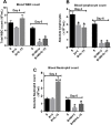
Fig 2. Burn wound infection caused decline in white blood cell and lymphocyte counts.
(A) Total white blood cell counts. (B) Blood lymphocyte counts. (C) Blood neutrophil counts. n = 8–10 in each group, *significantly different as compared to respective day 4 and day 6 sham groups; #significantly different from burn plus vehicle group (B+V) and P<0.05.
Fig 3
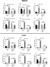
Fig 3. Effect of burn injury and wound infection with or without IL-15 SA treatment on CD4+, CD8+ T and CD19+ B lymphocyte cell counts.
Wound infection and sepsis was induced by inoculation of Pseudomonas aeruginosa on day 4 post burn injury. Upper half of the panel depicts cell counts in the spleen and lower half of the panel depicts cell counts in the liver. Spleen and liver cell counts were analyzed on day 4, and day 6 (equivalent to day 2 post wound infection) post burn injury. (A and G) Total immune cell counts among the respective groups of the single cell suspensions prepared from the spleen and liver. Flow cytometry was used to measure the individual T and B cell lymphocyte cell population in the spleen and liver. The bar graph represents the total cell counts of the CD4+ T cells (B and H), CD4+ CD44hi memory T cells (D and J), CD8+ T cells (C and I), CD8+ CD44hi T cells (E and K), and CD3-CD19+ B cells (F and L); in the spleen and liver. n = 8–12 in each group and P<0.05. *significantly different as compared to respective day 4 and day 6 sham groups; #significantly different from day 4 burn injury group (B+V) and $-significantly different from day 6 burn injury + vehicle treated wound infected group (B+WI+V).
Fig 4
Fig 4. Effect of burn injury and wound infection with or without IL-15 SA treatment on CD4+, CD8+ T and CD19+ B lymphocytes activation status.
CD69 surface expression was used as a marker of cellular activation and measured by flow cytometry. Upper half of the panel depicts data for spleen cells and lower half of the panel depicts data for liver tissue. Percentage of CD69 expression was measured on CD4+ T cells (A and D), CD8+ T cells (B and E), and CD3-CD19+ B cells (C and F); in the spleen and liver. n = 8–12 in each group and P<0.05. *significantly different as compared to respective day 4 and day 6 sham groups; #significantly different from day 4 burn injury group (B+V), and $—significantly different from day 6 burn injury + wound infection + vehicle treated group (B+WI+V).
Fig 5
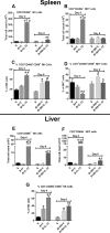
Fig 5. IL-15 SA treatment elicits expansion of NK and NKT cells post burn injury and wound infection causes their decline.
Upper half and lower half of the panel depicts cell counts and activation status (CD69+) of NK (CD3-CD49b+) and NKT (CD3+CD49b-) cells in the spleen and liver respectively. The graphs A and E depict NK cell counts in the spleen and liver respectively; and the graphs B and F show the NKT cell counts in the spleen and liver respectively. The graphs C and G depict NK cell CD69 expression in the spleen and liver respectively; and the graph D shows the NKT cell CD69 expression in the spleen. n = 8–10 in each group and P<0.05. *significantly different as compared to respective day 4 and day 6 sham groups; #significantly different from day 4 burn injury group (B+V), and $—significantly different from day 6 burn injury + wound infection + vehicle treated group (B+WI+V).
Fig 6
Fig 6. Plasma cytokine levels post burn injury and wound infection.
Plasma cytokines including (A) IL-6, (B) IL-10, (C) TNF-α, (D) IFN-γ, (E) KC and (F) MIP-2 were measured using Bio-Rad Bio-Plex assay. n = 6–8 in each group and P<0.05. *significantly different as compared to respective day 4 and day 6 sham groups.
Fig 7
Fig 7. Effect of IL-15 SA treatment on neutrophil recruitment and bacterial clearance upon intraperitoneal infection.
Pseudomonas aeruginosa (1x10⁸ CFU) was inoculated in the peritoneum of the mice via i.p injection. This model was used to assess if IL-15 SA treatment causes neutrophil recruitment and improves bacterial clearance. Mice were divided into four groups: 1) B+V = burn + vehicle treated non-infected mice; 2) B+IL-15 = burn + IL-15 SA treated non-infected mice; 3) B+V+I = burn + vehicle treated infected mice; 4) B+IL-15+I = burn + IL-15 SA treated infected mice. (A) At 6 hours post i.p infection, core body temperature was measured using a rectal temperature probe. Also, peritoneal lavage was performed using 2 mL sterile PBS solution and used for bacterial clearance (B); macrophage (C) and neutrophil (D) cell counts measurements using flow cytometry. n = 8–16 in each group and P<0.05. *significantly different as compared to B+V group; #significantly different from B+IL-15 group, significantly different as compared to B+V group $—significantly different from B+V+I group.
Fig 8
Fig 8. Plasma cytokine levels post intraperitoneal infection with Pseudomonas aeruginosa.
Plasma cytokines including (A) IL-6, (B) IL-10, (C) TNF-α, (D) IFN-γ, (E) KC and (F) MIP-2 were measured using Bio-Rad Bio-Plex assay. n = 6–8 in each group and P<0.05. *significantly different as compared to burn injury + vehicle treated group (B+V); #significantly different from burn injury + IL-15 SA treated group (B+IL-15); and $significantly different from burn injury + IL-15 SA treated infected group (B+IL-15+I).

References

    1. Williams FN, Herndon DN, Hawkins HK, Lee JO, Cox RA, Kulp GA, et al. (2009) The leading causes of death after burn injury in a single pediatric burn center. Crit Care 13: R183 10.1186/cc8170 - DOI - PMC - PubMed
    1. Mann EA, Baun MM, Meininger JC, Wade CE (2012) Comparison of mortality associated with sepsis in the burn, trauma, and general intensive care unit patient: a systematic review of the literature. Shock 37: 4–16. 10.1097/SHK.0b013e318237d6bf - V体育2025版 - DOI - PubMed
    1. Kraft R, Herndon DN, Al-Mousawi AM, Williams FN, Finnerty CC, Jeschke MG (2012) Burn size and survival probability in paediatric patients in modern burn care: a prospective observational cohort study. Lancet 379: 1013–1021. 10.1016/S0140-6736(11)61345-7 - DOI - PMC - PubMed
    1. Stanojcic M, Chen P, Xiu F, Jeschke MG (2015) Impaired Immune Response in Elderly Burn Patients: New Insights Into the Immune-senescence Phenotype. Annals of surgery. - PMC (V体育官网入口) - PubMed
    1. Jeschke MG, Patsouris D, Stanojcic M, Abdullahi A, Rehou S, Pinto R, et al. (2015) Pathophysiologic Response to Burns in the Elderly. EBioMedicine 2: 1536–1548. 10.1016/j.ebiom.2015.07.040 - DOI - PMC - PubMed

Publication types (VSports手机版)

MeSH terms