Skip to main page content
U.S. flag

An official website of the United States government

Dot gov

The . gov means it’s official. Federal government websites often end in . gov or . mil VSports app下载. Before sharing sensitive information, make sure you’re on a federal government site. .

Https

The site is secure. The https:// ensures that you are connecting to the official website and that any information you provide is encrypted and transmitted securely V体育官网. .

. 2016 Feb 5;11(2):e0148142.
doi: 10.1371/journal.pone.0148142. eCollection 2016.

Neutrophil Extracellular Traps Induce Organ Damage during Experimental and Clinical Sepsis

Affiliations

"V体育安卓版" Neutrophil Extracellular Traps Induce Organ Damage during Experimental and Clinical Sepsis

"VSports手机版" Paula Giselle Czaikoski et al. PLoS One. .

VSports手机版 - Abstract

Organ dysfunction is a major concern in sepsis pathophysiology and contributes to its high mortality rate. Neutrophil extracellular traps (NETs) have been implicated in endothelial damage and take part in the pathogenesis of organ dysfunction in several conditions. NETs also have an important role in counteracting invading microorganisms during infection. The aim of this study was to evaluate systemic NETs formation, their participation in host bacterial clearance and their contribution to organ dysfunction in sepsis. C57Bl/6 mice were subjected to endotoxic shock or a polymicrobial sepsis model induced by cecal ligation and puncture (CLP) VSports手机版. The involvement of cf-DNA/NETs in the physiopathology of sepsis was evaluated through NETs degradation by rhDNase. This treatment was also associated with a broad-spectrum antibiotic treatment (ertapenem) in mice after CLP. CLP or endotoxin administration induced a significant increase in the serum concentrations of NETs. The increase in CLP-induced NETs was sustained over a period of 3 to 24 h after surgery in mice and was not inhibited by the antibiotic treatment. Systemic rhDNase treatment reduced serum NETs and increased the bacterial load in non-antibiotic-treated septic mice. rhDNase plus antibiotics attenuated sepsis-induced organ damage and improved the survival rate. The correlation between the presence of NETs in peripheral blood and organ dysfunction was evaluated in 31 septic patients. Higher cf-DNA concentrations were detected in septic patients in comparison with healthy controls, and levels were correlated with sepsis severity and organ dysfunction. In conclusion, cf-DNA/NETs are formed during sepsis and are associated with sepsis severity. In the experimental setting, the degradation of NETs by rhDNase attenuates organ damage only when combined with antibiotics, confirming that NETs take part in sepsis pathogenesis. Altogether, our results suggest that NETs are important for host bacterial control and are relevant actors in the pathogenesis of sepsis. .


Conflict of interest statement

Competing Interests: The authors declare that the co-author José Carlos Alves-Filho is part of the editorial board of PLOS ONE. This does not alter the authors’ adherence to all the PLOS ONE policies on sharing data and materials V体育安卓版.

Figures (V体育官网)
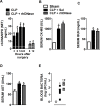
Fig 1
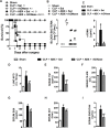
Fig 1. Degradation of systemic cf-DNA/NET by rhDNase treatment did not prevent organ damage during polymicrobial sepsis.
Mice were subjected to sham surgery or CLP-induced severe sepsis. (A) Blood samples were collected 3, 6, 12 and 24 hours after sepsis induction, and plasma concentrations of cf-DNA/MPO (NET) were determined (white horizontal bar represents the sham group at the indicated times). Blood samples were also collected 6 and 12 hours after sepsis induction in mice treated pre-sepsis (10 min) and post-sepsis (4 h) with rhDNase (10 mg/kg, sc.) * p < 0.05 compared with the sham group, white line; # p < 0.05 compared to the CLP- Sal 6 and 12 h groups (ANOVA followed by Tukey’s test, n = 5 per experimental group). (B) Animals were treated pre-sepsis (10 min) and post-sepsis (4 h) with Sal (control) or rhDNase (10 mg/kg, sc.). Six hours after sepsis induction, the serum levels of CK-MB, BUN (C) and AST (D) were quantified. Blood bacterial levels 6 h (E) were also quantified; # p < 0.05 compared with the CLP-Sal 6 h group (ANOVA followed by Tukey’s test, n = 5 per experimental group). The horizontal bars represent the median (Mann-Whitney U test, n = 5–7 per experimental group). The data are reported as the mean ± SEM. * p < 0.05 compared with the sham group; # p < 0.05 compared with the CLP+Sal group (ANOVA followed by Tukey’s test, n = 5 per experimental group).
Fig 2
Fig 2. NETs degradation by rhDNase treatment associated with antibiotic therapy improves the outcome of CLP-induced sepsis.
Mice were subjected to sham surgery or CLP-induced severe sepsis. (A) Mice were post-treated with saline or rhDNase (10 mg/kg, sc. - 1 h after the surgery and every 8 h thereafter), associated or not with ertapenem antibiotic (ABX—30 mg/kg, sc. - 1 h after the surgery and every 12 h thereafter for 48 h). The survival rates were evaluated over 7 days. * p < 0.05 compared with the sham group; δ p < 0.05 compared with the CLP + Sal group; θ p < 0.05 compared with the CLP + rhDNase group; # p < 0.05 compared with the CLP + ABX + Sal group (Mantel-Cox log-rank test, n = 10 per experimental group). (B-I) Mice were post-treated with saline or rhDNase (10 mg/kg, sc. - 1 and 8 h after the surgery), associated or not with ertapenem antibiotic (30 mg/kg, sc. - 1 h after the surgery). Twelve hours after sepsis induction, blood bacterial levels (B), serum concentration of cf-DNA, (C), MPO in lung tissue (D) and serum concentrations of TNF (E), CK-MB (F), BUN (G), AST (H) and endocan (I) were quantified. The data are reported as the mean ± SEM. * p < 0.05 compared with the sham group; # p < 0.05 compared with the CLP + ABX + Sal group (ANOVA followed by Tukey’s test, n = 5 per experimental group); bacteria: the horizontal bars represent the median (Mann-Whitney U test, n = 5–7 per experimental group).
Fig 3
Fig 3. Systemic degradation of NETs attenuated organ damage during LPS-induced endotoxic shock.
Endotoxic shock was induced by LPS injection (15 mg/kg, iv.) Mice were pre-treated (10 min) and post-treated (8 h) with saline or rhDNase (10 mg/kg, sc.). Twelve hours after endotoxic shock induction, the serum concentrations of cf-DNA (A) and TNF-α (B), serum levels of serum CK-MB (C), BUN (D), AST (E) and MPO in lung tissue (F) were determined. The data are reported as the mean ± SEM. * p < 0.05 compared with the sham group; # p < 0.05 compared with the LPS+Sal group (ANOVA followed by Tukey’s test, n = 5 per experimental group). (G) Survival rates of endotoxemic mice pre-treated (10 min) and post-treated (8/8 h) with saline or rhDNase (10 mg/kg, sc.) until the 48th hour. * p < 0.05 compared with the LPS+Sal group (Mantel-Cox log-rank test, n = 10 per experimental group).
Fig 4
Fig 4. Morphological changes in heart, lungs and liver tissues.
Animals were euthanized 12 h after endotoxic shock induction, and the heart, lungs, and liver were isolated, fixed by immersion in 10% paraformaldehyde, dehydrated and embedded in paraffin wax. Then, 5-μm-thick sections were stained with hematoxylin and eosin for histological examination. The images are representative of heart, lung and liver sections from the CTRL (A, D, G), LPS+Sal (B, E, H) and LPS+rhDNase (C, F, I) groups. Arrows indicate edema. Stars indicate leukocyte infiltration. Arrowheads indicate hyperplasia and hypertrophy of Kupffer cells. n = 5 per each experimental group.
Fig 5
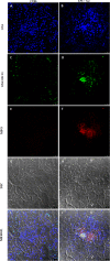
Fig 5. LPS-induced endotoxic shock leads to NETs deposition in kidney tissue.
Endotoxic shock was induced by LPS injection (15 mg/kg, iv.). In situ immunofluorescence analysis of frozen tissue sections of kidney harvested 12 h after endotoxic shock induction was conducted. (A-B) DNA stained. (C-D) DNA/histones stained. (E-F) MPO stained. (G-H) Tissue structure was visualized by differential interference contrast (DIC) microscopy. NETs were identified by the co-localization of DNA, histone and MPO markers. (I-J) Co-localization of DNA (blue), DNA/histone (green) and MPO (red) indicates intraglomerular NETs formation. n = 5 per experimental group.
Fig 6
Fig 6. Serum concentrations of cf-DNA are positively correlated with organ failure in septic patients.
(A) Cf-DNA was quantified in serum samples obtained from healthy control volunteers (n = 8) and septic patients (n = 31). * p < 0.05 compared with healthy control volunteers (Mann-Whitney U test). (B) Correlation of cf-DNA serum concentrations with SOFA scores. Serum cf-DNA concentration according to stages of (C) AKIN score and (D) ARDS, according to the Berlin Definition. *p < 0.05 compared with 0/1 stages (Mann-Whitney U test). (E) Patient serum samples incubated with rhDNase for 30 minutes. *p < 0.05 compared with serum+rhDNase (Paired t-test). AKIN—Acute Kidney Injury Network; ARDS—Acute Respiratory Distress Syndrome; no—no ARDS; rS–Spearman’s rho; SOFA–Sequential Organ Failure Assessment.

References

    1. Bone RC, Balk RA, Cerra FB, Dellinger RP, Fein AM, Knaus WA et al. Definitions for sepsis and organ failure and guidelines for the use of innovative therapies in sepsis. The ACCP/SCCM Consensus Conference Committee. American College of Chest Physicians/Society of Critical Care Medicine. Chest. 1992;101: 1644–55. - PubMed
    1. Remick DG. Pathophysiology of sepsis. Am J Pathol. 2007; 170: 1435–44. - PMC - PubMed
    1. Bachelerie F, Ben-Baruch A, Burkhardt AM, Combadiere C, Farber JM, Graham GJ, et al. International Union of Pharmacology. LXXXIX. Update on the Extended Family of Chemokine Receptors and Introducing a New Nomenclature for Atypical Chemokine Receptors. Pharmacol Rev. 2014; 66: 1–79. 10.1124/pr.113.007724 - VSports手机版 - DOI - PMC - PubMed
    1. Canetti C, Silva JS, Ferreira SH, Cunha FQ. Tumour necrosis factor-alpha and leukotriene B4 mediate the neutrophil migration in immune inflammation. Br J Pharmacol. 2001;134: 1619–28. - PMC (VSports app下载) - PubMed
    1. Kaplan MJ, Radic M. Neutrophil extracellular traps: double-edged swords of innate immunity. J Immunol. 2012;189: 2689–95. 10.4049/jimmunol.1201719 - DOI - PMC - PubMed

V体育官网 - Publication types

"VSports在线直播" Substances