"V体育ios版" Skip to main page content
U.S. flag

An official website of the United States government

Dot gov

The . gov means it’s official VSports app下载. Federal government websites often end in . gov or . mil. Before sharing sensitive information, make sure you’re on a federal government site. .

Https

The site is secure. The https:// ensures that you are connecting to the official website and that any information you provide is encrypted and transmitted securely V体育官网. .

. 2016 Mar;5(3):301-13.
doi: 10.5966/sctm.2015-0179. Epub 2016 Jan 27.

VSports app下载 - Soluble Tumor Necrosis Factor Receptor 1 Released by Skin-Derived Mesenchymal Stem Cells Is Critical for Inhibiting Th17 Cell Differentiation

Affiliations

Soluble Tumor Necrosis Factor Receptor 1 Released by Skin-Derived Mesenchymal Stem Cells Is Critical for Inhibiting Th17 Cell Differentiation

Fang Ke (V体育安卓版) et al. Stem Cells Transl Med. 2016 Mar.

Abstract

T helper 17 (Th17) cells play an important role in multiple sclerosis (MS) and its animal model experimental autoimmune encephalomyelitis (EAE). Th17 cell differentiation from naïve T cells can be induced in vitro by the cytokines transforming growth factor β1 and interleukin-6. However, it remains unclear whether other regulatory factors control the differentiation of Th17 cells. Mesenchymal stem cells (MSCs) have emerged as a promising candidate for inhibiting Th17 cell differentiation and autoimmune diseases. Despite the fact that several molecules have been linked to the immunomodulatory function of MSCs, many other key MSC-secreted regulators that are involved in inhibiting Th17 cell polarization are ill-defined. In this study, we demonstrated that the intraperitoneal administration of skin-derived MSCs (S-MSCs) substantially ameliorated the development of EAE in mice. We found that the proinflammatory cytokine tumor necrosis factor (TNF)-α, a key mediator in the pathophysiology of MS and EAE, was capable of promoting Th17 cell differentiation VSports手机版. Moreover, under inflammatory conditions, we demonstrated that S-MSCs produced high amounts of soluble TNF receptor 1 (sTNFR1), which binds TNF-α and antagonizes its function. Knockdown of sTNFR1 in S-MSCs decreased their inhibitory effect on Th17 cell differentiation ex vivo and in vivo. Thus, our data identified sTNFR1 and its target TNF-α as critical regulators for Th17 cell differentiation, suggesting a previously unrecognized mechanism for MSC therapy in Th17-mediated autoimmune diseases. .

Keywords: Autoimmune disease; Differentiation; T cells; Tissue-specific stem cells V体育安卓版. .


Figures

Figure 1.
Figure 1.
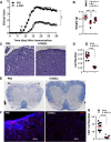
Amelioration of experimental autoimmune encephalomyelitis (EAE) by S-MSCs. (A): Clinical scores (means ± SEM) of mice with established EAE treated with PBS or S-MSCs at indicated time points (n = 10 mice per group). (B): Body weight of mice treated with PBS and S-MSCs or untreated control mice at day 30 after immunization (n = 7). (C): Spinal cord sections derived from PBS- or S-MSC-treated mice at day 18 after immunization were examined by H&E staining for infiltrating lymphocytes. (D): Infiltrating lymphocytes in the spinal cord of EAE mice treated with PBS or S-MSCs were counted. (E): Luxol fast blue staining of spinal cord sections from PBS- or S-MSC-treated mice at day 18 after immunization for visualizing demyelination. (F): Infiltrating CD4+ T cells of spinal cords from PBS- or S-MSC-treated EAE mice were analyzed by immunofluorescence staining. (G): The positively stained CD4+ T cells. For all measurements, the median of specifically stained cells counted in 12 high power fields is presented, and three separate cryosections of each mouse spinal cord were examined (four mice for each group). ∗∗, p < .01; ∗∗∗, p < .001. Representative data are shown that have been reproduced in two or three independent experiments. Abbreviations: DAPI, 4′,6-diamidino-2-phenylindole; PBS, phosphate-buffered saline; S-MSC, skin-derived mesenchymal stem cell.
Figure 2.
Figure 2.
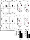
S-MSCs inhibited the Th17 cell differentiation in vivo. (A–D): Flow cytometric analyses of IL-17A+ CD4+ T cells derived from the spleen (A, C) and CNS (B, D) of healthy controls or EAE mice 18 days postimmunization (n = 3 mice per group). (E–H): Intracellular staining of IL-17A in CD4+T cells for splenocytes (E, G) and CNS (F, H) derived from EAE mice treated with PBS or S-MSCs (n = 3 mice per group, day 18 after immunization). (I, G): Protein levels of IL-17A and INF-γ in serum derived from EAE mice treated with PBS or S-MSCs were analyzed by ELISA (n = 7). ∗∗, p < .01; ∗∗∗, p < .001. The data are representative of three independent experiments. Abbreviations: CNS, central nervous system; Ctr, control; EAE, experimental autoimmune encephalomyelitis; IL, interleukin; PBS, phosphate-buffered saline; S-MSC, skin-derived mesenchymal stem cell; WT, wild type.
Figure 3.
Figure 3.
Secreted factors from S-MSCs inhibited Th17 cell differentiation ex vivo. Splenocytes (1 × 10⁶) isolated from experimental autoimmune encephalomyelitis mice 18 days postimmunization were restimulated with MOG35–55 (20 μg/ml) for 2 days in the presence or absence of S-MSCs (2 × 10⁵). (A): mRNA levels of Th17 cell factors (IL-1, IL-17A, IL-22, IL-23, and ROR-γt) were analyzed by quantitative polymerase chain reaction (qPCR). (B): Protein levels of IL-17A in supernatant were detected by enzyme-linked immunosorbent assay. (C, D): mRNA levels of Th17 cell factors (IL-17A and ROR-γt) were analyzed by qPCR. (E): mRNA levels of IL-17A were analyzed by qPCR when S-MSCs cocultured with splenocytes contact directly or separation by Transwell. ∗, p < .05; ∗∗, p < .01; ∗∗∗, p < .001. The data are representative of at least three independent experiments. Abbreviations: Control, restimulated splenocytes; IL, interleukin; NC, nonstimulated splenocytes; ROR-γt, retinoic acid receptor-related orphan receptor γt; S-MSC, restimulated splenocytes cocultured with skin-derived mesenchymal stem cells; Sup, restimulated splenocytes cultured in the presence of supernatant of S-MSCs at the indicated dilutions.
Figure 4.
Figure 4.
High levels of sTNFR1 were secreted by S-MSCs. (A): Mouse cytokine antibody array screening of supernatant derived from cultured cells indicated on the top of the figure (six mice were used for each group). (B): Splenocytes restimulated with MOG35–55 in the presence of S-MSCs for 24 hours and then splenocytes and S-MSCs were separated and subsequently cultured for another 24 hours. The supernatants were harvested, and the expression of sTNFR1 was examined by enzyme-linked immunosorbent assay (n = 4). The amounts of secreted sTNFR1 were listed. ∗∗∗, p < .001. The data in (B) are representative of at least two independent experiments. Abbreviations: MOG, myelin oligodendrocyte glycoprotein; S-MSC, skin-derived mesenchymal stem cell; sTNFR1, soluble tumor necrosis factor receptor 1.
Figure 5.
Figure 5.
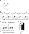
TNF-α promoted Th17 cell differentiation. (A): TNF-α expression in serum derived from healthy controls and EAE mice 18 days postimmunization was detected by enzyme-linked immunosorbent assay (n = 4). (B): Representative flow cytometry of IL-17A expression in Th17 cell differentiation in the presence of TNF-α with various concentrations for 3 days. (C, D): Flow cytometry of IL-17A expression in Th17 cell differentiation, and APCs treated with or without TNF-α (10 ng/ml) before coculture with Th17 polarized cells. Numbers adjacent to outlined areas indicate the percentage of cells in each. ∗, p < .05; ∗∗∗, p < .001. The data are representative of three independent experiments (means ± SEM). Abbreviations: APC, antigen presenting cell; Ctr, control; EAE, experimental autoimmune encephalomyelitis; IFN, interferon; IL, interleukin; TNF-α, tumor necrosis factor α.
Figure 6.
Figure 6.
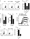
S-MSCs inhibited Th17 cell polarization through releasing sTNFR1. (A, B): Flow cytometry of IL-17A expression in Th17 cell differentiation in the absence or presence of sTNFR1 (100 ng/ml) or TNF-α (10 ng/ml) for 3 days. (C, D): The levels of IL-17A and sTNFR1 were evaluated by enzyme-linked immunosorbent assay. The supernatant derived from splenocytes restimulated with MOG35–55 in the presence of S-MSCs transfected with siNC or si894 (n = 4). (E): Experimental autoimmune encephalomyelitis clinical scores (means ± SEM) of mice treated with S-MSCs (shTNFR1) or S-MSCs (shNC) or PBS (n = 6). (F, G): Flow cytometry of IL-17A expression in Th17 cell polarization in the absence or presence of S-MSCs for 3 days. ∗, p < .05; ∗∗, p < .01; ∗∗∗, p < .001. The data in (E) are representative of two independent experiments. Abbreviations: IL, interleukin; ns, not significant; PBS, phosphate-buffered saline; sh, small hairpin; si, small interfering; S-MSC, skin-derived mesenchymal stem cell; sTNFR1, soluble tumor necrosis factor receptor 1; TNF-α, tumor necrosis factor α.

References

    1. Friedenstein AJ, Chailakhyan RK, Latsinik NV, et al. Stromal cells responsible for transferring the microenvironment of the hemopoietic tissues. Cloning in vitro and retransplantation in vivo. Transplantation. 1974;17:331–340. - PubMed
    1. da Silva Meirelles L, Chagastelles PC, Nardi NB. Mesenchymal stem cells reside in virtually all post-natal organs and tissues. J Cell Sci. 2006;119:2204–2213. - "VSports" PubMed
    1. Ke F, Zhang L, Liu Z, et al. Autocrine interleukin-6 drives skin-derived mesenchymal stem cell trafficking via regulating voltage-gated Ca(2+) channels. Stem Cells. 2014;32:2799–2810. - PubMed
    1. Sioud M, Mobergslien A, Boudabous A, et al. Evidence for the involvement of galectin-3 in mesenchymal stem cell suppression of allogeneic T-cell proliferation. Scand J Immunol. 2010;71:267–274. - PubMed
    1. Di Nicola M, Carlo-Stella C, Magni M, et al. Human bone marrow stromal cells suppress T-lymphocyte proliferation induced by cellular or nonspecific mitogenic stimuli. Blood. 2002;99:3838–3843. - PubMed

Publication types

MeSH terms

Substances

LinkOut - more resources