Skip to main page content
U.S. flag

An official website of the United States government

Dot gov

The . gov means it’s official. Federal government websites often end in . gov or . mil. Before sharing sensitive information, make sure you’re on a federal government site VSports app下载. .

Https

The site is secure. The https:// ensures that you are connecting to the official website and that any information you provide is encrypted and transmitted securely. V体育官网.

. 2016 Feb 9;113(6):1612-7.
doi: 10.1073/pnas.1518163113. Epub 2016 Jan 22.

Recruitment of A20 by the C-terminal domain of NEMO suppresses NF-κB activation and autoinflammatory disease

Affiliations

"VSports手机版" Recruitment of A20 by the C-terminal domain of NEMO suppresses NF-κB activation and autoinflammatory disease

V体育ios版 - Jevgenia Zilberman-Rudenko et al. Proc Natl Acad Sci U S A. .

Abstract

Receptor-induced NF-κB activation is controlled by NEMO, the NF-κB essential modulator. Hypomorphic NEMO mutations result in X-linked ectodermal dysplasia with anhidrosis and immunodeficiency, also referred to as NEMO syndrome. Here we describe a distinct group of patients with NEMO C-terminal deletion (ΔCT-NEMO) mutations. Individuals harboring these mutations develop inflammatory skin and intestinal disease in addition to ectodermal dysplasia with anhidrosis and immunodeficiency. Both primary cells from these patients, as well as reconstituted cell lines with this deletion, exhibited increased IκB kinase (IKK) activity and production of proinflammatory cytokines. Unlike previously described loss-of-function mutations, ΔCT-NEMO mutants promoted increased NF-κB activation in response to TNF and Toll-like receptor stimulation. Investigation of the underlying mechanisms revealed impaired interactions with A20, a negative regulator of NF-κB activation, leading to prolonged accumulation of K63-ubiquitinated RIP within the TNFR1 signaling complex. Recruitment of A20 to the C-terminal domain of NEMO represents a novel mechanism limiting NF-κB activation by NEMO, and its absence results in autoinflammatory disease VSports手机版. .

Keywords: A20; HED-ID; NF-κB; TLR; autoinflammatory disease. V体育安卓版.


Conflict of interest statement

The authors declare no conflict of interest.

"V体育官网" Figures

Fig. 1.
Fig. 1.
NEMO C-terminal truncation is associated with inflammatory disease and results in enhanced stimulation-induced proinflammatory gene expression and cytokine production in patient monocytes and T cells. (A) NEMO syndrome inflammatory disease phenotypes affecting the C terminus. Color-coded protein mutations depict disease phenotypes. Mutations not associated with inflammatory disease but included in this study are shown in black. (B) Healthy control and patient-purified CD14+ and CD4+ cells were cultured in media alone or with TNF or TLR ligands for 24 h and secreted cytokines were measured by capture assay. Four individual patients with the NEMO-E391X mutation and two individual patients with the NEMO-F312L mutation were studied. The mean value of three independent experiments (E391X) or two experiments (F312L) is shown by the horizontal black bar. Statistical significance is determined in relation to normal donor. (See Dataset S1 for absolute cytokine production values from patient and healthy control CD14+ monocytes and CD4+ T-cell experiments.) GVHD, graft versus host disease; HLH, hemophagocytic lymphohistiocytosis; PID, primary immunodeficiency.
Fig. S1.
Fig. S1.
Pedigrees, gene expression, and cytokine production in patient and healthy control cells. (A) Pedigrees of kindreds affected by ΔCT-NEMO and UBAN-NEMO mutants used in this study. (B) Expanded array of cytokine production indices from cells of patients harboring E391X-NEMO, F312L-NEMO, and healthy controls, as described in Fig. 1B. Cytokines measured by capture assay from cultured cells are depicted in columns, and individual stimuli are in rows. Data shown are mean values from six individual patients (four with E391X-NEMO and two with F312L-NEMO) normalized to six paired healthy-donor controls. The CPI is depicted by intensity of the shaded box. Results are mean values from four independent experiments. (C) E391X and F312L patient-derived PBMC were cultured in media alone or media supplemented with Flagellin or LPS for 3 h. Fold-induction represents the ratio of gene expression in stimulated cells to that in cells cultured in media alone. The mean value of three independent experiments (E391X) or two experiments (F312L) is shown by the horizontal black bar. Statistical significance is determined in relation to normal donor.
Fig. 2.
Fig. 2.
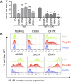
NEMO-truncation leads to increased NF-κB activity upon TNF-R1 or TLR5 stimulation and prevents TNF-induced apoptosis to the same degree as wild-type NEMO. (A) NEMO-deficient and reconstituted cell lines were cultured in media alone, TNF, or TNF + zVAD for 18 h. Apoptosis was measured by positivity for Annexin-V surface staining and mean percent-specific cell death was calculated as described in Materials and Methods, (n = 3). (B) Surface protein expression of the NF-κB reporter, rat Thy-1, in NEMO-deficient [NEMO(−)] or reconstituted Jurkat cells was detected by flow cytometry following stimulation in media alone (blue), TNF (green), or Flagellin (red) and quantified (n = 3) (Fig. S3A).
Fig. S2.
Fig. S2.
Stable expression of NEMO mutants in NEMO-deficient cell lines and patient samples. (A) NEMO intracellular stain as previously described of parental Jurkat cell line (3T8), NEMO-deficient (8321), and NEMO-reconstituted 8321 cells (rNEMO). (B) Cartoon of wild-type NEMO, NEMO syndrome mutants, and mutant forms of NEMO expressed in the NEMO-deficient Jurkat cell lines used in this study. (C) Western blot of cell lysates from patient and healthy control blasting T cells were probed with antibody specific for NEMO, IKKβ, and actin as a loading control.
Fig. S3.
Fig. S3.
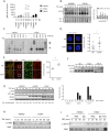
The canonical NF-κB pathway is activated in NEMO and ΔCT-mutant reconstituted cells following TNF stimulation. (A) Quantitation of surface protein expression (n = 3) of Thy1.1 NF-κB reporter as done in Fig. 2B. Average geometric mean fluorescence intensity (gMFI) and SEM appear beneath each condition. (B) Nuclear extracts were incubated with labeled NF-κB consensus sequence oligonucleotide or control (Oct-1) sequence and visualized by EMSA. Nuclear extracts loaded in lane labeled “cc” were preincubated with 100-fold excess of unlabeled consensus NF-κB or Oct-1 oligonucleotide before incubation with labeled oligonucleotide. Densitometry of the NF-κB/DNA complexes normalized to Oct-1 from a representative experiment (n = 3). (C) Reconstituted Jurkat T cells were stimulated with TNF and specificity of nuclear NF-κB complexes was determined by antibody-induced supershift of DNA-protein complexes. (D) Healthy control and E391X-NEMO patient PBMC were isolated. Nuclei were stained followed by intracellular p65 staining on cells following fixation. Images of two representative unstimulated cells are shown. Quantitation indicates nuclear fraction of p65 determined from images obtained by confocal microscopy at 63× magnification of ∼10 cells per condition. (E) Healthy control and E391X-NEMO patient iPS-derived fibroblast-like mesenchymal stem cells in culture. Nuclear and intracellular p65 staining was performed on fixed cells. Images at 4× magnification of a representative area of unstimulated cells are shown. The area bounded by the white box is shown enlarged at 10× magnification in panels on the right. For quantitation, each circle represents a single experimental replicate and each replicate consists of images containing ∼2,000 cells (n = 2). (F) Activated NEMO-E391X and NEMO-F312L patient T cells were stimulated with TNF. Western blot of whole cell lysates was probed with antibody specific to phosphorylated serine residues 32 and 36 of IκBα, NEMO was probed as a loading control. The result shown is representative of three independent experiments. (G) Reconstituted Jurkat T cells were stimulated with TNF for the indicated times and detection of p-IkBa and IkBa was performed by Western blot (n = 2); optical densitometry indicating the ratio of p-IkBa/IkBa was done (Right). (H) Reconstituted Jurkat T cells were stimulated with TNF and detection of IKK-b and IKK-a serine phosphorylation was performed by specific antibody following Western blot. (I) Reconstituted Jurkat T cells were stimulated with TNF followed by immunoprecipitation of IKK-β and subsequent detection of total phospho-serine using a specific antibody.
Fig. 3.
Fig. 3.
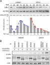
NEMO-truncation leads to increased TNF-induced IKK complex activity. (A) Western blot of IκBα following TNF stimulation with actin blot as a loading control. Quantitation of IκBα, normalized to actin, was performed by optical densitometry, below. (B) NEMO-deficient and reconstituted Jurkat cell lines were treated with TNF and an in vitro IKK kinase assay (KA) was performed using immunoprecipitated IKK complexes in the presence of ATP and substrate and Western blot analysis of coimmunoprecipitated phospho-IκBα; results are representative of three independent experiments.
Fig. 4.
Fig. 4.
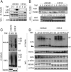
Impaired recruitment of A20 to a NEMO C terminus truncation mutant is associated with increased K63-polyubiquitinated RIP1 associated with NEMO and the TNFR. (A) Reconstituted Jurkat T Cells were stimulated with TNF for the indicated times, and NEMO was isolated by immunoprecipitation to detect NEMO/A20 association. NEMO was probed as an immunoprecipitation control. (B) Following TNF stimulation for the indicated times, the TNFR1 was immunoprecipitated (IP) and the associated A20 was detected by Western blot; TNFR1 was probed as a control. The asterisk denotes IP with IgG as a control. (C) Following TNF stimulation, NEMO was immunoprecipitated and the associated RIP1 was detected in addition to specific K63 ubiquitin linkages using K63-linkage–specific antibody, NEMO was probed as an IP control. (D) Following TNF stimulation, TNF-R1 was immunoprecipitated and the associated RIP1 was detected by Western blot; unmodified RIP1 is indicated by the black triangle. RIP1, phospho-IκBα, NEMO, and actin were detected in corresponding whole-cell lysates. Experiments were performed in NEMO-deficient Jurkat cells reconstituted with either wild-type or E391X NEMO.
Fig. S4.
Fig. S4.
The DUB CYLD can associate with DCT-NEMO in response to TNF. (A) 293 cells were transfected with CYLD, full-length NEMO, or E391X-NEMO vectors and treated with TNF for 10 min before immunoprecipitation of cell lysates with anti-NEMO followed by Western blot for CYLD and NEMO in IP and whole-cell lysates. (B) Reconstituted Jurkats were stimulated with TNF for 10 min, lysed, and CYLD was immunoprecipitated with anti-CYLD. The upper band in the NEMO blot of the IP is a combination of mouse heavy chain and full-length NEMO. High molecular-weight RIP1 is stabilized at the TNFR in cells that express E391X-NEMO. (C) Quantitation by densitometry of Western blot bands corresponding to RIP1 (unmodified and ubiquitinated) coimmunoprecipitated with TNFR1 as performed in Fig. 4D, (n = 3). The effect of genotype (NEMO or E391X) was determined by two-way ANOVA and was highly significant (P < 0.0001).
Fig. S5.
Fig. S5.
NEMO ubiquitin binding specificity and its effect on A20 recruitment and function in NF-κB activation and regulation. (Left) Full-length NEMO enables TLR and TNF-R signaling via combined K63- and M1-linked polyubiquitin recognition and recruitment of A20 that modifies RIP1, leading to its degradation. (Right) Loss of the C terminus leads to a decrease in K63 polyubiquitin binding, impaired association with A20, and enhanced TNF-induced IKK activity, in part because of enhanced stabilization of signaling components at the receptor, such as RIP1. Similarly, TLR stimulation leads to canonical IKK complex activation with absent recruitment of A20 and subsequently increased proinflammatory cytokine production.

Comment in

VSports最新版本 - References

    1. Lawrence T. The nuclear factor NF-kappaB pathway in inflammation. Cold Spring Harb Perspect Biol. 2009;1(6):a001651. - PMC - PubMed
    1. Ruland J. Return to homeostasis: Downregulation of NF-κB responses. Nat Immunol. 2011;12(8):709–714. - VSports - PubMed
    1. Rudolph D, et al. Severe liver degeneration and lack of NF-kappaB activation in NEMO/IKKgamma-deficient mice. Genes Dev. 2000;14(7):854–862. - PMC - PubMed
    1. Conze DB, Wu CJ, Thomas JA, Landstrom A, Ashwell JD. Lys63-linked polyubiquitination of IRAK-1 is required for interleukin-1 receptor- and Toll-like receptor-mediated NF-kappaB activation. Mol Cell Biol. 2008;28(10):3538–3547. - PMC - PubMed
    1. Wu CJ, Conze DB, Li T, Srinivasula SM, Ashwell JD. Sensing of Lys 63-linked polyubiquitination by NEMO is a key event in NF-kappaB activation [corrected] Nat Cell Biol. 2006;8(4):398–406. - PubMed

MeSH terms

Substances

LinkOut - more resources