Skip to main page content
U.S. flag

An official website of the United States government

Dot gov

The . gov means it’s official. Federal government websites often end in VSports app下载. gov or . mil. Before sharing sensitive information, make sure you’re on a federal government site. .

Https

The site is secure V体育官网. The https:// ensures that you are connecting to the official website and that any information you provide is encrypted and transmitted securely. .

. 2016 Mar 24;84(4):976-988.
doi: 10.1128/IAI.01389-15. Print 2016 Apr.

VSports注册入口 - Biological Activities of Uric Acid in Infection Due to Enteropathogenic and Shiga-Toxigenic Escherichia coli

Affiliations

Biological Activities of Uric Acid in Infection Due to Enteropathogenic and Shiga-Toxigenic Escherichia coli

John K Crane et al. Infect Immun. .

Abstract

In previous work, we identified xanthine oxidase (XO) as an important enzyme in the interaction between the host and enteropathogenic Escherichia coli(EPEC) and Shiga-toxigenic E. coli(STEC). Many of the biological effects of XO were due to the hydrogen peroxide produced by the enzyme. We wondered, however, if uric acid generated by XO also had biological effects in the gastrointestinal tract. Uric acid triggered inflammatory responses in the gut, including increased submucosal edema and release of extracellular DNA from host cells. While uric acid alone was unable to trigger a chloride secretory response in intestinal monolayers, it did potentiate the secretory response to cyclic AMP agonists. Uric acid crystals were formed in vivo in the lumen of the gut in response to EPEC and STEC infections VSports手机版. While trying to visualize uric acid crystals formed during EPEC and STEC infections, we noticed that uric acid crystals became enmeshed in the neutrophilic extracellular traps (NETs) produced from host cells in response to bacteria in cultured cell systems and in the intestine in vivo Uric acid levels in the gut lumen increased in response to exogenous DNA, and these increases were enhanced by the actions of DNase I. Interestingly, addition of DNase I reduced the numbers of EPEC bacteria recovered after a 20-h infection and protected against EPEC-induced histologic damage. .


Figures

FIG 1
FIG 1
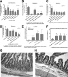
Effects of uric acid on indices of inflammation. (A to D) Effect of uric acid on HL-60 cell chemotaxis or spontaneous migration across an 8-μm-pore-size membrane from the upper to the lower chamber in Trevigen cell migration plates. (A) Lack of effect of uric acid on migration of HL-60 cells using uncoated membranes. (B) Coating the cell wells with 10 μg of DNA reduced the spontaneous migration of HL-60 cells across the membrane. In the presence of DNA, both fMet-Leu-Phe and DNase I (100 U/ml) increased the migration of HL-60 cells across the membrane. (C) In wells coated with 10 μg DNA, uric acid added to the lower chamber still failed to trigger chemotaxis across the membrane. (D) Effect of coating the migration wells with either DNA or uric acid or both. (E) Effects of PMA with or without uric acid on IL-1β production from rabbit peripheral blood leukocytes. (F to H) Effects of uric acid with or without Stx2 toxin on submucosal width in rabbit intestinal segments. (G and H) Normal control intestine (G) and rabbit intestinal loop treated with 600 μM uric acid alone (H). The double-headed arrows show the submucosal space; the arrow in panel H indicates a submucosal width of 74 μm. Magnification, ×100 (G and H). The error bars indicate SD.
FIG 2
FIG 2
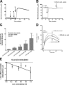
Effect of uric acid on chloride secretion in T84 cells in an Ussing chamber. All the tracings represent short-circuit current (Isc) tracings expressed in microamperes. (A) Lack of effect of adding 1 mM uric acid on the apical side (arrow 1) or on the basolateral side (arrow 2) of the monolayer. Arrow 3 shows that the tissue is still competent in secretion and responsive to 3 μM forskolin. (B) Additive effect of uric acid on secretion induced by forskolin. Forskolin (3 μM) was added first, and then uric acid was added apically, as indicated by the arrows. (C) Dose response of apical uric acid on ΔIsc, defined as the increase in Isc after adding uric acid. Each bar represents the mean and SD of 3 tracings. *, significantly greater than result with forskolin alone. (D) Effect of apical uric acid on Isc triggered by 3 μM guanylin, a cyclic GMP agonist. The top curve shows the response to 600 μM uric acid; the middle curve shows a small inhibitory effect of 1 mM uric acid, and the bottom curve shows the lack of effect of the sodium borate buffer. (E) Dose-dependent inhibition of guanylin-induced Isc by uric acid. Each point represents the mean ± SD of 3 tracings.
FIG 3
FIG 3
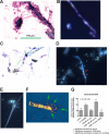
Incorporation of uric acid into DNA NETs. (A, B, and F) Formation of DNA NETs from rabbit peripheral blood leukocytes stimulated with 0.1 μM PMA. (C to E) Images obtained with differentiated HL-60 cells. (A) Bright-field microscopy of PMA-stimulated PBLs showing adherence of leukocytes to crystals and stringy filaments connecting the crystals (three-step stain; magnification, ×100). (B) Polarization microscope image of the same field as in panel A, demonstrating that the crystals show birefringence. The 100-μm scale bar in panel A also applies to panel B. (C) Bright-field microscope image showing that crystals are connected over long distances by stringy filaments, which appear to originate from the nuclei of PMA-treated HL-60 cells. (D) Fluorescent DH5α bacteria expressing green fluorescent protein (GFP) adhere to DNA NETs (arrows) (magnification, ×600; Hoechst stain for DNA). (E) Adherence of human EPEC E2348/69 bacteria to DNA NETs generated from PMA-stimulated HL-60 cells stained with Hoechst dye for DNA. Note that the EPEC bacteria demonstrate their typical localized adherence pattern even when adhering to DNA (magnification, ×600). (F) Merged image showing a birefringent uric acid crystal (photographed by polarization microscopy) superimposed on the same field photographed under fluorescence, with staining using 10 μM Sytox Orange. The orange-stained DNA seems to emanate from one end of the uric acid crystal (arrows). The uric acid crystal itself is 118 μM in length. (G) Uric acid-induced DNA release from HL-60 cells at 3 h of treatment. The error bars indicate SD.
FIG 4
FIG 4
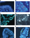
Formation of DNA NETs in vivo in the lumen of the GI tract in rabbit. (A) Hoechst stain of the ileum of a healthy uninfected rabbit. The intestinal segment was fixed in formaldehyde, embedded in paraffin, sectioned, and then deparaffinized and stained with 5 μg/ml Hoechst 33258 dye. (B) Hoechst stain of the ileum of a rabbit intestinal segment infected with rabbit STEC strain E22-stx2 for 20 h. The lumen of the gut is at the top right. STEC bacteria are adhering as a thick biofilm (double-headed arrow, which is 20.8 μm in length). The scale in panel B also applies to panels A, C, and D. (C) DNA NETs formed in vivo from rabbit intestine infected with rabbit EPEC strain E22. Cells that are being sloughed into the intestine are connected by DNA strands (red arrows) and surrounded by bacteria. A few individual EPEC bacteria are indicated by green arrows (magnification, ×600). (D) Another field from an intestine infected with EPEC E22 and stained with 10 μg/ml DAPI, showing that the DNA NETs can form a delicate, lacy network and not just linear strands. A few individual EPEC bacteria are again indicated by arrows. (E and F) Effect of DNase I on NETs formed during infection with EPEC E22 (magnification, ×1,000). (E) DNase-treated intestinal loops showed markedly fewer adherent bacteria; the arrows indicate a few E22 bacteria adhering to a villus tip. (F) Remnant bacteria observed in DNase-treated intestinal loops seemed to be adhering “at a distance” rather than in the typical intimate adherence pattern seen in panel B. Green bracket, 8 μm.
FIG 5
FIG 5
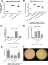
Effects of uricase and DNase I on infection with EPEC E22 in rabbit ileal loops in vivo. (A) Lack of effect of uricase on recovery of EPEC E22 bacteria from intestinal loops. (B) Effects of micrococcal nuclease and DNase I on recovery of E22 from rabbit ileal loops. †, micrococcal nuclease just missed statistical significance on E22 bacterial counts. (C) Effect of DNase I on myeloperoxidase activity in the fluid recovered from the ileal loop. (D) Effects of uricase and DNase I on myeloperoxidase in intestinal-loop fluids. (E) Effect of DNase I on histological damage in the ileum, as measured by the villus-to-crypt ratio, an index of villus blunting. (F) Effect of DNase I on the noncoliform aerobic bacteria recovered on MacConkey agar in intestinal loops treated with DNase I (right petri dish; the arrows indicate non-lactose-fermenting colonies) compared to intestinal loops infected with E22 alone (left petri dish; almost all the colonies are coliforms). The error bars indicate SD.
FIG 6
FIG 6
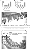
Evidence for positive-feedbacks loops in the intestine. (A) Effect of exogenous DNA with or without DNase I on uric acid formation in the lumen of the rabbit GI tract. Rabbit intestinal loops were left uninfected and were injected with 2 ml of DNase I alone, 2 ml of 100 μg/ml salmon DNA, or DNA plus DNase I. After 20 h, the uric acid concentration in the intestinal-loop fluid was assayed, showing that uric acid can be generated in vivo in the gut in the presence of DNA. (B) Feedback effects of uric acid on ATP release from HL-60 cells. Uric acid alone did not trigger a release of ATP from HL-60 cells, but it potentiated the ATP release triggered by exposure to hypotonic medium, which causes cell swelling. The hypotonic conditions used here were intended as a surrogate for physical stresses, such as mechanical stimulation, shear stress from liquid flow, or changes in pressure, as may occur in vivo. (C) Schematics showing positive-feedback loops that are hypothesized to occur in the GI tract. (Top) ATP released into the lumen as a result of infection is broken down to nucleosides, purines, and uric acid. However, since uric acid can itself enhance ATP release (B), a positive-feedback loop is formed. (Bottom) Uric acid can be formed from the breakdown of DNA, but uric acid also enhances the release of extracellular DNA from host cells (Fig. 3G), so another positive-feedback loop is created. The error bars indicate SD.

References (VSports注册入口)

    1. Hancock J, Salisbury V, Ovejero-Boglione M, Cherry R, Hoare C, Eisenthal R, Harrison R. 2002. Antimicrobial properties of milk: dependence on presence of xanthine oxidase and nitrite. Antimicrob Agents Chemother 46:3308–3310. doi:10.1128/AAC.46.10.3308-3310.2002. - DOI - PMC - PubMed
    1. Yamada Y, Saito H, Tomioka H, Jidoi J. 1987. Susceptibility of micro-organisms to active oxygen species: sensitivity to the xanthine-oxidase-mediated antimicrobial system. J Gen Microbiol 133:2007–2014. - PubMed
    1. Crane J, Naeher TM, Broome J, Boedeker E. 2013. Role of xanthine oxidase in infection due to enteropathogenic and Shiga-toxigenic Escherichia coli. Infect Immun 81:1129–1139. doi:10.1128/IAI.01124-12. - DOI - PMC - PubMed
    1. Behrens MD, Wagner WM, Krco CJ, Erskine CL, Kalli KR, Krempski J, Gad EA, Disis ML, Knutson KL. 2008. The endogenous danger signal, crystalline uric acid, signals for enhanced antibody immunity. Blood 111:1472–1479. - PMC (VSports手机版) - PubMed
    1. Martinon F, Petrilli V, Mayor A, Tardivel A, Tschopp J. 2006. Gout-associated uric acid crystals activate the NALP3 inflammasome. Nature 440:237–241. doi:10.1038/nature04516. - DOI (VSports在线直播) - PubMed

Publication types

MeSH terms

"V体育官网" LinkOut - more resources