Skip to main page content
U.S. flag

An official website of the United States government

Dot gov

The . gov means it’s official. Federal government websites often end in . gov or VSports app下载. mil. Before sharing sensitive information, make sure you’re on a federal government site. .

Https

The site is secure. The https:// ensures that you are connecting to the official website and that any information you provide is encrypted and transmitted securely. V体育官网.

. 2016 Jan 5:6:18822.
doi: 10.1038/srep18822.

"V体育平台登录" AGE-RAGE signal generates a specific NF-κB RelA "barcode" that directs collagen I expression

Affiliations

AGE-RAGE signal generates a specific NF-κB RelA "barcode" that directs collagen I expression

V体育2025版 - Yunqian Peng et al. Sci Rep. .

Abstract

Advanced glycation end products (AGEs) are sugar-modified biomolecules that accumulate in the body with advancing age, and are implicated in the development of multiple age-associated structural and functional abnormities and diseases. It has been well documented that AGEs signal via their receptor RAGE to activate several cellular programs including NF-κB, leading to inflammation VSports手机版. A large number of stimuli can activate NF-κB; yet different stimuli, or the same stimulus for NF-κB in different cellular settings, produce a very different transcriptional landscape and physiological outcome. The NF-κB barcode hypothesis posits that cellular network dynamics generate signal-specific post-translational modifications, or a "barcode" to NF-κB, and that a signature "barcode" mediates a specific gene expression pattern. In the current study, we established that AGE-RAGE signaling results in NF-κB activation that directs collagen Ia1 and Ia2 expression. We further demonstrated that AGE-RAGE signal induces phosphorylation of RelA at three specific residues, T254, S311, and S536. These modifications are required for transcription of collagen I genes and are a consequence of cellular network dynamics. The increase of collagen content is a hallmark of arterial aging, and our work provides a potential mechanistic link between RAGE signaling, NF-κB activation, and aging-associated arterial alterations in structure and function. .


VSports注册入口 - Figures
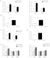
Figure 1
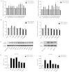
Figure 1. AGE-RAGE axis induces the expression of collagen 1a1 and 1a2 via NF-κB.
(A) AGEs induce transcription of col1a1 and col1a2 genes in murine macrophages. Peritoneal macrophages were isolated from C57BL/6 WT and RAGE-null mice and treated with AGEs for 2 h. Total RNAs were then extracted for qRT-PCR to measure col1a1 and col1a2 transcripts. (B) AGEs induce transcription of col1a1 and col1a2 genes in murine VSMCs. The cells were treated with AGEs for 3 h prior to total RNA extraction and qRT-PCR analyses for col1a1 and col1a2 transcripts. (C,D), ELISA analysis of collagen I protein in macrophages (C) and murine VSMCs (D).(E) AGE-RAGE signals to NF-κB for col1a1 and col1a2 transcription. WT, RAGE-null, and RelA-null MEFs were stimulated with 2 doses of AGEs (20 and 50 μg/ml) for 3 h prior to RNA extraction and qRT-PCR analysis for col1a1 and col1a2 transcripts. Data from qRT-PCR and ELISA were from three independent experiments (n = 3), and each sample was measured in triplicate. Readings from cells treated with BSA were used as the negative control. The results were expressed as mean ± SEM. *p < 0.05; **p < 0.01; ***p < 0.001.
Figure 2
Figure 2. ChIP assays of AGEs-induced RelA binding to 5′ regulatory loci of col1a1 and col1a2 genes.
MEF cells were treated with either BSA or AGEs for 3 h prior to harvest for ChIP assays. The location of each putative NF-κB binding motif relative to the transcription initiation site was marked underneath each group. All qPCR data were from three independent experiments (n = 3), and each sample was measured in triplicate. The results were expressed as mean ± SEM. *p < 0.05; **p < 0.01.
Figure 3
Figure 3. Deciphering AGEs-induced RelA barcode required for transcription of col1a1 and col1a2 genes.
(A) qRT-PCR assessment of AGEs-induced transcription of col1a1 and col1a2 genes in RelA mutant-reconstituted MEFs. (B) qRT-PCR assessment of AGEs-induced transcription of col1a1 and col1a2 genes in reconstituted MEFs harboring RelA(S311A/S536A), (marked as SSAA) and RelA(T254A/S311A/S536A, marked as TSSAAA) composite mutants. (C) Western blots using anti-RelA antibodies to assess the expression of RelA/mutants in reconstituted RelA-null MEFs. (D) ChIP analyses of the binding of RelA phosphorylation mutants to col1a1 –8618 locus in reconstituted MEF cells: RelA WT, T254A and S311A AGEs- treated vs. BSA-treated: p < 0.01; RelA S536A, SSAA, and TSSAAA AGEs-treated vs. BSA-treated, p > 0.05. Among AGEs-treated groups: RelA T254A and S311A vs. WT, p > 0.05; RelA S536A, SSAA, and TSSAAA vs. WT, p < 0.05. (E) ChIP analyses of the binding of RelA phosphorylation mutants to col1a2 –5906 locus in reconstituted MEF cells: RelA WT and S311A AGEs-treated vs. BSA-treated: p < 0.01; RelA T254A, S536A, SSAA and TSSAAA vs. BSA-treated: p > 0.05; Among AGEs-treated groups: RelA S311A vs. WT, p > 0.05; RelA T254A, S536A, and SSAA vs. WT, p < 0.05; RelA TSSAAA vs. WT, p < 0.01. For both (D,E), the data in BSA-treated group shown in the figure include both WT and RelA mutants (n = 18), and data in individual RelA group treated with AGEs were from 3 independent experiments (n = 3) and each sample was measured in triplicate. All results were expressed as mean ± SEM.
Figure 4
Figure 4. AGEs activate the MAPK/ERK–Rsk-1 pathway to generate a RelA barcode.
(A) Sequential activation of ERK1/2 and Rsk-1, and phosphorylation of RelA S536 residue. Total cell lysates (10 μg) were analyzed and β-actin was used as overall loading control. Left panels are the representative western blots, and right panels are the densitometric analyses of three western blots. (B) Pharmacological inhibition of the MAPK/ERK pathway blocks RelA S536 phosphorylation. MEF cells were pre-treated with ERK inhibitor PD98058 (10 nM), or Rsk-1 inhibitor BI-D1870 (10 μM) for 1 h prior to AGEs stimulation. Western blots were performed using 10 μg of total cell lysates. (C) Pharmacological blockers to ERK1/2 and Rsk-1 down-regulate AGEs-induced transcription of col1a1 and col1a2 genes in MEF cells. Specific transcripts were analyzed with qRT-PCR (n = 3). The results were expressed as mean ± SEM. *p < 0.05; **p < 0.01.

References

    1. Ott C. et al. Role of advanced glycation end products in cellular signaling. Redox Biol 2, 411–429 (2014). - PMC - PubMed
    1. Semba R. D., Nicklett E. J. & Ferrucci L. Does accumulation of advanced glycation end products contribute to the aging phenotype? J Gerontol A Biol Sci Med Sci 65, 963–975 (2010). - PMC - PubMed
    1. Brownlee M. The pathological implications of protein glycation. Clin Invest Med 18, 275–281 (1995). - PubMed (V体育官网入口)
    1. Yan S. F., Ramasamy R. & Schmidt A. M. The RAGE axis: a fundamental mechanism signaling danger to the vulnerable vasculature. Circ Res 106, 842–853 (2010). - "V体育安卓版" PMC - PubMed
    1. Ibrahim Z. A., Armour C. L., Phipps S. & Sukkar M. B. RAGE and TLRs: relatives, friends or neighbours? Mol Immunol 56, 739–744 (2013). - PubMed

Publication types

MeSH terms

Substances

LinkOut - more resources