Skip to main page content
U.S. flag

An official website of the United States government

Dot gov

The . gov means it’s official VSports app下载. Federal government websites often end in . gov or . mil. Before sharing sensitive information, make sure you’re on a federal government site. .

Https

The site is secure V体育官网. The https:// ensures that you are connecting to the official website and that any information you provide is encrypted and transmitted securely. .

. 2016 Jan 4;12(1):e1005359.
doi: 10.1371/journal.ppat.1005359. eCollection 2016 Jan.

A Highly Conserved Bacterial D-Serine Uptake System Links Host Metabolism and Virulence (V体育官网入口)

Affiliations

"VSports手机版" A Highly Conserved Bacterial D-Serine Uptake System Links Host Metabolism and Virulence

James P R Connolly et al. PLoS Pathog. .

"V体育官网入口" Abstract

The ability of any organism to sense and respond to challenges presented in the environment is critically important for promoting or restricting colonization of specific sites. Recent work has demonstrated that the host metabolite D-serine has the ability to markedly influence the outcome of infection by repressing the type III secretion system of enterohaemorrhagic Escherichia coli (EHEC) in a concentration-dependent manner VSports手机版. However, exactly how EHEC monitors environmental D-serine is not understood. In this work, we have identified two highly conserved members of the E. coli core genome, encoding an inner membrane transporter and a transcriptional regulator, which collectively help to "sense" levels of D-serine by regulating its uptake from the environment and in turn influencing global gene expression. Both proteins are required for full expression of the type III secretion system and diversely regulated prophage-encoded effector proteins demonstrating an important infection-relevant adaptation of the core genome. We propose that this system acts as a key safety net, sampling the environment for this metabolite, thereby promoting colonization of EHEC to favorable sites within the host. .


Conflict of interest statement (VSports手机版)

The authors have declared that no competing interests exist.

VSports - Figures
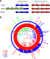
Fig 1
Fig 1. Genomic and phylogenomic context of the yhaOMKJ locus.
(A) Genomic context of the D-serine tolerance locus (blue) in three distinct E. coli isolates–CFT073 (UPEC), EDL933 (EHEC) and MG1655 (K-12). The system encodes DsdC (a LysR type transcriptional regulator), DsdX (a D-serine outer membrane transporter) and DsdA (a D-serine deaminase). In EDL933 the D-serine tolerance locus is truncated and replaced with the sucrose utilization locus (cscRAKB highlighted in green). (B) Genomic context of the second putative D-serine sensory locus (red) in CFT073, EDL933 and MG1655. The system encodes YhaJ (a putative LysR type transcriptional regulator), YhaK (a redox-sensitive bicupin), YhaM (a putative deaminase) and YhaO (a putative inner membrane D-serine transporter). (B) The yhaOMKJ locus is highly conserved across the E. coli phylogeny. Circularized phylogenomic tree of 1591 E. coli and Shigella isolates overlaid with gene carriage for the dsdCXA locus and the yhaOMKJ locus. The yhaOMKJ genes are indicated by red blocks and the dsdCXA locus by blue blocks. Ordering of the genes is numbered and corresponds to the gene in the legend labeled *. Presence of a gene is determined by > 80% identity over > 80% of the coding sequence. Pseudogenes are indicated as yellow blocks. E. coli phylogroups are subdivided by color with the branch point labeled on the tree. Phylogroup A = Blue; Phylogroup B1 = Green; Phylogroup B2 = Red; Phylogroup C = Magenta; Phylogroup D = Purple; Phylogroup E = Cyan; Phylogroup F = Brown; Shigella = Gold. The position of prototypical strains is indicated on the outside of the figure.
Fig 2
Fig 2. Identification of YhaO and YhaJ as potential virulence determinants.
(A) Screening of the yhaOMKJ locus for a role in virulence. SDS-PAGE profile of secreted proteins from TUV93-0, yhaO, yhaM, yhaK and yhaJ cultured in MEM-HEPES. Arrows indicate the location of the major LEE-encoded secreted effectors Tir, EspD and EspA as identified by mass-spectrometry. Samples were normalized according to cellular OD600 to normalize loading into each well. Immunoblot analysis of EspD levels from secreted (Sec) and whole cell lysate (WCL) fractions confirmed the SDS-PAGE results. Anti-GroEL was used to verify equal concentrations of WCL, which corresponded to OD600 normalized culture samples, loaded into each well (B) SDS-PAGE analysis highlighting complementation of the ΔyhaO and ΔyhaJ phenotypes by plasmids pyhaO and pyhaJ. SDS PAGE and immunoblot analysis of secreted protein profiles and EspD cytoplasmic expression confirmed the results. Protein secretion experiments were performed on multiple occasions.
Fig 3
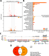
Fig 3. RNA-seq analysis of ΔyhaO and ΔyhaJ under LEE-inducing conditions.
(A) Volcano plots illustrating the identification of differentially expressed genes (DEGs) in the ΔyhaO (red) and ΔyhaJ (orange) mutants when grown in MEM-HEPES to induce expression of the LEE. Grey bars indicate the cutoffs for DEG identification (corrected P-values on the Y-axis and fold change on the X-axis). (B) Functional grouping of DEGs identified according to broad GO terms. Read and orange bars correspond to wild type TUV93-0 versus ΔyhaO and ΔyhaJ respectively. The numbers of DEGs for each group are indicated below and the graph is separated according to downregulated genes and upregulated genes. (C) RNA-seq read coverage mapping across the LEE pathogenicity island illustrating downregulation of the LEE in the ΔyhaO and ΔyhaJ mutants under inducing conditions. Coverage is color coded as grey (TUV93-0), red (ΔyhaO) and orange (ΔyhaJ). Maximum height of the read peaks has been scaled according to the wild type TUV93-0. Genetic organization of the LEE has been indicated below so as to correspond to coverage peaks. Individual ORFs are color coded according to the legend and the operon structure of the LEE is indicated in black. (D) Venn diagram indicating the overlap between the ΔyhaO and ΔyhaJ regulons. Upward or downward arrows indicate upregulated and downregulated DEGs respectively. The DEGs found in both datasets are annotated on the right of the figure.
Fig 4
Fig 4. YhaO and YhaJ are required for attaching and effacing lesion formation on host cells.
(A) Wide-field fluorescence microscopy images of HeLa cells incubated with TUV93-0, ΔyhaO, ΔyhaO + pyhaO, ΔyhaJ and ΔyhaJ + pyhaJ in MEM-HEPES (LEE-inducing conditions). Host cells were stained with FITC-Phalloidin to fluorescently label actin green (488) and bacterial cells were either transformed with a plasmid constitutively expressing RFP (ΔyhaO and ΔyhaJ) or stained with Alexafluor 555 (pyhaO and pyhaJ) to label them red. Merged channels clearly show the areas of localized actin condensation beneath colonized bacterial cells, which corresponds to A/E lesion and pedestal formation as indicated by a white arrow. (B) Quantification of the average percentage of colonized host cells in the ΔyhaO and ΔyhaJ mutants and corresponding complementation backgrounds relative to TUV93-0. (C) Quantification of the average percentage of attached bacteria forming A/E lesions on bound host cells. Data was calculated from three biological replicates with at least twenty-five random fields of view taken per replicate. ***, ** and * denote P ≤ 0.001, P ≤ 0.01 and P ≤ 0.05 respectively.
Fig 5
Fig 5. Biochemical analyses of YhaO functionality in a CFT073 ΔdsdXΔcycA background.
(A) Assessment of the ability of YhaO to transport D-serine. Comparison of the UPEC wild type CFT073 (1) and the D-serine transporter mutant ΔdsdXΔcycA (2) for the ability to grow on MOPS minimal agar plates containing D-serine as a sole carbon source. Complementation of ΔdsdXΔcycA with either pdsdX from CFT073 (3) or pyhaO from TUV93-0 (4) restored the ability to grow on D-serine as a carbon source. (B) Concentration dependent uptake of D-[3H]-serine. A concentration range of 0 to 200 μM D-[3H]-serine was tested and uptake was represented as nmol*mg-1*min-1. (C) Addition of 10 μM CCCP to the reaction (+) at 100% relative uptake of D-[3H]-serine was used to determine the effects of membrane potential on D-[3H]-serine uptake by YhaO. (D) Competitive uptake of L-serine and L-threonine by YhaO. A concentration range (0 to 100 mM) L-serine or L-threonine were added to the reaction at 100% relative uptake of D-[3H]-serine to determine the specificity of YhaO for D-serine. Bars representing L-threonine and L-serine are indicated in dark and light grey respectively.
Fig 6
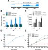
Fig 6. Transcriptional regulation of D-serine uptake in EHEC and UPEC.
(A) Coverage of RNA-seq reads across the yhaOMKJ gene cluster. Data was mapped from previously obtained RNA-seq data that investigated the response of TUV93-0 gene expression to D-serine [21]. The height of coverage peaks has been scaled to that of TUV93-0 using EasyFig [35]. TUV93-0 transcript expression is highlighted in grey whereas TUV93-0 plus D-serine is highlighted in blue. (B) yhaO expression is responsive to environmental D-serine. TUV93-0 and CFT073 were transformed with a plasmid containing a GFP-yhaO promoter fusion (pyhaO:GFP). Activity of the yhaO promoter was measured during growth in relative fluorescence units (RFU) using TUV93-0 alone in MEM-HEPES (grey), TUV93-0 supplemented with 1 mM D-serine (blue) and CFT073 alone (dark grey) or supplemented with 1 mM D-serine (green). Data was calculated from three biological replicates and plotted at increasing OD600 values. ** and *** denote P ≤ 0.01 and P ≤ 0.001 respectively. (C) dsdC and dsdX expression in response to D-serine in UPEC. CFT073 was transformed with pdsdC:GFP (light grey) and pdsdX:GFP (dark grey) transcriptional reporters and activity of measured during growth in MEM-HEPES with and without 1 mM D-serine (clear or dashed bars respectively) as relative fluorescence units (RFU). Data was calculated from three biological replicates. * denotes P ≤ 0.05. (D) Growth curves of EHEC TUV93-0 in MEM-HEPES alone (grey) and supplemented with 1 mM D-serine (blue). (E) Growth curves of UPEC CFT073 and UPEC ΔdsdA in MEM-HEPES alone (black, grey) and supplemented with 1 mM D-serine (green, red). Growth experiments were performed in biological triplicate.
Fig 7
Fig 7. YhaJ directly regulates yhaO expression in EHEC.
(A) Purified YhaJ was tested for its ability to bind the yhaO promoter region (pyhaO; ~300 bp region upstream of the yhaO coding sequence) by EMSA. DIG-labeled pyhaO was incubated with increasing concentrations of YhaJ that corresponded to a shift in free-DNA indicating a YhaJ-DNA complex. Specificity of the binding reaction was tested by the addition of a 100-fold excess (+) of unlabeled pyhaO probe to the binding reaction to outcompete binding of the DIG-labeled probe to YhaJ. These reaction conditions were carried out using a fragment of the kan gene as a negative control. Additionally, the unlabeled kan probe in 100-fold excess was used as a non-specific competitor for YhaJ binding to the DIG-labeled yhaO probe (pyhaO vs kan). (B) Activity of the yhaO promoter in the ΔyhaJ mutant background. A plasmid containing a GFP-yhaO promoter fusion was transformed into TUV93-0 and ΔyhaJ to monitor transcription of yhaO in RFU during growth in MEM-HEPES. Data was calculated from three biological replicates and plotted at increasing OD600 values. * denotes P ≤ 0.05.
Fig 8
Fig 8. YhaJ directly regulates the LEE in EHEC.
(A) Schematic representation of the LEE pathogenicity island. The master regulator ler upstream regulatory region is expanded to illustrate the rationale behind the design of the nested deletion series to monitor LEE1 promoter activity as described by Islam et al. Promoters P1 and P2 as well as corresponding -10 and -35 elements are indicated. (B) Monitoring the impact of YhaJ on LEE1 expression in TUV93-0. LEE10 and LEE20 plasmids were transformed into TUV93-0 (grey) and ΔyhaJ (orange) and LacZ activity was measured in Miller units at an OD600 of approximately 0.7 during growth in MEM-HEPES. The presence of promoters P1 and P2 in each assay is indicated above the graph. * and NS denote P ≤ 0.05 and no significance respectively and the data was calculated from three biological replicates. (C) Purified YhaJ was tested for its ability to bind the LEE1 P1 and P2 promoter regions by EMSA. DIG-labeled LEE1 P1 and P2 specific DNA probes were incubated with increasing concentrations of YhaJ. A shift in free-DNA that corresponds to a YhaJ-DNA complex was only observed for LEE1 P1 and this was in agreement with the data presented in panel B. Specificity of the binding reaction was tested by the addition of a 100-fold excess (+) of unlabeled P1 or P2 probe to the binding reaction to outcompete binding of the DIG-labeled probe to YhaJ. A 100-fold excess of unlabeled kan probe was also used as a non-specific competitor for YhaJ binding to the P1 region (LEE1 P1 vs kan) to ensure specify of the band shift pattern. EMSA experiments were performed in triplicate to confirm the results.
Fig 9
Fig 9. Schematic model of LEE regulation by the YhaO/YhaJ D-serine sensory system.
Summary of small molecule signals that are encountered by EHEC in the intestinal (red) and extraintestinal (blue) environments [9]. In the intestinal environment LEE expression is affected by signals such as fucose, ethanolamine and quorum sensing molecules (epinephrine, norepinephrine and AI-3). YhaJ constitutively regulates yhaO as well as stimulating the LEE (+) helping to promote A/E lesion formation and colonization of host tissue. In the extraintestinal environment D-serine can be encountered in high concentrations leading to repression (-) of the LEE by an unknown (?) direct mechanism [21]. Expression of yhaO is also increased resulting in further uptake of D-serine and thus a greater transcriptional response to this signal (+++) promoting inhibition of colonization in unfavorable environments. The outer membrane (OM), peptidoglycan layer (PG) and inner membrane (IM) of EHEC are indicated.

Comment in

References

    1. Kaper JB, Nataro JP, Mobley HL (2004) Pathogenic Escherichia coli. Nat Rev Microbiol 2: 123–140. 10.1038/nrmicro818 - "V体育官网" DOI - PubMed
    1. Touchon M, Hoede C, Tenaillon O, Barbe V, Baeriswyl S, et al. (2009) Organised genome dynamics in the Escherichia coli species results in highly diverse adaptive paths. PLoS Genet 5: e1000344 10.1371/journal.pgen.1000344 - DOI - PMC - PubMed
    1. Van Elsas JD, Semenov A V, Costa R, Trevors JT (2011) Survival of Escherichia coli in the environment: fundamental and public health aspects. ISME J 5: 173–183. 10.1038/ismej.2010.80 - DOI (V体育官网入口) - PMC - PubMed
    1. Croxen MA, Law RJ, Scholz R, Keeney KM, Wlodarska M, et al. (2013) Recent advances in understanding enteric pathogenic Escherichia coli. Clin Microbiol Rev 26: 822–880. 10.1128/CMR.00022-13 - DOI - PMC - PubMed
    1. Croxen MA, Finlay BB (2010) Molecular mechanisms of Escherichia coli pathogenicity. Nat Rev Microbiol 8: 26–38. 10.1038/nrmicro2265 - DOI (V体育平台登录) - PubMed

Publication types

MeSH terms

LinkOut - more resources (VSports)