Skip to main page content
U.S. flag

An official website of the United States government

Dot gov

The . gov means it’s official. Federal government websites often end in VSports app下载. gov or . mil. Before sharing sensitive information, make sure you’re on a federal government site. .

Https

The site is secure. The https:// ensures that you are connecting to the official website and that any information you provide is encrypted and transmitted securely. V体育官网.

. 2015 Dec 22;6(41):43182-201.
doi: 10.18632/oncotarget.6568.

"VSports最新版本" Integrative omics reveals MYCN as a global suppressor of cellular signalling and enables network-based therapeutic target discovery in neuroblastoma

Affiliations

"VSports在线直播" Integrative omics reveals MYCN as a global suppressor of cellular signalling and enables network-based therapeutic target discovery in neuroblastoma

David J Duffy et al. Oncotarget. .

Abstract

Despite intensive study, many mysteries remain about the MYCN oncogene's functions. Here we focus on MYCN's role in neuroblastoma, the most common extracranial childhood cancer. MYCN gene amplification occurs in 20% of cases, but other recurrent somatic mutations are rare. This scarcity of tractable targets has hampered efforts to develop new therapeutic options. We employed a multi-level omics approach to examine MYCN functioning and identify novel therapeutic targets for this largely un-druggable oncogene. We used systems medicine based computational network reconstruction and analysis to integrate a range of omic techniques: sequencing-based transcriptomics, genome-wide chromatin immunoprecipitation, siRNA screening and interaction proteomics, revealing that MYCN controls highly connected networks, with MYCN primarily supressing the activity of network components. MYCN's oncogenic functions are likely independent of its classical heterodimerisation partner, MAX. In particular, MYCN controls its own protein interaction network by transcriptionally regulating its binding partners. Our network-based approach identified vulnerable therapeutically targetable nodes that function as critical regulators or effectors of MYCN in neuroblastoma. These were validated by siRNA knockdown screens, functional studies and patient data. We identified β-estradiol and MAPK/ERK as having functional cross-talk with MYCN and being novel targetable vulnerabilities of MYCN-amplified neuroblastoma. These results reveal surprising differences between the functioning of endogenous, overexpressed and amplified MYCN, and rationalise how different MYCN dosages can orchestrate cell fate decisions and cancerous outcomes VSports手机版. Importantly, this work describes a systems-level approach to systematically uncovering network based vulnerabilities and therapeutic targets for multifactorial diseases by integrating disparate omic data types. .

Keywords: 4sU-seq; MYC (c-MYC); mRNA sequencing (mRNA-seq); neuroblastoma; transcriptional regulation. V体育安卓版.


Conflict of interest statement

CONFLICTS OF INTERESTS

The authors disclose no potential conflicts of interest.

Figures

Figure 1
Figure 1. An integrative omics approach for analysing MYCN networks
A. Schematic outline of the experimental and analysis approach. B. MYCN mRNA expression in each mRNA-seq sample. CPMkb: counts per million adjusted by gene length in kilobases. Single-read runs are denoted by (sr), with the remainder of the samples being generated by paired-end runs. The time after the SY5Y-MYCN samples indicates the duration of MYCN induction, with 0h being un-induced. C. Schematic overview of how the inferred transcriptional regulators (ITRs) of a set of differentially expressed (DE) genes are identified by Ingenuity Pathway Analysis (IPA). Here, transcription of genes A, C and E is inhibited by the ITR, while B is activated by it, and D is DE but not regulated by the ITR.
Figure 2
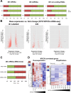
Figure 2. MYCN can repress or activate gene expression dependent on MYCN gene amplification status
A. Proportions of up- and down-regulated mRNAs (mRNA-seq), miRNAs (miRNA-seq) and non-coding RNAs (mRNA-seq) upon MYCN overexpression (SY5Y-MYCN). All data are normalised to relevant control un-induced samples. The 4h LAB time-point is from 4sU labelled cells. B. All significant differentially expressed (DE) genes, with expression level in the un-induced state plotted against the fold change after MYCN induction. 4h Labelled, 24h and 48h MYCN overexpression time-points shown. Each significant DE gene is denoted by a red dot. C. Proportions of up- and down-regulated mRNAs (mRNA-seq), pair-wise comparisons between each of the metastatic MNA lines and KCN (edgeR). D. Heat maps of the MYCN overexpression time-course (as compared by edgeR) and the cell line MYCN correlated genes (as compared by DESeq), from mRNA-seq. A, B denote the biological duplicates. Samples ordered by MYCN expression level.
Figure 3
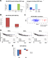
Figure 3. Global MYCN DNA binding profile and MYCN-MAX divergent functions
A. MYCN genome binding (ChIP-seq) is predominantly to enhancers (left) and intragenic regions (right). Abbreviations: Immed. = immediate, Down. = downstream. B. MYCN and MAX ITR activation at 48h of MYCN induction, as revealed by IPA analysis of the mRNA-seq data. Activation status is based on differential regulation of known target genes. C. MYCN and MAX mRNA levels are not correlated with each other in the patient data (microarray). D. MAX mRNA expression levels measured by microarray do not correlate to neuroblastoma patient outcome. Three MAX probes shown (MAX1-3). E. RNAi screen viability results for MYCN and MAX knockdown in SY5Y-MYCN cells. In the induced samples MYCN overexpression was activated 24h prior to and maintained throughout the subsequent 72h RNAi treatment.
Figure 4
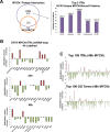
Figure 4. MYCN protein-protein interactors, and global repression of cellular networks by overexpressed MYCN
A. Overlap between MYCN binding proteins, as assessed by CoIP-MS analysis, in MYCN amplified KCN and 24h overexpressed MYCN in SY5Y-MYCN (left). The top 5 ITRs, identified with IPA, of the 415 proteins which only bound amplified MYCN, KCN cells but not overexpressed MYCN (SY5Y-MYCN) (right). Number of the proteins regulated per ITR shown on the y-axis and the p-value of overlap with all known genes associated with that ITR shown above each bar. B. Activation/inhibition z-scores of the top ITRs (IPA) of the genes differentially expressed at 48h, 24h and 4h LAB (4sU labelled) of MYCN overexpression. C. Global view of the activation/inhibition z-scores of the top 100 ITRs (top) and disease and functions GO terms (bottom) for 48h MYCN overexpression (IPA). Ranked by p-value of overlap, of the number of ITR/term component genes which were differentially expressed. GO terms and ITR names are shown in Table S2, the majority of both were downregulated.
Figure 5
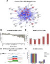
Figure 5. Identification of therapeutically targetable nodes through ITR and network analysis: MAPK
A. The 53 common ITRs (from top 100) between the 3 metastatic MNA lines (IMR32, Kelly, KCNR) were identified. Then a protein interaction map of previously known connections between them was generated with String. MAPK associated nodes, as defined by String's KEGG pathway enrichment tool, are shaded red. B. 72h RNAi screen viability results for the genes from the MNA protein interaction map which increased or reduced cell viability by at least +/− 1 log2FC. In induced samples MYCN overexpression was induced 24h prior to and maintained throughout the RNAi treatment. C. Activation z-scores of MAPK1 in the mRNA-seq samples where it was an ITR, p-value of overlap (between the total number of known MAPK1 ITR component genes and those which were differentially expressed) shown above each bar. D. MYCN binding peaks, located in the 1st intron of the MAPK1 gene, as detected by MYCN ChIP-seq in the SY5Y-MYCN (48h of MYCN overexpression), KCN and KCNR cell lines. Track visualisation was performed using SeqMonk. E. Relative viability of neuroblastoma cells treated with the MEK inhibitor CI-1040, as assed by MTS assay and relative to control cell viability. SY5Y-MYCN+ indicates that MYCN expression was induced for 72 hours.
Figure 6
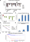
Figure 6. Identification of therapeutically targetable nodes through ITR and network analysis: β-estradiol
A. Activation/inhibition z-score plot of the top 10 ITRs (IPA) for each of the 4 MNA cell lines compared with SY5Y (mRNA-seq). * denotes regulators that were in the top 10 ITRs for all lines. B. Inhibition z-scores of β-estradiol ITR in the MNA (versus SY5Y), Retinoic Acid (RA) treated (un-induced) SY5Y-MYCN and MYCN overexpressing SY5Y-MYCN (both versus un-induced SY5Y-MYCN) mRNA-seq samples. C. Number of MYCN bound genes (ChIP-seq) that are also known β-estradiol targets according to IPA analysis. Rank in ITR analysis and p-value of overlap (between the bound genes and all known β-estradiol targets) shown above each bar. D. Viability of neuroblastoma cells relative to control cells when treated with β-estradiol, as measured by MTS assay. SY5Y-MYCN+ overexpressed MYCN, while SY5Y-MYCN- were un-induced. SY5Y Differenti. Are SY5Y cells pre-treated with 1μM RA for 8 days to induce differentiation into neurons, they were also cultured in the presence of RA for the duration of the 48h β-estradiol treatment. E. The number of ER signalling associated proteins which bound the MYCN protein (CoIP) in SY5Y-MYCN, MYCN un-induced (0h) and overexpressing (24h) and MNA KCN. F. The DE genes contributing to the identification of β-estradiol as an ITR of the mRNA-seq data was used to generate a gene signature. Only genes which were DE in at least 2 cell lines were used. This signature for β-estradiol (bottom) was predictive of neuroblastoma patient outcome.
Figure 7
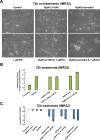
Figure 7. Retinoic Acid, CI-1040 and β-estradiol combination treatments
A. Imaging of IMR32 cells treated for 72h with MAPK inhibitor (CI-1040) or β-estradiol singly, or in combination with Retinoic Acid (RA). All panels are imaged at 40x magnification. B. The differentiation ratio of IMR32 cells treated for 72h with individual agents or combination treatments with RA, was calculated by dividing the length of the longest neurite of a cell by the cell's width. Measurements made using ImageJ v1.44p (http://imagej.nih.gov/ij). Range of measured cells (N) per treatment group is 132-209. Error bars depict the standard error of the mean. C. Cell viability analysis of IMR32 cells treated for 72h with individual agents or combination treatments with RA, as detected by MTS assay.

References

    1. Schwab M, Alitalo K, Klempnauer KH, Varmus HE, Bishop JM, Gilbert F, Brodeur G, Goldstein M, Trent J. Amplified DNA with limited homology to myc cellular oncogene is shared by human neuroblastoma cell lines and a neuroblastoma tumor. Nature. 1983;305:245–248. - PubMed
    1. Malynn BA, de Alboran IM, O'Hagan RC, Bronson R, Davidson L, DePinho RA, Alt FW. N-myc can functionally replace c-myc in murine development, cellular growth, and differentiation. Genes & Development. 2000;14:1390–1399. - PMC - PubMed
    1. Hatton BA, Knoepfler PS, Kenney AM, Rowitch DH, de Alborán IM, Olson JM, Eisenman RN. N-myc Is an Essential Downstream Effector of Shh Signaling during both Normal and Neoplastic Cerebellar Growth. Cancer Research. 2006;66:8655–8661. - PubMed
    1. Barone G, Anderson J, Pearson ADJ, Petrie K, Chesler L. New Strategies in Neuroblastoma: Therapeutic Targeting of MYCN and ALK. Clinical Cancer Research. 2013;19:5814–5821. - PMC - PubMed
    1. Cohn SL, Pearson ADJ, London WB, Monclair T, Ambros PF, Brodeur GM, Faldum A, Hero B, Iehara T, Machin D, Mosseri V, Simon T, Garaventa A, Castel V, Matthay KK. The International Neuroblastoma Risk Group (INRG) Classification System: An INRG Task Force Report. Journal of Clinical Oncology. 2009;27:289–297. - PMC - PubMed

Publication types

MeSH terms

"V体育安卓版" LinkOut - more resources