V体育平台登录 - Skip to main page content
U.S. flag

An official website of the United States government

Dot gov

The . gov means it’s official. Federal government websites often end in . gov or . mil VSports app下载. Before sharing sensitive information, make sure you’re on a federal government site. .

Https

The site is secure. The https:// ensures that you are connecting to the official website and that any information you provide is encrypted and transmitted securely. V体育官网.

. 2015 Dec 10;528(7581):218-24.
doi: 10.1038/nature15749.

"V体育安卓版" The histone chaperone CAF-1 safeguards somatic cell identity

Affiliations

The histone chaperone CAF-1 safeguards somatic cell identity

Sihem Cheloufi et al. Nature. .

Abstract (V体育2025版)

Cellular differentiation involves profound remodelling of chromatic landscapes, yet the mechanisms by which somatic cell identity is subsequently maintained remain incompletely understood. To further elucidate regulatory pathways that safeguard the somatic state, we performed two comprehensive RNA interference (RNAi) screens targeting chromatin factors during transcription-factor-mediated reprogramming of mouse fibroblasts to induced pluripotent stem cells (iPS cells). Subunits of the chromatin assembly factor-1 (CAF-1) complex, including Chaf1a and Chaf1b, emerged as the most prominent hits from both screens, followed by modulators of lysine sumoylation and heterochromatin maintenance. Optimal modulation of both CAF-1 and transcription factor levels increased reprogramming efficiency by several orders of magnitude and facilitated iPS cell formation in as little as 4 days VSports手机版. Mechanistically, CAF-1 suppression led to a more accessible chromatin structure at enhancer elements early during reprogramming. These changes were accompanied by a decrease in somatic heterochromatin domains, increased binding of Sox2 to pluripotency-specific targets and activation of associated genes. Notably, suppression of CAF-1 also enhanced the direct conversion of B cells into macrophages and fibroblasts into neurons. Together, our findings reveal the histone chaperone CAF-1 to be a novel regulator of somatic cell identity during transcription-factor-induced cell-fate transitions and provide a potential strategy to modulate cellular plasticity in a regenerative setting. .


Figures (V体育安卓版)

Extended Data Figure 1
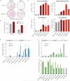
Extended Data Figure 1. Validation of hits from chromatin-focused shRNA screens
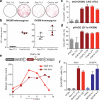
(a) Quantitative RT-PCR analysis to confirm suppression of Chaf1a and Chaf1b expression with miR-30-based vectors from arrayed screen. Sh Chaf1a pool, sh Chaf1b pool and sh CAF-1 pool denote pools of shRNAs targeting either Chaf1a, Chaf1b or both. (b) Western blot analysis to confirm knockdown of CAF-1 components using the top-scoring miR-30-based shRNAs from arrayed screen (see Supplementary Figure 1 for full scans). (c) Quantification of data shown in Fig. 1f. (d) Quantitative RT-PCR analysis confirming knockdown with top-scoring miR-E-based shRNAmiRs targeting Chaf1a, Chaf1b or Ube2i from multiplexed screen. Error bars show standard deviation from biological triplicates. RNA and protein were extracted from reprogrammable MEFs 72 hours after dox induction in panels a-d. (e) Suppression of CAF-1 components, Ube2i and Setdb2 enhances reprogramming in the presence or absence of ascorbic acid (AA) as well as in serum replacement media containing LIF (SR-LIF). Oct4-GFP+ cells were scored by flow cytometry on day 11 after 7 days of OKSM induction and 4 days of transgene-independent growth. Error bars show standard deviation from biological triplicates. (f) Number of dox-independent, alkaline phosphatase (AP)-positive colonies emerging 2 weeks after plating 10,000 reprogrammable MEFs carrying shRNA vectors against indicated targets and cultured in serum replacement media containing 2i (SR-2i), n=1 experiment. (g) Effect of suppressing SUMO E2 ligase Ube2i, E1 ligases Sae1 and Uba2 on iPSC formation. Shown is fraction of Oct4-GFP+ cells at day 11 (7 days of OKSM induction, 4 days of transgene-independent growth). Error bars depict standard deviation from biological triplicates.
Extended Data Figure 2
Extended Data Figure 2. Germline transmission of iPSCs, genetic interaction of shRNA hits and effect of CAF-1 or Ube2i suppression on reprogramming dynamics
(a) Germline transmission of agouti chimeras generated from iPSCs using dox-inducible shRNA vectors targeting Chaf1a, Chaf1b, or Ube2i. Germline transmission was determined by scoring for agouti coat color offspring upon breeding chimeras with albino females. Germline transmission was observed in 8/8, 4/4 and 6/8 cases for Chaf1a iPSC-derived chimeras, in 7/7, 4/4, 7/7 and 9/9 cases for Chaf1b iPSC-derived chimeras, and in 5/5, 7/7 and 5/5 cases for Ube2i iPSC-derived chimeras. (b) Table summarizing effects of co-suppressing pairs of targets on emergence of Oct4-GFP+ cells, shown as the ratio of Oct4-GFP+ to Oct4-GFP cells relative to an empty vector control. Experiment equivalent to Fig. 2b except that second shRNAs were transduced two days after induction of reprogramming. (c) Representative FACS plots showing effects of Chaf1a/b or Ube2i suppression on emergence of Oct4-GFP+ cells at days 7, 9, and 11 of OKSM expression. Histogram plots show fraction of Nanog+ cells within Oct4-GFP+ cells.
Extended Data Figure 3
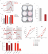
Extended Data Figure 3. Effect of CAF-1 suppression on OKSM levels and cellular growth, and shRNA rescue experiment
(a) Quantitative RT-PCR for transgenic OKSM expression using reprogrammable MEFs transduced with indicated shRNA vectors. Error bars show standard deviation from biological triplicates. (b) RNA-seq analysis of OKSM transgene expression in reprogrammable MEFs transduced with Renilla and Chaf1a shRNAs and exposed to dox for 0, 3 or 6 days. Error bars indicate standard deviation from biological triplicates. (c) Western blot analysis for Sox2 and Tbp (loading control) in reprogrammable MEFs transduced with shRNA vectors targeting Renilla (Ren.713) or different CAF-1 components and exposed to dox for 3 days (see Supplementary Figure 1 for full scans). The same membrane was probed with anti-CAF-1 p150 and anti-CAF-1 p60 antibody to confirm knockdown (data not shown). (d) Rescue experiment to demonstrate specificity of Chaf1b.367 shRNA vector. Reprogrammable MEFs carrying Oct4-tomato knock-in reporter were infected with lentiviral vectors expressing either EGFP or human CAF-1 p60 (CHAF1B) before transducing cells with Renilla or Cha1fb.367 shRNAs and applying dox for 6 days. Colonies were counted at day 11. Note that CAF-1 p60 overexpression attenuates enhanced reprogramming elicited by Chaf1b suppression. (e,f) Competitive proliferation assay between shRNA vector-infected and non-infected reprogrammable cells using indicated shRNAs in the presence or absence of dox (OKSM expression). Note that CAF-1 suppression does not substantially affect the proliferation potential of reprogrammable MEFs after 1-3 days of dox (OKSM) induction while it impairs the long-term growth potential of uninduced MEFs. Data were normalized to cell counts in “no OKSM” condition for (e) and “day 2” time point for (f). Error bars show standard deviation from biological triplicates.
Extended Data Figure 4
Extended Data Figure 4. Confirmation of CAF-1 reprogramming phenotype with alternative transgenic and non-transgenic vector systems
(a) Alkaline phosphatase (AP)-positive, transgene-independent iPSC colonies at day 14 following transduction of R26-M2rtTA MEFs with tetO-STEMCCA lentiviral OKSM expression vector and either Chaf1a.164 or Ren.713 shRNA vectors and treatment with high (2 μg/ml) or low (0.2 μg/ml) doses of dox for 10 days. (b) Quantification of data shown in (a). Experiment was performed at 3 different plating densities (n=1 experiment per density), representative data are shown. (c) Comparison of reprogramming efficiencies between Col1a1::tetOP-OKSM; R26-M2rtTA reprogrammable MEFs and wild type MEFs infected directly with OKSM-expressing lentiviral vectors containing either a strong Ef1a full-length promoter (Ef1a-OKSM long) or a weaker truncated promoter (Ef1a-OKSM short). TRE3G-OKSM is a lentiviral vector with a strong promoter, whose activity is downregulated over time upon infection of CAGS-rtTA3 transgenic MEFs (see below). Error bars show standard deviation from biological triplicates. (d) Quantitative RT-PCR data showing variability in OKSM expression levels over time using different vector systems. Cells were analyzed after 3 and 6 days of infection (lentiviral vectors) or dox exposure (reprogrammable MEFs). Error bars show standard deviation from biological triplicates. OGR MEF, transgenic MEFs carrying Oct4-GFP and CAGS-rtTA3 alleles. (e) Quantification of Oct4 protein levels by intracellular flow cytometry (top) and cellular granularity/complexity by side scatter (SSC) analysis of indicated samples (bottom). Error bars show standard deviation from biological triplicates.
Extended Data Figure 5
Extended Data Figure 5. Effects of CAF-1 dose on NIH3T3 growth and reprogramming potential
(a) Competitive proliferation assay to determine effect of indicated Chaf1a and Chaf1b shRNA vectors on long-term growth potential of immortalized NIH3T3 cell line. Cells were infected with indicated constructs and the fraction of shRNA vector-positive cells was measured by flow cytometry at different time points. Data were normalized to cell counts at day 2 post transduction. Rpa3.455, control shRNA that induces apoptosis. Error bars show standard deviation from biological triplicates. (b) Histogram plots of MEFs harboring R26-M2rtTA allele and either Col1a1::tetOP-miR30-tRFP-Ren.713 or Col1a1::tetOP-miR30-tRFP-Chaf1a.164 shRNA knock-in allele after transduction with pHAGE (Ef1a-OKSM) lentiviral vector and exposure of cells to different doses of dox for 2, 4 and 6 days. Low doses of dox result in lower expression of the shRNAmiR cassettes than high doses of dox. (c) Quantification of data shown in (b) using the geometric mean (n=1 experiment for 3 indicated time points). d) Reprogramming efficiency of Col1a1::tetOP-miR30-tRFP-Chaf1a.164; R26-M2rtTA MEFs infected with pHAGE (Ef1a-OKSM) vector and induced with high (2 μg/ml) or low (0.2 μg/ml) doses of dox for indicated number of days before scoring for Nanog+ iPSCs by immunocytochemistry on day 9. (e) Classification of CRISPR/Cas9-induced mutations by sequence analysis of representative iPSC clones (wt, wild type; indel, insertion/deletion; fs, frameshift; *, point mutation). (f) Western blot analysis for CAF-1 subunits p150 and p60 in 6 representative iPSC clones after CRISPR/Cas9-induced modifications of the Chaf1a locus (see Supplementary Figure 1 for full scans). Wt/wt samples shows unmodified wild type control samples.
Extended Data Figure 6
Extended Data Figure 6. Effect of CAF-1 suppression on HSPC reprogramming and transdifferentiation
(a) Gating strategy for determining Pecam+ fraction (shaded area) in panel (b); data identical to Fig. 4c. (b) Quantification of the fraction of Pecam+ cells at day 4 and day 6 of reprogramming. Data obtained from one experiment using 2 different Chaf1 shRNAs. (c) Transgene dependence assay during the reprogramming of hematopoietic stem and progenitor cells (HSPCs) into iPSCs in the presence of Chaf1a or Renilla shRNAs. Dox pulses were given for 3 or 6 days and alkaline phosphatase (AP)-positive colonies were scored at day 10. (d) Quantitative RT-PCR analysis of Chaf1a expression to confirm knockdown after 3 days of dox induction, i.e. coexpression of shRNAmiR and Ascl1 (n=4 independent infections of the same Col1a1::tetOP-Chaf1a.164 shRNA MEF line; mean value +/− standard deviation). (e) Gating strategy for determining Cd14+ and Mac1+ fractions (shaded area) shown in (f); data identical to Fig. 4g. Positive gates were based on untreated (0 hour) control cells. (f) Quantification of the fraction of Cd14+ and Mac1+ cells at 0, 24 and 48 hours of transdifferentiation using indicated CAF-1 shRNA or empty control vector (n=2 independent infections; rep, replicate). (g) Quantitative RT-PCR analysis of Chaf1a and Chaf1b expression to confirm knockdown in transduced pre-B cell line prior to induction of transdifferentiation (kd/ctrl, knockdown/empty vector control; n=1 experiment, representative of 2 independent infections).
Extended Data Figure 7
Extended Data Figure 7. CAF-1 suppression promotes chromatin accessibility at enhancer elements
(a) Experimental outline and assays (SONO-seq, ATAC-seq, Sox2 ChIP-seq, H3K9me3 ChIP-seq, microarrays and RNA-seq) to dissect effect of CAF-1 suppression on chromatin accessibility, transcription factor binding, heterochromatin patterns and gene expression. Assays were performed either in early reprogramming intermediates (day 3) or throughout the reprogramming time course (ATAC-seq and gene expression). (b) SONO-seq analysis of CAF-1 knockdown and control cells at day 3 of reprogramming to determine accessible chromatin regions across promoters (n=5, 513) and ESC-specific enhancers (n=14, 265). CAF-1 shRNA vectors Chaf1a.164, Chaf1a.2120, Chaf1b.365 and Chaf1b.1221 were pooled for this experiment. (c) ATAC-seq peak distribution across different genomic features. Shown is classification of peaks that are gained in CAF-1 knockdown cells compared to Renilla. (d) ATAC-seq analysis of Chaf1a and Renilla control cells at day 3 of reprogramming to measure global chromatin accessibility over pluripotency-specific super-enhancer elements. ATAC-seq data from Chaf1a.164 shRNA- and Chaf1a.2120 shRNA-transduced cells were merged for this analysis. (e) ATAC-seq accessibility maps at super-enhancer elements associated with the Sall4 locus. Shaded grey bars highlight more accessible sites in Chaf1a knockdown samples at days 3 and 6 of reprogramming compared to Renilla shRNA controls. (f) ATAC-seq analysis of Chaf1a and control cells at day 3 of reprogramming to measure global chromatin accessibility over lineage-specific super-enhancer elements (C2C12, myoblast cell line; proB, progenitor B cells; Th, T helper cells). N denotes number of examined enhancer elements for each cell type. ATAC-seq data from Chaf1a.164 shRNA- and Chaf1a.2120 shRNA-transduced cells were merged for this analysis.
Extended Data Figure 8
Extended Data Figure 8. CAF-1 suppression facilitates Sox2 binding to chromatin
(a) Sox2 ChIP-seq enrichment across pluripotency-specific super-enhancer elements at day 3 of reprogramming in the presence of indicated shRNA vectors. (b) Venn diagram depicting shared and unique Sox2 targets in Chaf1a and Renilla knockdown cells. (c) Bar graph shows the number and fraction of ESC-specific Sox2 targets (blue color) among Sox2-bound sites that are unique to Chaf1a or Renilla knockdown cells at day 3 of OKSM expression. (d) Sox2 ChIP-seq analysis of Chaf1a and control shRNA-infected cells at day 3 of reprogramming to determine enrichment of Sox2 binding across lineage-specific super-enhancer elements (C2C12, myoblast cell line; proB, progenitor B cells; Th, T helper cells; P value < 10−15 for all comparisons between Chaf1a knockdown cells and control).
Extended Data Figure 9
Extended Data Figure 9. CAF-1 suppression induces specific depletion of H3K9me3 at somatic heterochromatin domains
(a) Scatter plots comparing H3K9me3 enrichment nearby ATAC-seq sensitive and super-enhancer regions between control (Ren.713) and Chaf1a knockdown cells (Chaf1a.164 and Chaf1a.2122) at day 3 of reprogramming. Values reflect normalized H3K9me3 ChIP signal (IP/Input) for 5 kb genomic regions overlapping ATAC-seq sensitive regions (red), super-enhancer regions (orange) and regions within 50 kb upstream and downstream of super-enhancers (black). (b) Scatter plots comparing H3K9me3 enrichment over transposable element (TE) families in control and Chaf1a knockdown cells at day 3 of reprogramming. Values reflect normalized H3K9me3 ChIP-seq signal (IP/Input) over families of TEs in the mouse genome. (c) Heatmap shows the relative changes (z-normalized) of TE family expression as estimated by RNA sequencing in control and Chaf1a knockdown cells at day 0, 3 and 6 of reprogramming. Data is clustered using the k-means algorithm. (d) Cumulative histogram showing the relative fraction of RRRs (x-axis) that display negative or positive enrichment (fold-change) of average H3K9me3 signal at day 3 of reprogramming in control and Chaf1a knockdown cells. Note that more RRR regions exhibit depletion of H3K9me3 in Chaf1a knockdown samples. (e) H3K9me3 ChIP-seq analysis of reprogramming-resistant regions (RRRs) after 0 and 3 days of reprogramming. Box plots depict representative RRRs on chromosome 7 (p<0.05 for both shRNAs). See also Fig. 5d. (f) Histogram plot showing activation of UAS-Oct4-GFP transgene upon suppression of Chaf1b (shRNA+ line) in the presence of Gal4-VP16 fusion protein. See Fig. 5f for quantification.
Figure 1
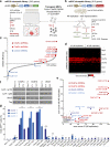
Figure 1. Arrayed and multiplexed shRNAmiR screening strategies to identify suppressors of reprogramming
(a,b) Schematic of arrayed (a) and multiplexed (b) RNAi screens. (c) Results from arrayed screen, depicting average reprogramming efficiency ratios of two biological replicates normalized to Renilla (Ren.713) shRNA control. (d) Heatmap depicting enrichment of selected shRNAs (shown in rows, ordered by gene symbol) over all 96 replicates (columns). (e) Scatter plot representing sum score of enriched shRNAs across all replicates. (f) Western blot analysis confirming shRNA suppression of CAF-1 p150 (Chaf1a), CAF-1 p60 (Chaf1b) and Ube2i at day 3 of reprogramming (see Supplementary Figure 1 for full scans). (g) Validation of hits from multiplex screen. Error bars indicate standard deviation (SD) from biological triplicates (*, p<0.05; **, p<0.01).
Figure 2
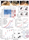
Figure 2. CAF-1 suppression accelerates reprogramming and yields developmentally competent iPSCs
(a) Generation of iPSC-derived chimeras using indicated shRNAmiRs. (b) Effect of co-suppression of indicated targets on reprogramming potential of MEFs, shown as ratio of Oct4-GFP+ to Oct4-GFP cells at day 11 relative to an empty vector control. (c) Flow cytometry plots of representative samples used for panel (b). (d,e) Time course analysis of Oct4-GFP (d) and Nanog (e) expression upon suppression of indicated targets in MEFs undergoing reprogramming. (f) Establishment of transgene-independent Oct4-GFP+ iPSCs in the presence of depicted shRNAs vectors. Samples were induced with dox for indicated number of days before dox withdrawal and analysis at day 13. (g) Expression dynamics of reprogramming markers Epcam and Oct4-tomato after 4 and 6 days of OKSM expression (media supplemented with 2i, ascorbate and Dot1l inhibitor). (h) Alkaline phosphatase (AP)-positive, transgene-independent iPSC colonies scored at day 11 after 4 or 6 days of OKSM expression (representative example from 2 biological replicates and 3 technical replicates).
Figure 3
Figure 3. Reprogramming phenotype depends on optimal CAF-1 and OKSM dose
(a) Comparison of reprogramming efficiency upon Chaf1a knockdown using MEFs carrying one or two copies of Col1a1::tetOP-OKSM and R26-M2rtTA. Colonies were scored at day 10 after 6 days of dox exposure and 4 days of dox-independent growth. Error bars indicate standard deviation from biological triplicates. (b) Quantification of data shown in (a). (c,d) Effect of CAF-1 suppression on reprogramming efficiency when directly infecting MEFs with lentiviral vectors achieving medium (c) or high (d) OKSM expression levels, as determined by flow cytometry for Oct4-GFP at day 11. Error bars indicate standard deviation from biological triplicates. (e) Influence of duration and degree of Chaf1a suppression on reprogramming potential of MEFs carrying doxinducible shRNA cassette (top), as determined by immunocytochemistry for Nanog at day 9. Datapoints represent single experiment. (f) Comparison of reprogramming efficiencies when using shRNAs or sgRNAs targeting Chaf1a, as determined by flow cytometry for Oct4-GFP after 7 days of dox exposure and 4 days of dox-independent growth. Error bars indicate standard deviation from biological triplicates.
Figure 4
Figure 4. CAF-1 suppression enhances reprogramming in different cell conversion systems
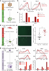
(a) Reprogramming of fetal hematopoietic stem and progenitor cells (HSPCs) into iPSCs. (b) Flow cytometric analysis of Pecam expression during HSPC reprogramming using indicated shRNAs. (c) Quantification of data shown in (b). Values represent fold-change expression differences between experimental and control samples using geometric mean. Data were obtained from one experiment using 2 different Chaf1 shRNAs. (d) Transdifferentiation of MEFs into induced neurons (iNs). (e) Representative image of MAP2+ iNs after 13 days of transdifferentiation. Scale bars: 100 μm. (f) Quantification of transdifferentiation efficiency (n=5 independent experiments; values are mean +/− standard deviation; **, unpaired t-test; p=0.0075). (g) Transdifferentiation of pre-B cells into macrophages. (h) Activation of macrophage markers Cd14 and Mac1 in representative samples at indicated time points. (i) Cd14 and Mac1 expression levels in indicated samples (values represent fold-change expression differences between experimental and control samples using geometric mean; n=2 independent viral transductions).
Figure 5
Figure 5. CAF-1 suppression facilitates chromatin accessibility, Sox2 binding and transcriptional activation of pluripotency genes
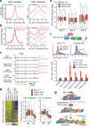
(a) ATAC-seq analysis of ESC-specific enhancers and promoters at day 3 of reprogramming. Shown are merged data for Chaf1a.164 and Chaf1a.2120 shRNA-infected cells (p>0.5 and p<10−15 between Chaf1a and Renilla shRNAs for promoters and enhancers, respectively; n denotes number of examined promoter and enhancer elements). (b) Sox2 ChIP-seq analysis of ESC-specific enhancers and promoters at day 3 of reprogramming using weak (2120) and strong (164) Chaf1a hairpin (p<1e-15 for both shRNAs; see panel (a) for definition of n). (c) Representative ATAC-seq and Sox2 ChIP-seq peaks at the Sall1 super-enhancer (y axis: tag density profiles). (d) H3K9me3 ChIP-seq analysis of reprogramming-resistant regions (RRRs) after 0 and 3 days of OKSM expression. Heatmap shows all RRRs (rows); box plots show individual RRRs between day 0 and 3 in Chaf1a knockdown cells (p<0.05 for both shRNAs). (e) Chromatin accessibility at day 0, 3 and 6 for genes that become transcriptionally upregulated in Chaf1a shRNA-treated cells by day 6 (*, p<0.05; **, p<0.01). (f) Chromatin in-vivo assay (CiA) to directly measure effect of CAF-1 suppression on transcriptional activity of endogenous Oct4 locus in fibroblasts upon overexpression of Gal4-VP16 fusion protein targeted to Oct4 promoter. (g) Summary and model. TF, transcription factor; pol-II, RNA polymerase II.

References

    1. Lee TI, Young RA. Transcriptional regulation and its misregulation in disease. Cell. 2013;152:1237–1251. doi:10.1016/j.cell.2013.02.014. - PMC - PubMed
    1. Takahashi K, Yamanaka S. Induction of pluripotent stem cells from mouse embryonic and adult fibroblast cultures by defined factors. Cell. 2006;126:663–676. doi:10.1016/j.cell.2006.07.024. - PubMed
    1. Vierbuchen T, Wernig M. Molecular roadblocks for cellular reprogramming. Molecular cell. 2012;47:827–838. doi:10.1016/j.molcel.2012.09.008. - VSports最新版本 - PMC - PubMed
    1. Yang CS, Chang KY, Rana TM. Genome-wide functional analysis reveals factors needed at the transition steps of induced reprogramming. Cell reports. 2014;8:327–337. doi:10.1016/j.celrep.2014.07.002. - PMC (VSports app下载) - PubMed
    1. Mikkelsen TS, et al. Dissecting direct reprogramming through integrative genomic analysis. Nature. 2008;454:49–55. doi:10.1038/nature07056. - PMC - PubMed

Publication types

MeSH terms

Associated data