Skip to main page content
U.S. flag

An official website of the United States government

Dot gov

The . gov means it’s official. Federal government websites often end in . gov or . mil. Before sharing sensitive information, make sure you’re on a federal government site VSports app下载. .

Https

The site is secure. The https:// ensures that you are connecting to the official website and that any information you provide is encrypted and transmitted securely V体育官网. .

. 2016 Jan;126(1):349-64.
doi: 10.1172/JCI82720. Epub 2015 Dec 14.

MicroRNA-31 initiates lung tumorigenesis and promotes mutant KRAS-driven lung cancer (VSports)

V体育平台登录 - MicroRNA-31 initiates lung tumorigenesis and promotes mutant KRAS-driven lung cancer

Mick D Edmonds (V体育平台登录) et al. J Clin Invest. 2016 Jan.

Abstract

MicroRNA (miR) are important regulators of gene expression, and aberrant miR expression has been linked to oncogenesis; however, little is understood about their contribution to lung tumorigenesis. Here, we determined that miR-31 is overexpressed in human lung adenocarcinoma and this overexpression independently correlates with decreased patient survival. We developed a transgenic mouse model that allows for lung-specific expression of miR-31 to test the oncogenic potential of miR-31 in the lung VSports手机版. Using this model, we observed that miR-31 induction results in lung hyperplasia, followed by adenoma formation and later adenocarcinoma development. Moreover, induced expression of miR-31 in mice cooperated with mutant KRAS to accelerate lung tumorigenesis. We determined that miR-31 regulates lung epithelial cell growth and identified 6 negative regulators of RAS/MAPK signaling as direct targets of miR-31. Our study distinguishes miR-31 as a driver of lung tumorigenesis that promotes mutant KRAS-mediated oncogenesis and reveals that miR-31 directly targets and reduces expression of negative regulators of RAS/MAPK signaling. .


Figures

Figure 10
Figure 10. Decreased levels of negative regulators of the RAS/MAPK pathway in vivo with miR-31 overexpression.
(A and B) qRT-PCR analysis of the indicated mRNA from the lungs of miR-31/CCSP and littermate CCSP transgenic mice that received dox for (A) 2 months or (B) 12 months from Figure 4. Values are normalized to β-actin levels. The number of mice evaluated is denoted by n values. (C) qRT-PCR for the indicated mRNA and (D) Western blot analysis of 2 tumors from each group of H1993 xenograft tumors from mice in Figure 3. Samples are from the same gel and exposure; noncontiguous samples are indicated by a black line. The number of tumors evaluated is denoted by n values. Data represented by bar graphs in AC are mean values. Error bars represent SEM. *P < 0.05; #P < 0.05; §P < 0.05, t tests.
Figure 9
Figure 9. Reduced expression of negative regulators of the RAS/MAPK pathway with miR-31 overexpression in human lung adenocarcinoma.
(A) qRT-PCR analysis of the indicated miR-31 target mRNA in normal (N) human lung tissue and lung adenocarcinoma (Ad) (normal, n = 8; stage I, n = 7; stage II, n = 6; stage III, n = 4; stage IV, n = 6). Values were normalized to β-actin levels. *P < 0.05, t tests. (B) Regression analysis for miR-31 and the indicated target mRNA in human lung tissue evaluated in A. P values were determined by Spearman’s rank correlation. (C) Expression of the 6 miR-31 targets in the lung adenocarcinoma TCGA RNA sequencing data set. Data are presented as log2 transformation of gene expression. P values were calculated by Wilcoxon rank-sum test for normal versus tumor and Kruskal-Wallis rank-sum test for normal versus stages I–IV (normal n = 58; stage I n = 279; stage II n = 124; stage III n = 84; stage IV n = 27). In box-and-whisker plots, horizontal bars indicate the mean values, boxes indicate 25th to 75th percentiles, and whiskers indicate 10th and 90th percentiles. Data represented by bar graphs in A are mean values. Error bars represent SEM.
Figure 8
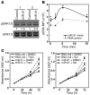
Figure 8. RAS/MAPK signaling is altered by miR-31 and essential for miR-31–regulated lung cell growth.
(A) Western blots for phospho-ERK1/2 (pERK1/2) and total ERK1/2 in Beas-2B cells transfected with miR-31 mimic or RNA control at two concentrations (200 and 400 nM, referred to in the figure as 1 and 2, respectively). Densitometry values are indicated. Data are representative of 2 independent experiments. (B) Beas-2B cells transfected (performed in triplicate) with miR-31 mimic or RNA control were evaluated by intracellular phospho-flow cytometry for phospho-ERK1/2 and total ERK1/2. The MFI of pERK is relative to total ERK (*P ≤ 0.04, t tests). Data are representative of 3 independent experiments. (C) 16HBE cells were transfected with miR-31 mimic or RNA control and treated with 100 nM trametinib (Trem), selumetinib (Sel), or vehicle (DMSO) control. MTT assays were performed (performed in triplicate). Data are representative of 2 independent experiments.
Figure 7
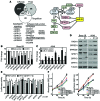
Figure 7. miR-31 directly targets multiple negative regulators of the RAS/MAPK pathway.
(A) Shared putative miR-31 target mRNA identified from 3 prediction algorithms are listed. Asterisks denote mRNA that were manually aligned and determined to have a putative miR-31–binding region. Schematic of the RAS/MAPK signaling pathway. (B and C) qRT-PCR (performed in triplicate) for putative miR-31 target mRNA of (B) Beas-2B cells transfected with miR-31 mimic or RNA control or (C) A549 cells transfected with miR-31 inhibitor or inhibitor control. Values are normalized to β-actin levels. *P < 0.04, t tests. Data are representative of 3 independent experiments. (D) Beas-2B cells infected with miR-31–encoded (31) lentivirus or an empty lentivirus (–) and A549 cells transfected with miR-31 inhibitor (31 inh) or inhibitor control (–) were Western blotted. Densitometry results are shown in Supplemental Figure 7C. The data presented are representative of at least 2 independent experiments, and the data for both Beas-2B and A549 cells were from 3 separate experiments run on different gels. (E) 293T cells were transfected with luciferase vectors encoding all or a region of the 3′-UTR from each gene indicated (two parts of SPRED1 3′-UTR) and miR-31 mimic, miR-17-5p mimic, or RNA control. Luciferase assays were performed (performed in triplicate); expression is relative to β-galactosidase activity. *P < 0.03, t tests. Data are representative of 4 independent experiments. (F) MTT assays of H1993 cells infected with a miR-31–encoded retrovirus or empty retrovirus (vector) and transfected with vectors encoding the indicated miR-31 target cDNA. Experiments are representatives of 2 independent experiments. Error bars represent SEM.
Figure 6
Figure 6. miR-31 enhances urethane-induced lung tumor growth.
A cohort of miR-31/CCSP and littermate CCSP control mice was injected with urethane once and given dox for 6 weeks. (A) Surface lung nodules were counted; error bars represent SEM. *P = 0.03, t test. (B) Representative H&E-stained sections (original magnification: ×10). (C) The number of adenomas per mouse was determined from H&E-stained sections. *P = 0.047, t test. The number of mice evaluated is denoted by n values.
Figure 5
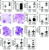
Figure 5. miR-31 promotes KRASG12D-driven lung tumorigenesis.
Cohorts of miR-31/CCSP/KRASG12D and littermate CCSP/KRASG12D control mice were given dox in their drinking water for (AG) 2 months or (HK) 10 months. (A) Ki67 IHC was performed on lung sections (*P = 0.00208, t test). (B) Surface lung nodules were counted; error bars represent SEM. (C, D, and IK) The number of hyperplasia lesions, adenomas, and carcinomas in each mouse was determined from H&E-stained lung sections (*P = 0.02; #P = 0.0006; §P = 0.0024; **P = 0.0016; ##P = 0.037, t tests). AAH, atypical adenomatous hyperplasia. (E and H) Representative H&E-stained sections of lungs (original magnification: ×0.5 [H]; ×10 [E]). Larger histology pictures are shown in Supplemental Figure 6B. (F) The mean tumor diameter was determined from H&E-stained sections (*P = 0.01, t test). (G) Tumor burden was determined by comparing the area of tumor to that of normal lung from H&E-stained lung sections (*P = 0.01, t test).
Figure 4
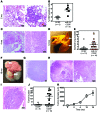
Figure 4. miR-31 initiates benign and malignant lung tumor formation in vivo.
Cohorts of miR-31/CCSP and littermate CCSP control mice were given dox in their drinking water for (A and B) 2 months, (C) 4 months, (D and E) 12 months, or (FJ) 18 months. (A, C, and GI) Representative photographs of H&E-stained lung sections from miR-31/CCSP mice (original magnification: ×2 [H, left]; ×4 [G]; ×10 [A and C, left, and H, right]; ×20 [I]; ×40 [A and C, right]). Larger histology pictures are shown in Supplemental Figure 5. (B) Ki67 IHC was performed on lung sections (*P = 0.00124, t test). The number of mice evaluated is denoted by n values. (D and F) Representative photographs of (D) Bouin’s fixed lungs and (F) gross lungs, with arrows pointing to tumors. (E) Lung surface tumor nodules were counted on Bouin’s fixed lungs (*P > 0.03, t test). The number of mice evaluated is denoted by n values. (I) Representative photograph of an H&E-stained section of lung adenocarcinoma showing invasion into a blood vessel. (J) Tumor burden was determined by comparing the area of tumor to that of normal lung from H&E-stained lung sections (*P = 0.0145, t test). The number of mice evaluated is denoted by n values. (K) The mean tumor diameter for lung tumors in miR-31/CCSP mice given dox for the indicated intervals was determined from H&E-stained sections.
Figure 3
Figure 3. Elevated miR-31 increases adenocarcinoma cell growth in vivo.
H1993, H1437, and H460 cells were infected with miR-31–encoded retrovirus (miR-31) or empty retrovirus (vector [vec]) control. Cells were injected subcutaneously into the flanks of nude mice, and (A) tumor volume was measured at intervals (*P < 0.04, t tests) and (B) tumor weight was measured (*P = 0.034, #P = 0.039, and §P = 0.042 respectively, t tests) at time of sacrifice (day 22, 24, and 18, respectively). Tumor number indicated by n values. Error bars indicate SEM. Representative pictures of tumors from A are shown in B.
Figure 2
Figure 2. miR-31 promotes lung epithelial cell proliferation.
(A and B) Beas-2B and 16HBE cells transfected with miR-31 mimic or RNA control (performed in triplicate). (A) MTT assays were performed, and (B) viable cells were counted at intervals. Data are representative of 3 independent experiments. (C and D) A549 and H1993 adenocarcinoma cells transfected with miR-31 inhibitor or inhibitor control (performed in triplicate). (C) MTT assays were performed, and (D) viable cells were counted at intervals. Data are representative of 3 independent experiments. (E and F) The cells indicated were infected with a miR-31–encoded retrovirus or empty retrovirus (vector) control. (E) Clonogenic and (F) soft agar assays were performed in triplicate. Data are representative of 2 independent experiments. Colonies were counted after 14 days. *P = 0.0032; **P = 0.0067; #P = 0.023; ##P = 0.027, t tests.
Figure 1
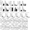
Figure 1. miR-31 is overexpressed and correlates with poor survival in lung adenocarcinoma.
(A) qRT-PCR analysis (performed in triplicate) of miR-31 expression in human lung epithelial and adenocarcinoma cells lines. ΔCt values graphed are relative to the endogenous control RNU6B small RNA with SEM. *P ≤ 0.0012, t test. Data are representative of 3 independent experiments. (B and C) qRT-PCR analysis of miR-31 expression in normal human lung tissue and (B) all human lung adenocarcinoma samples or (C) samples separated by stage of disease. miR-31 expression relative to RNU6B; mean values are indicated by solid bars, and values are shown as mean ± SEM. *P = 0.004; #P = 0.0009; **P = 0.0017; ##P = 0.0001, t test. The number of samples is shown. (D) miR-31 expression in normal lung and lung adenocarcinomas from TCGA miR expression profiles displayed as log2-transformed values normalized to reads per million. *P = 4.99 × 10⁻¹⁴, t test. (E) Data from D separated by stage. *P = 4.8 × 10⁻¹³; #P = 5.3 × 10⁻¹⁰; §P = 3.4 × 10⁻¹⁰; **P = 9.3 × 10–06, t tests. The number of samples is shown. (F and G) Kaplan-Meier analysis with (F) median (158 samples for each) and (G) lowest (n = 79) and highest (n = 79) quartiles of miR-31 expression for lung adenocarcinoma from TCGA expression data. P values were determined by log-rank test.

References

    1. Siegel RL, Miller KD, Jemal A. Cancer statistics, 2015. CA Cancer J Clin. 2015;65(1):5–29. doi: 10.3322/caac.21254. - DOI - PubMed
    1. Vidigal JA, Ventura A. The biological functions of miRNAs: lessons from in vivo studies. Trends Cell Biol. 2015;25(3):137–147. doi: 10.1016/j.tcb.2014.11.004. - DOI - PMC - PubMed
    1. Costinean S, et al. Pre-B cell proliferation and lymphoblastic leukemia/high-grade lymphoma in E(mu)-miR155 transgenic mice. Proc Natl Acad Sci U S A. 2006;103(18):7024–7029. doi: 10.1073/pnas.0602266103. - DOI - PMC - PubMed
    1. Medina PP, Nolde M, Slack FJ. OncomiR addiction in an in vivo model of microRNA-21-induced pre-B-cell lymphoma. Nature. 2010;467(7311):86–90. doi: 10.1038/nature09284. - DOI - PubMed
    1. Sandhu SK, et al. B-cell malignancies in microRNA Emu-miR-17~92 transgenic mice. Proc Natl Acad Sci U S A. 2013;110(45):18208–18213. doi: 10.1073/pnas.1315365110. - DOI - PMC - PubMed

Publication types

MeSH terms