Skip to main page content
U.S. flag

An official website of the United States government

Dot gov

The . gov means it’s official. Federal government websites often end in . gov or VSports app下载. mil. Before sharing sensitive information, make sure you’re on a federal government site. .

Https

The site is secure. The https:// ensures that you are connecting to the official website and that any information you provide is encrypted and transmitted securely V体育官网. .

. 2015 Dec 17;528(7582):413-7.
doi: 10.1038/nature16140. Epub 2015 Dec 9.

Neutrophils support lung colonization of metastasis-initiating breast cancer cells

Affiliations

Neutrophils support lung colonization of metastasis-initiating breast cancer cells

Stefanie K Wculek et al. Nature. .

"V体育ios版" Erratum in

Abstract

Despite progress in the development of drugs that efficiently target cancer cells, treatments for metastatic tumours are often ineffective. The now well-established dependency of cancer cells on their microenvironment suggests that targeting the non-cancer-cell component of the tumour might form a basis for the development of novel therapeutic approaches. However, the as-yet poorly characterized contribution of host responses during tumour growth and metastatic progression represents a limitation to exploiting this approach. Here we identify neutrophils as the main component and driver of metastatic establishment within the (pre-)metastatic lung microenvironment in mouse breast cancer models. Neutrophils have a fundamental role in inflammatory responses and their contribution to tumorigenesis is still controversial. Using various strategies to block neutrophil recruitment to the pre-metastatic site, we demonstrate that neutrophils specifically support metastatic initiation. Importantly, we find that neutrophil-derived leukotrienes aid the colonization of distant tissues by selectively expanding the sub-pool of cancer cells that retain high tumorigenic potential. Genetic or pharmacological inhibition of the leukotriene-generating enzyme arachidonate 5-lipoxygenase (Alox5) abrogates neutrophil pro-metastatic activity and consequently reduces metastasis. Our results reveal the efficacy of using targeted therapy against a specific tumour microenvironment component and indicate that neutrophil Alox5 inhibition may limit metastatic progression. VSports手机版.


Figures
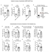
<b>Extended Data Figure 1</b>.
Extended Data Figure 1.. Mammary tumour-bearing MMTV-PyMT+ mice show specifically neutrophilia in the metastatic lung
ac, Flow cytometric quantification of CD11b+Ly6G+ neutrophils in the bone marrow (n = 6 (wild-type), n = 5 (MMTV-PyMT+)) (a), liver (n = 4 (wild-type), n = 5 (MMTV-PyMT+)) (b) and spleen (n = 6 (wild-type), n = 5 (MMTV-PyMT+)) (c) of wild-type (WT) and tumour-bearing MMTV-PyMT+ mice. d, Quantification of neutrophils in the tumour and metastatic lung of MMTV-PyMT+ mice (n = 2 per group), pre-metastatic lung neutrophil levels depicted in Fig. 1a are shown for comparison in dashed lines. Met., metastatic. el, Flow cytometric quantification of immune cell frequencies in wild-type and metastatic lungs of MMTV-PyMT+ mice (n = 4 (wild-type), n = 7 (metastatic) if not otherwise indicated) including CD45+ total immune cells (e), total CD11b+F4/80+ macrophages (f) (n = 4 (wild-type), n = 4 (metastatic)), the CD11blow F4/80high alveolar macrophage subpopulation (n = 4 (wild-type), n = 4 (metastatic)) (g), the CD11bhigh F4/80low interstitial macrophage subpopulation (n = 4 (wild-type), n = 4 (metastatic)) (h), CD45+CD11c+ dendritic cells (i), CD45+CD49b+ NK cells (j), CD45+CD19+ B lymphocytes (k) and CD45+CD3+ T lymphocytes (l). Statistical analysis by two-sided t-test. Data are represented as mean ± s.e.m. NS, not significant, *P < 0.05, **P < 0.01, ***P < 0.001.
<b>Extended Data Figure 2</b>.
Extended Data Figure 2.. Analysis of MMTV-PyMT+Gcsf−/− mice, G-CSF-deficient MMTV-PyMT cancer cells and MMTV-PyMT+Ela2-Cre-DTA+ mice
a, Representative flow cytometric analysis of CD11b+Ly6G+ neutrophils in the lung of wild-type and tumour-bearing MMTV-PyMT+Gcsf+/+ and MMTV-PyMT+Gcsf−/− mice. b, Primary mammary tumour burden of MMTV-PyMT+Gcsf+/+ (n = 13) or MMTV-PyMT+Gcsf−/− (n = 24) mice. c, Flow cytometric quantification of frequencies of total CD11b+F4/80+ macrophages (left), the CD11blowF4/80high alveolar macrophage subpopulation (middle) and the CD11bhighF4/80low interstitial macrophage subpopulation (right) in the lung of tumour-bearing MMTV-PyMT+Gcsf+/+ (n = 4) and MMTV-PyMT+Gcsf−/− (n = 7) mice. d, MMTV-PyMT+Gcsf−/− primary cancer cells were freshly isolated and grafted onto two mammary glands of Rag1-null mice (106 cells per injection) and analysed 5 weeks thereafter. CD11b+Ly6G+ neutrophil presence in the lung was assessed by flow cytometry (left), primary tumour burden was assessed by weighing (middle) and spontaneous lung metastasis incidence was assessed by quantification of visible surface lung metastases relative to tumour load (right) (n = 3 (Gcsf+/+), n = 4 (Gcsf−/−)). eg, Analysis of tumour-bearing MMTV-PyMT+ control and MMTV-PyMT+Ela2-Cre-DTA+ mice. Representative flow cytometric analysis of CD11b+Ly6G+ neutrophils in the lung (e). Lung neutrophil quantification (n = 8 (wild-type), n = 7 (PyMT+ control), n = 5 (PyMT+Ela2-Cre-DTA+)) (f, left) and primary mammary tumour burden (n = 14 (PyMT+ control), n = 6 (PyMT+Ela2-Cre-DTA+)) (f, right) with representative H&E-stained histological lung sections (g). Scale bar, 500μm. h, Flow cytometric quantification of frequencies of total CD11b+F4/80+ macrophages (left), the CD11blowF4/80high alveolar macrophage subpopulation (middle) and the CD11bhighF4/80low interstitial macrophage subpopulation (right) in the lung of tumour-bearing MMTV-PyMT+ control (n = 7) and MMTVPyMT+Ela2-Cre-DTA+ (n = 5) mice. i, Frequencies of bone marrow (top) and blood (bottom) CD11b+Ly6G+ neutrophils (left; blood n = 3 (wild-type), n = 6 (PyMT+ control)), CD11b+F4/80+ macrophages (middle; blood n = 3 (wild-type), n = 6 (PyMT+ control)) and CD11b+CD115+ monocytes (right; blood n = 3 (wild-type), n = 5 (PyMT+ control)) in wild-type, MMTV-PyMT+ control and MMTV-PyMT+Ela2-Cre-DTA+ mice analysed by flow cytometry (n = 4 (wild type), n = 6 (PyMT+ control), n = 2 (PyMT+Ela2-Cre-DTA+) if not otherwise indicated). j, Exclusion of immune responses against DTA expression in the bone marrow by analysis of NK-cell (left) and cytotoxic T-cell (right) activation. Flow cytometric quantification of activated CD69+ among total CD45+CD49b+ NK cells as well as activated CD44+ or CD69+ among total CD45+CD3+CD8+ cytotoxic T cells in the bone marrow of wild-type (n = 4), MMTV-PyMT+ control (n = 6) and MMTV-PyMT+Ela2-Cre-DTA+ (n = 2) mice. Statistical analysis by two-sided t-test. Data are represented as mean ± s.e.m. NS, not significant, *P < 0.05, **P < 0.01, ***P < 0.001.
<b>Extended Data Figure 3</b>.
Extended Data Figure 3.. Comparison of wild-type lung neutrophils with tumour-induced, pre-metastatic lung neutrophils
a, Representation of timing and dynamics of neutrophil and cancer cell infiltration into the lung of mice grafted with two mammary tumours by orthotopic injection of 106 MMTV-PyMT tumour cells. b, Flow cytometric analysis for cell size (forward scatter (FSC)), granularity (side scatter (SSC)) and expression of surface markers CXCR2, CD31, MHC-I, MHC-II, ICAM1 and Fas (n is indicated) as well as mRNA expression analysis of Tnfa, arginase 1, Vegfa, Ccl2, Ccl3, iNOS (also known as Nos2) and Ccl5 by quantitative polymerase chain reaction (PCR) of CD11b+Ly6G+ wild-type (WT) or pre-metastatic (Pre-met.) lung neutrophils 3 weeks after primary tumour graft (n = 3 (pre-metastatic compared with one normal lung reference)). Statistical analysis by two-sided t-test (flow cytometry) and one-sample t-test (mRNA). Data are represented as mean ± s.e.m. *P < 0.05, **P < 0.01, ***P < 0.001.
<b>Extended Data Figure 4</b>.
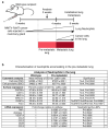
Extended Data Figure 4.. Immune cell frequencies and activation in the pre-metastatic lung of MMTV-PyMT tumour-bearing mice is not dependent on neutrophil presence (part 1)
a, Representation of timing and dynamics of neutrophil and cancer cell infiltration into the lung of mice grafted with two mammary tumours by orthotopic injection of 106 MMTV-PyMT tumour cells. bo, Flow cytometric quantification and representative analysis of the following immune cell types in wild-type (WT) or pre-metastatic (Pre-met.) lungs treated daily with either control IgG or anti-Ly6G (1A8) neutrophil-blocking antibody from tumour onset onwards (n = 4 per group if not otherwise indicated): b, f, total CD45+ immune cells (n = 12 per group); c, g, CD11b+Ly6G+ neutrophils (n = 8 per group); d, g, CD11b+SiglecF+ eosinophils; e, g, CD11blowF4/80high alveolar macrophages and CD11bhighF4/80low interstitial macrophages; h, j, CD45+CD11c+ dendritic cells; i, k, MHC-II+CD86+ activated dendritic cells; l, n, CD45+CD19+ B cells; and m, o, MHC-II+CD86+ activated B cells. Statistical analysis by two-sided t-test. Data are represented as mean ± s.e.m. NS, not significant, *P < 0.05, **P < 0.01, ***P < 0.001.
<b>Extended Data Figure 5</b>.
Extended Data Figure 5.. Immune cell frequencies and activation in the pre-metastatic lung of MMTV-PyMT tumour-bearing mice is not dependent on neutrophil presence (part 2)
ai, Flow cytometric quantification and representative analysis of the following immune cell types in wild-type (WT) or pre-metastatic (Pre-met.) lungs treated daily with either control IgG or anti-Ly6G (1A8) neutrophil-blocking antibody from tumour onset onwards (n = 4 per group if not otherwise indicated): a, c, CD45+CD49b+ NK cells; b, c, CD69+ activated NK cells; d, e, CD45+CD3+CD8+ cytotoxic T cells (n = 8 per group); f, g, CD44+ or CD69+ activated T cells; and h, i, the ratio of CD4+CD25+Foxp3+ regulatory T cells per activated T cell. Statistical analysis by two-sided t-test. Data are represented as mean ± s.e.m. NS, not significant.
<b>Extended Data Figure 6</b>.
Extended Data Figure 6.. Neutrophil isolation from the lung of MMTVPyMT+ mice and effect of neutrophil-derived factors on tumour formation potential
a, Representative flow cytometric analysis of neutrophil purity after isolation from the pre-metastatic lung compared to total lung tissue. Only neutrophil purity of ≥90% was used for further experiments. b, Neutrophil viability was assessed by flow cytometry for propidium iodide (PI) negativity after isolation (n = 10). c, d, MMTVPyMT cells grown in control or LuN medium for 3 days in adherent conditions were plated in non-attachment conditions followed by sphere quantification at day 10 post-seeding (technical replicate n = 17 (control), n = 21 (LuN) of biological triplicates) (c) or 104 cells grafted onto the mammary gland of Rag1-null mice for analysis of tumour formation potential (d). Tumour burden was determined by weighing about 3 weeks after (n = 12 per group), complementary to Fig. 2d. eh, Flow cytometric quantification of frequencies of total present GFP-labelled MMTV-PyMT cells (e, g) and frequencies of CD24+CD90+ MICs among total GFP-labelled MMTV-PyMT cells (f, h) in the lung of Rag1-null mice 3 days after intravenous injection of 5×10⁵ total GFP-labelled MMTV-PyMT cells followed by either three intravenous injections with control or LuN medium (n = 6 (PyMT+control), n = 8 (PyMT+LuN)) (e, f) or by one intravenous injection with 25×106 neutrophils freshly isolated from a pre-metastatic lung (n = 7 (PyMT control), n = 8 (PyMT+neutrophils) (g, h). f, h, Two independent experiments are shown to complement Fig. 2h, i. Exp, experiment. ik, Experimental setup (i): Rag1-null mice were intravenously injected with 1–10×105 (j) or 0.5×10⁶ total GFP-labelled MMTV-PyMT cells (k) followed by either 3-5 intravenous injections with 200μl control or LuN medium (j) or by three intravenous injections with 25×106 neutrophils (k) freshly isolated from a pre-metastatic lung. Quantification of experimental metastatic incidence by determination of bioluminescence intensity (n = 7 (control), n = 9 (LuN)) (j) or flow cytometric analysis of GFP+ cancer cells in the lung (n = 5 (control), n = 4 (neutrophil)) (k) is shown. Statistical analysis by two-sided t-test (c, j, k) and two-way ANOVA (dh). Data are represented as mean ± s.e.m. NS, not significant, *P < 0.05, **P < 0.01, ***P < 0.001.
<b>Extended Data Figure 7</b>.
Extended Data Figure 7.. LTRs are expressed on mouse and human breast cancer cells and enriched on metastasis-initiating and highly tumorigenic cancer cell sub-pools
a, Sphere formation potential of MMTV-PyMT cells under presence of LTB4 or LTC/D/E4 (technical replicate n = 8 per group of biological triplicates). b, c, Three-day LTB4 and LTC/D/E4-treated MMTVPyMT cells in adherent culture were analysed for primary tumour initiation potential by orthotopic transplantation of 104 cells in Rag1-null mice (n = 14 per group) (b). Exp, experiment; TC, tumour cell isolation. Representative image of tumours is shown (c). d, e, Flow cytometric analysis of primary MMTV-PyMT cancer cells, the mouse mammary cancer cell line 4T1 and the human breast cancer cell line MDA-MB-231 for expression of BLT1 or BLT2 (d) as well as CysLT1 or CysLT2 (e). f, Representative flow cytometric analysis of BLT2+ and CysLT2+ cells among MMTV-PyMT non-MICs and MICs. gi, Flow cytometric quantification of LTR expression on Aldefluor (ALD)+ (n = 3 per group) (g) or CD44high MDA-MB-231 cells (n = 4 per group) (h) as well as CD49f+/high 4T1 cells (n = 4 per group) (i). jl, Sorted LTR+ or LTR MMTV-PyMT tumour cells were plated in non-attachment conditions followed by sphere quantification at day 10 post-seeding (technical replicate n = 10 per group of biological duplicates) (j) or 103 cells grafted onto the mammary gland of Rag1-null mice for analysis of tumour formation potential. Tumour burden was determined by weighing (n = 8 per group) after 3 weeks (k) and representative image of tumours is shown (l). Statistical analysis by two-sided t-test (a, hk) and two-way ANOVA (b). Data are represented as mean ± s.e.m. NS, not significant, *P < 0.05, **P < 0.01, ***P < 0.001.
<b>Extended Data Figure 8</b>.
Extended Data Figure 8.. LTs promote stemness within the total cancer cell population by specifically promoting proliferation of MICs
a, In vitro passaging (P indicates passage number) in non-adherent conditions of sorted CD24+CD90+ MICs and CD24+CD90 non-MICs (n = 4 per group for P0+P1 and n = 3 per group for P2+P3). Quantification was performed by determination of percentage of remaining cell number after 7–10 days. b, Flow cytometric quantification of 3-day LT-treated 4T1 cells for frequency of highly tumorigenic CD49fhigh cells (n = 6). c, Quantification of western blots for ERK1/2 phosphorylation of MMTV-PyMT cells following LTB4 (left) or LTC/D/E4 (right) stimulation relative to α-vinculin as shown in Fig. 3i (n = 2 per time point except n = 9 (30 min LTB4)). d, Dot blot and quantification of ERK1/2 phosphorylation in MDA-MB-231 cells after 3 h stimulation with LTB4 measured by R&D Proteome Profiler Human Phospho-Kinase Array (ARY003B; one-membrane array). e, Flow cytometric quantification of LTR expression of sorted LTR-reduced 4T1 cells (n = 3 per group). f, g, Representative analysis and quantification of western blots for total ERK1/2 and ERK1/2 phosphorylation relative to α-vinculin of unsorted 4T1 cells or 4T1 cells sorted for LTR negativity (n = 2 per group). hk, Analysis and quantification of western blot for total ERK1/2 and ERK1/2 phosphorylation relative to α-vinculin of 4T1 cells following LTB4 (h, i) or LTC/D/E4 (j, k) stimulation in the presence of BLT2 inhibitor LY255283 or CysLT2 inhibitor BAY-u9773, respectively (one time series). Dotted lines in indicate the control level of ERK1/2 phosphorylation. The decrease of ERK1/2 phosphorylation observed after 5–15 min when adding both leukotrienes and their receptor inhibitors is due to the increase in ethanol concentration. Data are shown as ERK1/2 phosphorylation recovery and increase from 5 to 45 min after stimulation (i, k). l, Flow cytometric quantification of 3-day LTC/D/E4-treated MDA-MB-231 cells for frequency of LTR+ cells (n = 4 per group). m, Three-day LT-treated MMTV-PyMT cells in adherent culture were analysed for BrdU incorporation of CD24+CD90 non-MICs in the additional presence of PD0325901 MEK inhibitor (MEKi; n = 3 per group). DMSO, dimethylsulfoxide treated; EtOH, ethanol treated. Statistical analysis by two-sided t-test (l, m) and one-sided t-test (b). Data are represented as mean ± s.e.m. NS, not significant, *P < 0.05. Blot source data are shown in Supplementary Fig. 1.
<b>Extended Data Figure 9</b>.
Extended Data Figure 9.. Analysis of Alox5-null bone marrow chimaeric mice transplanted with primary mammary MMTV-PyMT tumours and failure of Alox5-null neutrophils to support cancer cell metastatic initiation potential
a, Efficiency of chimaeric mice generation was determined by semi-quantitative PCR analysis of DNA isolated from the bone marrow of lethally irradiated wild-type mice reconstituted with wild-type or Alox5-null bone marrow. A calibration curve of the ratio between the PCR band amplified from the wild-type (WT) and Alox5-null (KO) allele was used to calculate the percentage of bone marrow reconstitution efficiency. Tests of 8 representative Alox5−/− chimaeric mice and 10 controls are shown. Only mice with >80% Alox5-null bone marrow reconstitution were used for further experiments. bd, Analysis of wild-type and Alox5-null bone marrow chimaeric mice 1.5 months after transplantation with 2 mammary MMTV-PyMT tumours (106 PyMT cells) or tumour-free controls. Representative flow cytometric analysis (b) and quantification of CD11b+Ly6G+ neutrophil presence in the lung (c) (n = 4 (wild-type), n = 4 (Alox5−/−), n = 5 (wild-type PyMT), n = 7 (Alox5−/− PyMT) as well as primary mammary tumour burden (n = 6 (wild-type PyMT), n = 9 (Alox5−/− PyMT)) (d). e, f, 5×10⁵ luciferase-expressing MMTV-PyMT cells treated with control, wild-type LuN (LuN-WT) or Alox5-deficient neutrophil-derived LuN (LuN-Alox5ko) medium for 3 days in adherent culture were intravenously injected into Rag1-null mice. Quantification of cancer-cell-derived bioluminescence in the lung over time (n = 5 (control), n = 5 (LuN-WT), n = 4 (LuNAlox5ko)) (e) and representative image is shown (f). Statistical analysis by two-sided t-test. Data are represented as mean ± s.e.m. NS, not significant, *P < 0.05, **P < 0.01. Blot source data are shown in Supplementary Fig. 2.
<b>Extended Data Figure 10</b>.
Extended Data Figure 10.. Breast cancer cell growth, proliferation and self-renewal are not directly affected by treatment with the Alox5 inhibitor Zil
a, b, Neutrophils were isolated from the lungs of MMTVPyMT mammary tumour-bearing mice treated daily with Zil and used to condition culture medium (LuN-Zil) (a). Enzyme-immunoassay analysis of LTB4 levels in control, LuN or LuN-Zil medium (n = 4 (control), n = 4 (LuN), n = 3 (LuN-Zil)) (b). c, d, fi, Analysis of CD11b+Ly6G+ neutrophils in the lung by flow cytometry (c, f, h) and primary tumour burden (d, g, i) at the time of analysis of Rag1-null mice orthotopically transplanted and intravenously injected with GFP-labelled 105 primary MMTV-PyMT cancer cells (n = 3 (DMSO), n = 9 (PyMT DMSO), n = 7 (PyMT Zil)) (c, d), 105 mouse 4T1 cancer cells (n = 4 (DMSO), n = 5 (4T1 DMSO), n = 7 (4T1 Zil)) (f, g) or 106 human MDA-MB-231 cancer cells (n = 4 (DMSO), n = 6 (MDA231 DMSO), n = 5 (MDA231 Zil)) (h, i), and treated with Zil to complement Fig. 4d–k. e, Determination of in vivo cancer cell proliferation 18 h after intravenous injection of 105 GFP-labelled MMTV-PyMT cancer cells into MMTV-PyMT tumour-bearing, Zil-treated mice by 6 h BrdU pulse and flow cytometric quantification of BrdU+ among GFP+ cancer cells in the lung (n = 3 (PyMT DMSO), n = 4 (PyMT Zil). j, Quantification of mammary tumour load of control (DMSO) or Zil-treated wild-type mice 4–6 weeks after orthotopic transplantation with 106 MMTV-PyMT cells onto the mammary gland. Daily Zil treatment started 1 day prior to mammary tumour engraftment (n = 11 (DMSO), n = 8 (Zil)). k, Flow cytometric quantification of BrdU incorporation after a 3 h pulse of two primary MMTV-PyMT cell preparations and one culture of the mouse 4T1 cell line treated with 1μM Zil for 24 h in adherent conditions. l, Flow cytometric quantification of frequency of CD24+CD90+ MICs in total MMTV-PyMT cells after 3-day treatment with 1μM Zil or control DMSO in adherent culture (n = 3 per group). m, Sphere formation of MMTV-PyMT cancer cells in the presence of 1μM Zil after 7 days (technical replicate n = 8 per group of biological duplicates). Statistical analysis by two-sided t-test (bd, fm) and one sided t-test (e). Data are represented as mean ± s.e.m. NS, not significant, *P < 0.05, ***P < 0.001.
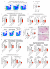
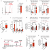
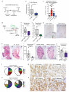
Figure 1
Figure 1. Neutrophils infiltrate pre-metastatic lungs and favour metastasis
a, b, Analysis of wild-type (WT) or MMTV-PyMT+ mice. a, Lung neutrophils frequencies determined by flow cytometry (n = 5 (wild-type), n = 4 (pre-metastatic lung), n = 4 (metastatic lung)). Met., metastatic. b, Lung neutrophils or cancer cells determined by histology staining for S100A9 or PyMT (brown). Scale bars, 100 μm. Magnifications in inserts. c, Haematoxylin & eosin (H&E)-stained neutrophil. Scale bar, 5 μm. d, Lung neutrophil quantification by flow cytometry (n = 5 (wild-type), n = 4 (PyMT+ Gcsf+/+), n = 7 (PyMT+ Gcsf−/−)). e, f, Spontaneous metastasis of MMTV-PyMT+ Gcsf+/+ (n = 13) or MMTV-PyMT+ Gcsf−/− (n = 24) (e) and MMTV-PyMT+ control (n = 14) or MMTV-PyMT+Ela2-Cre-DTA+ (n = 6) mice (f). g, Representative H&E-stained sections of lung. Scale bar, 500 μm. h, Experimental setup for neutrophil depletion. i, Flow cytometric lung neutrophil quantification (n = 4 (tumour-free), n = 12 (IgG tumour), n = 11 (Ly6G tumour)). j, k, Spontaneous (n = 8 per group) (j) and experimental metastasis (n = 12 per group) (k). Lin, CD45 CD31 TER119. l, Histological GFP-stained lung sections including close-up on spontaneous (arrow) and experimental metastases (brown). Scale bar, 500μm. Statistical analysis by two-sided t-test. Data are represented as mean ± standard error of the mean (s.e.m.). *P < 0.05, **P < 0.01, ***P < 0.001.
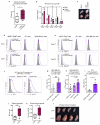
Figure 2
Figure 2. Neutrophil-derived signals promote tumorigenicity and increase the metastatic cell sub-pool
a, b, Images and quantification (technical replicate n = 14 (control), n = 9 (LuN) of biological triplicates) of primary MMTV-PyMT spheres in indicated medium. SFI, sphere formation index. Scale bar, 10μm. cf, Medium pre-treated luciferase+MMTV-PyMT cells (c) grafted onto the mammary gland (d) or intravenously injected (e, f) into Rag1-null mice. Lung metastases quantified by histological sectioning (n = 5 (control), n = 4 (LuN)). f, Representative bioluminescence signal. g, Experimental setup. h, i, Flow cytometric quantification of MICs in lungs of LuN-treated (n = 3 (PyMT control), n = 4 (PyMT+LuN)) (h) or neutrophil-treated mice (n = 3 (PyMT control) n = 4 (PyMT+neutrophils)) (i). j, Representation of cell heterogeneity change. Statistical analysis by two-sided t-test (b), Mann–Whitney test (e) and one representative experiment of two analysed by analysis of variance (ANOVA) (h, i). Data are represented as mean ± s.e.m. *P < 0.05, **P < 0.01.
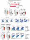
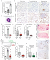
Figure 3
Figure 3. LTs enrich for MICs and tumorigenicity
a, b, Enzyme immunoassay detecting LTB4 (n = 4 per group) (a) or LTC/D/E4 (n = 2 per group) (b). c, Overview of LTs and LTRs. d, e, Flow cytometric quantification of BLT2+ (n = 4 tumours) (d) and CysLT2+ cells (n = 2 tumours) (e) among indicated sub-pools. fh, Representation of LT treatment (f): frequency of MICs (n = 8 per group) (g); and experimental lung metastasis (n = 6 per group) with representative images of GFP+ colonies. Scale bar, 3 mm. Lin, CD45 CD31 TER119. i, Western blot of ERK1/2 phosphorylation and total ERK1/2 levels of LTB4- or LTC/D/E4-treated cells for indicated minutes. Loading control: α -vinculin. jk, 5-Bromodeoxyuridine (BrdU) incorporation comparing LT-treated MICs with non-MICs (n = 3 (non-MICs), n = 4 (MICs)) (j) or MICs treated with LTs and/or PD0325901 MEK inhibitor (MEKi; n = 3 per group) (k) DMSO, dimethylsulfoxide treated; EtOH, ethanol treated. Statistical analysis by two-sided t-test (a, d, h, j, k) and one-sample t-test (g). Data are represented as mean ± s.e.m. NS, not significant. *P < 0.05, **P < 0.01. Blot source data are in Supplementary Fig. 1.
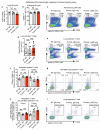
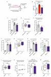
Figure 4
Figure 4. Alox5 inhibition decreases lung metastasis initiation
a, b, Alox5-null bone marrow (BM) chimaera experimental setup (a) and spontaneous metastasis (n = 6 (wild-type bone marrow), n = 9 (Alox5−/− bone marrow)) (b). WT, wild-type. c, Surface metastases of medium pre-treated cancer cells (n = 10 (control), n = 9 (LuN wild-type), n = 5 (LuN Alox5ko), n = 3 (LuN-Zil)). d, Experimental setup for Zil treatment. ek, Spontaneous (e) and experimental (f, i, k) metastasis of MMTVPyMT cells (n = 9 (PyMT DMSO), n = 7 (PyMT Zil)) (eg), 4T1 cells (n = 5 (4T1 DMSO), n = 7 (4T1 Zil)) (h, i) or MDA-MB-231 cells (n = 6 (MDA231 DMSO), n = 5 (MDA231 Zil)) (j, k). Lin, CD45 CD31 TER119. Representative histological lung sections GFP stained with close-up on spontaneous (arrows) and experimental metastases (brown) (g) or H&E stained (h, k). Scale bars, 500μm. lo, BLT2 (l, m) or CysLT2 (n, o) staining (brown) of human breast cancer and matched lymph node (LN) metastases (n ≥ 30 per group). Quantification of staining intensity and frequency (l, n) and representative images (m, o). Scale bar, 50μm. Statistical analysis by two-sided t-test (b, e, f, i), Mann–Whitney test (c) and one-sided t-test (k). Data are represented as mean ± s.e.m. NS, not significant, *P < 0.05, ***P < 0.001.

Comment in

References

    1. Quail DF, Joyce JA. Microenvironmental regulation of tumor progression and metastasis. Nature medicine. 2013;19:1423–1437. - PMC - PubMed
    1. Bald T, et al. Ultraviolet-radiation-induced inflammation promotes angiotropism and metastasis in melanoma. Nature. 2014;507:109–113. - PubMed
    1. Galdiero MR, et al. Tumor associated macrophages and neutrophils in cancer. Immunobiology. 2013;218:1402–1410. - PubMed
    1. Finisguerra V, et al. MET is required for the recruitment of anti-tumoural neutrophils. Nature. 2015;522:349–353. - PMC - PubMed
    1. Hiratsuka S, Watanabe A, Aburatani H, Maru Y. Tumour-mediated upregulation of chemoattractants and recruitment of myeloid cells predetermines lung metastasis. Nature cell biology. 2006;8:1369–1375. - PubMed

Publication types

MeSH terms

Substances