Skip to main page content
U.S. flag

An official website of the United States government

Dot gov

The . gov means it’s official. Federal government websites often end in . gov or . mil. Before sharing sensitive information, make sure you’re on a federal government site. VSports app下载.

Https

The site is secure. The https:// ensures that you are connecting to the official website and that any information you provide is encrypted and transmitted securely V体育官网. .

. 2016 Jun;54(6):872-81.
doi: 10.1165/rcmb.2015-0119OC.

Temporal and Spatial Expression of Transforming Growth Factor-β after Airway Remodeling to Tobacco Smoke in Rats

Affiliations

V体育官网 - Temporal and Spatial Expression of Transforming Growth Factor-β after Airway Remodeling to Tobacco Smoke in Rats

Laura L Hoang et al. Am J Respir Cell Mol Biol. 2016 Jun.

Abstract

Airway remodeling is strongly correlated with the progression of chronic obstructive pulmonary disease (COPD). In this study, our goal was to characterize progressive structural changes in site-specific airways, along with the temporal and spatial expression of transforming growth factor (TGF)-β in the lungs of male spontaneously hypertensive rats exposed to tobacco smoke (TS). Our studies demonstrated that TS-induced changes of the airways is dependent on airway generation and exposure duration for proximal, midlevel, and distal airways. Stratified squamous epithelial cell metaplasia was evident in the most proximal airways after 4 and 12 weeks but with minimal levels of TGF-β-positive epithelial cells after only 4 weeks of exposure. In contrast, epithelial cells in midlevel and distal airways were strongly TGF-β positive at both 4 and 12 weeks of TS exposure. Airway smooth muscle volume increased significantly at 4 and 12 weeks in midlevel airways. Immunohistochemistry of TGF-β was also found to be significantly increased at 4 and 12 weeks in lymphoid tissues and alveolar macrophages. ELISA of whole-lung homogenate demonstrated that TGF-β2 was increased after 4 and 12 weeks of TS exposure, whereas TGF-β1 was decreased at 12 weeks of TS exposure VSports手机版. Airway levels of messenger RNA for TGF-β2, as well as platelet-derived growth factor-A, granulocyte-macrophage colony-stimulating factor, and vascular endothelial growth factor-α, growth factors regulated by TGF-β, were significantly decreased in animals after 12 weeks of TS exposure. Our data indicate that TS increases TGF-β in epithelial and inflammatory cells in connection with airway remodeling, although the specific role of each TGF-β isoform remains to be defined in TS-induced airway injury and disease. .

Keywords: airway epithelium; chronic obstructive pulmonary disease; spontaneously hypertensive rats; tobacco smoke; transforming growth factor-β V体育安卓版. .


Figures
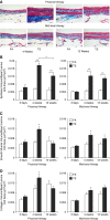
Figure 1.
Figure 1.
Histopathological evaluation of tobacco smoke (TS)–associated injuries in pulmonary airways. (A) Representative lung sections depicting changes in volume of airway epithelium, smooth muscle, and collagen; quantitative analysis of (B) epithelium volume, (C) airway smooth muscle volume, and (D) collagen volume at 3 days, 4 weeks, and 12 weeks of TS exposure in proximal and midlevel airways. Data are expressed as mean (±SEM) for six animals per group. Scale bar: 50 μm. *P < 0.05, **P < 0.001, ANOVA with Bonferroni’s post hoc test. FA, filtered air.
Figure 2.
Figure 2.
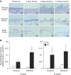
Mucosubstance in the airway epithelium. (A) Representative lung sections depicting changes in mucosubstance in the epithelial cells of proximal and midlevel airways. Distal airway epithelium does not stain positive for Alcian blue (AB)/periodic acid–Schiff (PAS). Arrows indicate epithelium stained positive for mucosubstance using AB/PAS. (B) Volume fraction analyses for mucosubstance with AB/PAS. Data are expressed as mean (±SEM). Scale bars: 50 μm. *P < 0.05, ANOVA with Bonferroni’s post hoc test.
Figure 3.
Figure 3.
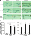
Transforming growth factor (TGF)-β expression in the airway epithelium. (A) Representative lung sections depicting TGF-β expression in the epithelium of proximal, midlevel, and distal airways after progressive smoke exposure for 3 days, 4 weeks, and 12 weeks. (B) Quantitative measurement of TGF-β expression in proximal, midlevel, and distal airway epithelium from paraffin-embedded sections. Data are expressed as mean (±SEM) for six animals per group. Scale bar: 50 μm. *P < 0.001, ANOVA with Bonferroni’s post hoc test.
Figure 4.
Figure 4.
TGF-β expression in the lymphoid aggregates. (A) Representative lung sections depicting TGF-β expression in the lymphoid aggregates after progressive smoke exposure for 3 days, 4 weeks, and 12 weeks. Arrows indicate areas of TGF-β–positive staining. After 4 and 12 weeks of TS exposure, lymphoid aggregates of spontaneously hypertensive (SH) rats demonstrated increased levels of TGF-β compared with lymphoid cells of rats exposed to FA. (B) Staining score for TGF-β expression in lymphoid aggregates from paraffin-embedded sections. Data are expressed as mean (±SEM) for four to six animals per group. Scale bars: 50 μm. *P < 0.05, **P < 0.0005, ***P < 0.001, ANOVA with Bonferroni’s post hoc test.
Figure 5.
Figure 5.
TGF-β expression in the alveolar macrophages. (A) Representative lung sections depicting TGF-β expression in the alveolar macrophages after progressive smoke exposure for 3 days, 4 weeks, and 12 weeks. Arrows indicate TGF-β–negative macrophage. Arrowheads indicate TGF-β–positive macrophages. Terminal bronchiole and adjacent alveolar structures in rats exposed to smoke for 4 and 12 weeks contained numerous clusters of activated macrophages in the airspaces that stained strongly for TGF-β. Compared with rats exposed to TS, rats exposed to FA had fewer numbers of alveolar macrophages. (B) Measurement of frequency of TGF-β–positive alveolar macrophages from each total field in paraffin-embedded sections for the TS group at 4 weeks and 12 weeks. Data are expressed as mean (±SEM) for six animals per group. Scale bars: 50 μm. *P < 0.05, **P < 0.005, ANOVA with Bonferroni’s post hoc test.
Figure 6.
Figure 6.
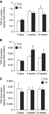
Whole accessory lobe–specific TGF-β1, -β2, and -β3 protein levels. (A) TGF-β1, (B) TGF-β2, and (C) TGF-β3 protein levels measured by ELISA in accessory lobe homogenates are reported. TGF-β1 protein was significantly reduced after 12 weeks of TS exposure. By contrast, TGF-β2 protein in lung homogenates was significantly elevated after 4 and 12 weeks of exposure to TS. TGF-β3 protein was not significantly changed in response to TS exposure at any time point. Data are expressed as mean (±SEM) for five to six animals per group.*P < 0.01, **P < 0.001, ANOVA with Bonferroni’s post hoc test.
Figure 7.
Figure 7.
Airway-specific gene expression of growth factors in 12-week smoke-exposed SH rats. Effects of 12-week TS exposure on the gene expression of growth factors that may be implicated in the airway remodeling process. mRNA levels of growth factors measured by quantitative real-time RT-PCR in isolated airways of caudal lobes are reported. Data are expressed as mean (±SEM) for five animals per group. *P < 0.05, **P < 0.005, ***P < 0.0005, ANOVA with Bonferroni’s post hoc test. CTGF, connective tissue growth factor; EGF, epidermal growth factor; FGF, fibroblast growth factor; GF, growth factor; GM-CSF, granulocyte-macrophage colony-stimulating factor; PDGF-A, platelet-derived growth factor α chain; VEGF, vascular endothelial growth factor.

References

    1. Balkissoon R, Lommatzsch S, Carolan B, Make B. Chronic obstructive pulmonary disease: a concise review. Med Clin North Am. 2011;95:1125–1141. - PubMed
    1. Barnes PJ. Mediators of chronic obstructive pulmonary disease. Pharmacol Rev. 2004;56:515–548. - PubMed
    1. Baraldo S, Turato G, Saetta M. Pathophysiology of the small airways in chronic obstructive pulmonary disease. Respiration. 2012;84:89–97. - V体育ios版 - PubMed
    1. Hogg JC, Chu F, Utokaparch S, Woods R, Elliott WM, Buzatu L, Cherniack RM, Rogers RM, Sciurba FC, Coxson HO, et al. The nature of small-airway obstruction in chronic obstructive pulmonary disease. N Engl J Med. 2004;350:2645–2653. - PubMed
    1. Kamato D, Burch ML, Piva TJ, Rezaei HB, Rostam MA, Xu S, Zheng W, Little PJ, Osman N. Transforming growth factor-β signalling: role and consequences of Smad linker region phosphorylation. Cell Signal. 2013;25:2017–2024. - PubMed

Publication types

MeSH terms