Skip to main page content
U.S. flag

An official website of the United States government

Dot gov

The . gov means it’s official VSports app下载. Federal government websites often end in . gov or . mil. Before sharing sensitive information, make sure you’re on a federal government site. .

Https

The site is secure V体育官网. The https:// ensures that you are connecting to the official website and that any information you provide is encrypted and transmitted securely. .

. 2015 Oct 22;11(10):e1005187.
doi: 10.1371/journal.ppat.1005187. eCollection 2015 Oct.

The Fungal Exopolysaccharide Galactosaminogalactan Mediates Virulence by Enhancing Resistance to Neutrophil Extracellular Traps (V体育2025版)

Affiliations

The Fungal Exopolysaccharide Galactosaminogalactan Mediates Virulence by Enhancing Resistance to Neutrophil Extracellular Traps

"V体育官网" Mark J Lee et al. PLoS Pathog. .

Abstract

Of the over 250 Aspergillus species, Aspergillus fumigatus accounts for up to 80% of invasive human infections. A. fumigatus produces galactosaminogalactan (GAG), an exopolysaccharide composed of galactose and N-acetyl-galactosamine (GalNAc) that mediates adherence and is required for full virulence. Less pathogenic Aspergillus species were found to produce GAG with a lower GalNAc content than A. fumigatus and expressed minimal amounts of cell wall-bound GAG. Increasing the GalNAc content of GAG of the minimally pathogenic A. nidulans, either through overexpression of the A. nidulans epimerase UgeB or by heterologous expression of the A. fumigatus epimerase Uge3 increased the amount of cell wall bound GAG, augmented adherence in vitro and enhanced virulence in corticosteroid-treated mice to levels similar to A. fumigatus. The enhanced virulence of the overexpression strain of A. nidulans was associated with increased resistance to NADPH oxidase-dependent neutrophil extracellular traps (NETs) in vitro, and was not observed in neutropenic mice or mice deficient in NADPH-oxidase that are unable to form NETs. Collectively, these data suggest that cell wall-bound GAG enhances virulence through mediating resistance to NETs VSports手机版. .


Conflict of interest statement

The authors have declared that no competing interests exist.

V体育官网入口 - Figures

Fig 1
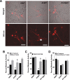
Fig 1. Production of GalNAc-rich GAG correlates with reported virulence of Aspergillus spp.
(A) Scanning electron micrograph of hyphae of indicated species at 20,000X magnification. Arrows indicate surface decorations associated with cell wall-bound GAG. The GAG deficient A. fumigatus Δuge3 mutant [10] and the A. fumigatus ΔstuA mutant [49] which produces only minimal amounts of GAG are included for comparison purposes. (B) Cell wall GalNAc staining with FITC-conjugated soybean agglutinin (SBA). SBA binding to mature hyphal mats of the indicated species was quantified by fluorometry. Data are represented as mean +/- SEM. * indicates a significant difference between A. fumigatus and other species, p<0.05 by ANOVA and pairwise comparison.
Fig 2
Fig 2. nidulans produces GalNAc-poor GAG which is associated with non-adherence.
A. (A) Galactose and GalNAc content of secreted GAG from either A. fumigatus or A. nidulans as identified by gas chromatography and quantified by hexose or hexosamine assays. (B) Formation of adherent biofilms on tissue culture-treated polystyrene plates by A. fumigatus and A. nidulans. After 24 hours growth, biofilms were washed and visualized by staining with 0.1% crystal violet. (C) Detection of β-1,3-glucan exposure on the surface of hyphae by immunostaining with Fc-dectin-1 antibody by fluorometry. (D) Relative expression of ugeB in A. nidulans and uge3 in A. fumigatus, during growth in Brian medium as measured by real-time RT-PCR. Expression of tef1 from each respective species was used as an internal reference gene. Primer efficiency was verified, and was not different between species (S2E Fig). For all panels: data are represented as mean +/- SEM. AfWT indicates A. fumigatus, and AnWT indicates A. nidulans. * indicates a significant difference between A. fumigatus and A. nidulans, p<0.05 by Student t test or ANOVA with Tukey’s test for pairwise comparison, where applicable.
Fig 3
Fig 3. Overexpression of uge3 or ugeB in A. nidulans increases the GalNAc content of GAG and enhances the formation of adherent biofilms.
(A) Relative expression of uge3 in the An-Uge3 strain and ugeB in the An-UgeB strain compared to the expression level of ugeB in wild-type A. nidulans grown in Brian medium and as measured by real-time RT-PCR. (B) Total amount of secreted GAG from the indicated strains. (C) GalNAc content of secreted GAG from the indicated strains as determined by gas chromatography and quantified by hexose or hexosamine assays. (D) Cell wall GalNAc staining with FITC-conjugated soybean agglutinin (SBA). SBA binding to mature hyphal mats of the indicated strains was quantified by fluorometry. (E) Scanning electron micrograph of hyphae of indicated species at 20,000X magnification. Arrows indicate surface decorations associated with cell wall-bound GAG. (F) Formation of adherent biofilms on tissue culture treated polystyrene plates by the indicated strains. After 24 hours growth, biofilms were washed and visualized by staining with 0.1% crystal violet. (G) Detection of β-1,3-glucan exposure on the surface of hyphae by immunostaining with Fc-dectin-1 antibody labeled with FITC secondary antibody and quantified by fluorometry at 495 nm. For all panels: An-Uge3 indicates the A. nidulans overexpressing uge3 strain; An-UgeB indicates the A. nidulans overexpressing ugeB strain; and AnWT indicates wild type A. nidulans. Data are represented as mean +/- SEM and * indicates a significant difference between A. nidulans, and both overexpression strains, p<0.05 by ANOVA with Tukey’s test for pairwise comparison.
Fig 4
Fig 4. Overexpression of uge3 in A. nidulans increases virulence in mice.
(A) Survival of corticosteroid treated BALB/c mice infected with the indicated conidial species and strains. Data represent the results from two independent experiments for total N = 20 for A. nidulans or An-Uge3; N = 10 for A. fumigatus or An-UgeB; N = 8 for PBS control. (B) Survival of corticosteroid treated C57BL/6 mice infected with the indicated conidial species and strains. Five or more mice were infected for each group. * indicates a significant difference in survival of A. nidulans compared with A. fumigatus, An-Uge3, and An-UgeB overexpression strains as determined by the Mantel-Cox log-rank test with pairwise comparison applying Bonferroni correction.
Fig 5
Fig 5. Increase in virulence of the A. nidulans strain overexpressing uge3 is associated with increase in fungal burden and pulmonary tissue invasion.
(A) Pulmonary fungal burden measured by relative galactomannan content in the lungs of mice infected with the indicated strains, N = 10 Balb/c mice for each fungal strain. (B) Total fungal lesion size as determined by morphometric analysis of lung histopathology for the indicated strains, N = 4 Balb/c mice for each fungal strain. (C) Pulmonary histopathology sections from Balb/c mice infected with indicated strains and stained with PAS for visualization of fungi. The yellow dotted line indicates the limit of the airway used for morphometric analysis. White arrow indicates fungal elements outside the airway and invading into pulmonary tissues. Scale bar represents 100 μm (black) or 50 μm (white). (D) Lesion invasion beyond the airway as determined by morphometric analysis of infected mouse lung histopathology for the indicated strains, N = 4 Balb/c mice for each fungal strain. (E) Pulmonary histopathology sections from Balb/c mice infected for 4 days with the indicated strains and labeled with an anti-GAG antibody to visualize GAG on hyphae. Arrow indicates hyphae. Scale bar represents 80 μm (black) or 30 μm (white). (F) Total pulmonary leukocytes from mice 4 days after infection with the indicated strains as measured by CD45 detection by flow cytometry. N = 10 Balb/c mice for each fungal strain, N = 5 for PBS control. (G) Total pulmonary neutrophils from of mice 4 days after infection with the indicated strains as measured by Ly6G+, CD11bhigh, and CD11clow detection using flow cytometry. N = 10 Balb/c mice for each fungal strain, N = 5 Balb/c mice for PBS control. (H) Pulmonary histopathology sections from Balb/c mice 4 days after infection with the indicated strains and labeled with anti-caspase-3 antibody to visualize host cells undergoing apoptosis. Arrow indicates hyphae. Scale bar represents 100 μm (black) or 50 μm (white). For all panels: An-Uge3 indicates the A. nidulans overexpressing uge3 strain and AnWT indicates wild type A. nidulans. For panel A, B, D, F, and G, data are represented as median with interquartile ranges and * indicates a significant difference between A. nidulans and the An-Uge3 strain, p<0.05 by Kruskal-Wallis test with Dunn’s test for pairwise comparison.
Fig 6
Fig 6. Increasing cell wall-associated GAG enhances the resistance of A. nidulans to NADPH oxidase-dependent neutrophil extracellular traps (NETs) but not reactive oxygen species.
(A) Fungal injury by primary human neutrophils (PMN) either untreated (black bars) or treated with the NADPH oxidase inhibitor diphenyleneiodonium (DPI) (gray bars). (B) Susceptibility of the indicated fungal strains to injury after treatment with 3.2 mM hydrogen peroxide. * indicates a significant difference between A. nidulans and A. fumigatus or An-Uge3 strains, p<0.05 by ANOVA with Tukey’s test for pairwise comparison. § indicates a significant difference in killing of A. nidulans between DPI-treated and untreated neutrophils, p<0.05 by ANOVA with Tukey’s test for pairwise comparison.
Fig 7
Fig 7. Inhibition or disruption of NETs attenuates the susceptibility of A. nidulans to killing by human neutrophil-mediated killing.
(A) Neutrophil extracellular traps formation by primary human PMN as visualized by the DNA intercalating agent propidium iodide. Arrows indicate the increased binding of propidium iodide stained NETs on the surface of wild-type A. nidulans hyphae. Images were acquired using a 543 nm laser and detected by confocal microscopy at 600X magnification with 4X digital zoom. Scale bar represents 10 μm. (B) Susceptibility of fungal strains to injury by PMNs in the presence (gray bars) or absence (black bars) of micrococcal nuclease (MNase). (C) Susceptibility of fungal strains to injury by PMNs from healthy donor (black bars) or from a CGD patient (grey bars). (D) Susceptibility of fungal strains to injury by PMNs pre-treated with 10 μM dexamethasone (grey bars) or untreated (black bars). * indicates a significant difference between A. nidulans and A. fumigatus or An-Uge3 strains, p<0.05 by ANOVA with Tukey’s test for pairwise comparison. § indicates significant difference treatment groups of PMNs co-incubated with A. nidulans, p<0.05 by ANOVA with Tukey’s test for pairwise comparison. All data are represented as mean +/- SEM.
Fig 8
Fig 8. GAG-mediated resistance to neutrophil killing is dependent on neutrophil lysate content.
Susceptibility of fungal strains to injury by lysates derived from, (A) primary human neutrophils, (B) primary human neutrophils treated with DPI., (C) primary C57BL/6 mouse neutrophils or gp91 phox deficient (CGD) mouse neutrophils. * indicates a significant difference between A. nidulans and A. fumigatus or An-Uge3 strains, p<0.05 by ANOVA with Tukey’s test for pairwise comparison.
Fig 9
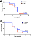
Fig 9. GAG mediated enhancement of A. nidulans virulence requires functional leukocyte NADPH oxidase.
(A) Survival of leukopenic mice infected with either A. nidulans or An-Uge3 conidia. N = 9 per infection group. (B) Survival of gp91 phox deficient mice lacking functional NADPH oxidase infected with either A. nidulans or An-Uge3 conidia. N = 17 per infection group. For all panels: An-Uge3 indicates the A. nidulans overexpressing uge3 strain; AnWT indicates wild type A. nidulans; and AfWT indicates wild type A. fumigatus. n.s. indicates no significant difference in survival of A. nidulans compared with the An-Uge3, strain as determined by the Mantel-Cox log-rank test with pairwise comparison applying Bonferroni correction.

References

    1. Garcia-Vidal C, Upton A, Kirby KA, Marr KA (2008) Epidemiology of invasive mold infections in allogeneic stem cell transplant recipients: biological risk factors for infection according to time after transplantation. Clinical infectious diseases: an official publication of the Infectious Diseases Society of America 47: 1041–1050. - PMC - PubMed
    1. Geiser DM, Klich MA, Frisvad JC, Peterson SW, Varga J, et al. (2007) The current status of species recognition and identification in Aspergillus. Studies in Mycology 59: 1–10. 10.3114/sim.2007.59.01 - DOI - PMC - PubMed
    1. Lucas GM, Tucker P, Merz WG (1999) Primary Cutaneous Aspergillus nidulans Infection Associated with a Hickman Catheter in a Patient with Neutropenia. Clinical Infectious Diseases 29: 1594–1596. - PubMed
    1. Henriet SS, Verweij PE, Warris A (2012) Aspergillus nidulans and chronic granulomatous disease: a unique host-pathogen interaction. J Infect Dis 206: 1128–1137. 10.1093/infdis/jis473 - V体育安卓版 - DOI - PubMed
    1. Song E, Jaishankar GB, Saleh H, Jithpratuck W, Sahni R, et al. (2011) Chronic granulomatous disease: a review of the infectious and inflammatory complications. Clin Mol Allergy 9: 10 10.1186/1476-7961-9-10 - DOI - PMC - PubMed

Publication types