Skip to main page content
U.S. flag

An official website of the United States government

Dot gov

The VSports app下载. gov means it’s official. Federal government websites often end in . gov or . mil. Before sharing sensitive information, make sure you’re on a federal government site. .

Https

The site is secure. The https:// ensures that you are connecting to the official website and that any information you provide is encrypted and transmitted securely. V体育官网.

. 2015 Oct 13;112(41):12782-7.
doi: 10.1073/pnas.1513532112. Epub 2015 Sep 28.

Generation of Th17 cells in response to intranasal infection requires TGF-β1 from dendritic cells and IL-6 from CD301b+ dendritic cells

Affiliations

Generation of Th17 cells in response to intranasal infection requires TGF-β1 from dendritic cells and IL-6 from CD301b+ dendritic cells

Jonathan L Linehan et al. Proc Natl Acad Sci U S A. .

Abstract

Intranasal (i. n. ) infections preferentially generate Th17 cells VSports手机版. We explored the basis for this anatomic preference by tracking polyclonal CD4(+) T cells specific for an MHC class II-bound peptide from the mucosal pathogen Streptococcus pyogenes. S. pyogenes MHC class II-bound peptide-specific CD4(+) T cells were first activated in the cervical lymph nodes following i. n. inoculation and then differentiated into Th17 cells. S. pyogenes-induced Th17 formation depended on TGF-β1 from dendritic cells and IL-6 from a CD301b(+) dendritic cell subset located in the cervical lymph nodes but not the spleen. Thus, the tendency of i. n. infection to induce Th17 cells is related to cytokine production by specialized dendritic cells that drain this site. .

Keywords: CD301b+; Streptococcus pyogenes; Th17; dendritic cells V体育安卓版. .


Conflict of interest statement

The authors declare no conflict of interest.

Figures

Fig. 1.
Fig. 1.
Infection with Sp-2W induces the clonal expansion and Th17 differentiation of 2W:I-Ab–specific cells. (A) CD4+ T cells within a CD11c CD11b B220 CD3+ population from the spleen and lymph nodes of a B6 mouse 7 d after i.n. infection with Sp-2W bacteria following enrichment with 2W:I-Ab tetramer and antibody staining as described in Materials and Methods. (B and C) 2W:I-Ab–specific T cells from 2W:I-Ab tetramer-enriched samples from mice that were infected i.n. with Sp-2W bacteria 7 d before analysis. (B) IL-17A and CD69 production 3 h after i.v. challenge with heat-killed S. pyogenes (Left) or Sp-2W (Right) bacteria. (C) IL-17A or IFN-γ production 3 h after i.v. challenge with heat-killed S. pyogenes (Left) or Sp-2W (Right) bacteria. Numbers indicate percent of cells in the gate or quadrant. Data are representative of five (A and C) or three (B) independent experiments.
Fig. 2.
Fig. 2.
2W:I-Ab–specific T cells differentiate into Th17 cells in the CLNs after i.n. Sp-2W inoculation. Plots represent 2W:I-Ab–specific T cells in 2W:I-Ab tetramer-enriched samples from the indicated organs and at the indicated times after i.n. infection with Sp-2W bacteria. For NALT and CLNs, three mice were pooled per sample; for spleen, one mouse was used per sample. Numbers over each gate indicate the total number of 2W:I-Ab–specific T cells in that organ. Numbers in the lower right corner of each gate indicate the percentage of 2W:I-Ab–specific T cells in the sample. Data are representative of three independent experiments.
Fig. 3.
Fig. 3.
Dendritic cell-derived IL-6 is necessary for 2W:I-Ab–specific Th17 differentiation after i.n. Sp-2W infection. (A, C, and D) Cytokine production by 2W:I-Ab–specific T cells from 2W:I-Ab tetramer-enriched samples from WT B6, Il6−/−, or chimeric mice infected i.n. 7 d earlier with Sp-2W bacteria, 3 h after i.v. challenge with heat-killed Sp-2W bacteria. In C, the bone marrow donor is indicated before the -> symbol and the recipient after. The chimeras in D were produced with a 1:1 mixture of bone marrow from the indicated donors and treated with DT on day –1, 2, and 5 relative to infection. Contour plots from representative samples and scatter plots with values from individual mice are shown. Numbers on the contour plots indicate the percentage of cells in each quadrant. (B) Foxp3 expression by 2W:I-Ab–specific T cells in 2W:I-Ab tetramer-enriched samples from WT B6 or Il6−/− mice infected i.n. 7 d earlier with Sp-2W bacteria. Numbers indicate the percentage of cells in the indicated gate. ***P < 0.0005, *P < 0.05; ns, not significant (unpaired two-tailed Student’s t test). Data are representative of three independent experiments.
Fig. 4.
Fig. 4.
Hematopoietic cell-derived TGF-β1 is necessary for 2W:I-Ab–specific Th17 cell differentiation after i.n. Sp-2W infection. (A and B) Cytokine production by 2W:I-Ab–specific T cells from 2W:I-Ab tetramer-enriched samples from mice infected i.n. 7 d earlier with Sp-2W bacteria, 3 h after i.v. challenge with heat-killed Sp-2W bacteria. (A) Contour and scatter plots from chimeras containing a 2:1 mixture of congenically marked CD4-Cre (CD45.1/CD45.2) and CD4-Cre/Tgfbr2f/f (CD45.2) T cells, showing 2W:I-Ab–specific T cells derived from the CD4-Cre bone marrow or the CD4-Cre/Tgfbr2f/f bone marrow donor. (B) Contour and scatter plots from chimeras produced by transplanting bone marrow cells from the donors indicated before the -> symbol into irradiated recipients of the type indicated after the -> symbol. The scatter plots show the percentage of the 2W:I-Ab–specific T cells from the indicated source that produced the indicated cytokine in individual animals. Numbers on the contour plots indicate the percentage of cells in each quadrant. ***P < 0.0005, **P < 0.005, *P < 0.05 (unpaired two-tailed Student’s t test).
Fig. 5.
Fig. 5.
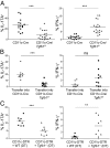
Dendritic cell-derived TGF-β1 is necessary for 2W:I-Ab–specific Th17 cell differentiation after i.n. Sp-2W infection. (A and C) IL-17A and IFN-γ production by 2W:I-Ab–specific T cells from 2W:I-Ab tetramer-enriched samples from individual CD11-Cre–expressing WT B6, CD11-Cre–expressing Tgfb1f/f, or chimeric mice infected i.n. 7 d earlier with Sp-2W bacteria, 3 h after i.v. challenge with heat-killed Sp-2W bacteria. The chimeras in C were produced with a 1:1 mixture of bone marrow from the indicated donors and treated with DT on days –1, 2, and 5 relative to infection. (B) Cytokine production by transferred 2W:I-Ab–specific T cells in individual CD11-Cre–expressing B6 or CD11-Cre–expressing Tgfb1f/f mice infected i.n. 7 d earlier with Sp-2W bacteria, 3 h after i.v. challenge with heat-killed Sp-2W bacteria. ***P < 0.0005, **P < 0.005, *P < 0.05 (unpaired two-tailed Student’s t test).
Fig. 6.
Fig. 6.
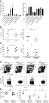
CD301b+ migratory dendritic cell-derived IL-6 is necessary for 2W:I-Ab–specific Th17 cell differentiation after i.n. Sp-2W inoculation. (A) Quantitative RT-PCR for Il6 (Left) or Tgfb1 (Right) mRNA relative to Hprt mRNA from magnetically enriched, then FACS-sorted CD11c+ cells from the indicated tissues of uninfected mice or mice infected i.n. with Sp-2W 3 d earlier. Each sample was pooled from three mice. (B and C) Cytokine production by 2W:I-Ab–specific T cells from 2W:I-Ab tetramer-enriched samples from (B) WT or Batf3−/− or (C) CD11c-Cre or CD11c-Cre/Notch2f/f mice infected i.n. 7 d earlier with Sp-2W bacteria, 3 h after i.v. challenge with heat-killed Sp-2W bacteria. (D) Contour plots of CD19 CD90 CD45.1 cells from CLNs of 1:1 Mgl2DTR-eGFP:WT or Mgl2DTR-eGFP:Il6−/− chimeric mice inoculated i.n. with heat-killed Sp-2W bacteria 7 d earlier and treated with or without DT. Gates used to identify CD11c+ MHCIIhigh migratory dendritic cells along with the percentage of cells in these gates are shown in the Top row. The Bottom row shows CD301b (Mgl2) expression on CD11c+ MHCIIhigh migratory dendritic cells in each group. (E) Total number of 2W:I-Ab–specific T cells in the spleen and lymph node (Left panel) of DT-treated chimeric mice produced with a 1:1 mixture of Mgl2DTR-eGFP and WT or Il6−/− bone marrow and inoculated i.n. 7 d earlier with heat-killed Sp-2W bacteria and cytokine production by CLN cells 3 h after in vitro culture with phorbol 12-myristate 13-acetate and ionomycin (Middle and Right panels). Pharmacologic stimulation of T cells was used for cytokine recall in this case because heat-killed bacteria do not reach the CLNs after i.v. injection. **P < 0.005; ns, not significant (unpaired two-tailed Student’s t test).

References

    1. Korn T, Bettelli E, Oukka M, Kuchroo VK. IL-17 and Th17 Cells. Annu Rev Immunol. 2009;27:485–517. - PubMed
    1. Bettelli E, et al. Reciprocal developmental pathways for the generation of pathogenic effector TH17 and regulatory T cells. Nature. 2006;441(7090):235–238. - PubMed
    1. Mangan PR, et al. Transforming growth factor-beta induces development of the T(H)17 lineage. Nature. 2006;441(7090):231–234. - PubMed
    1. Veldhoen M, Hocking RJ, Atkins CJ, Locksley RM, Stockinger B. TGFbeta in the context of an inflammatory cytokine milieu supports de novo differentiation of IL-17-producing T cells. Immunity. 2006;24(2):179–189. - PubMed
    1. Li MO, Wan YY, Flavell RA. T cell-produced transforming growth factor-beta1 controls T cell tolerance and regulates Th1- and Th17-cell differentiation. Immunity. 2007;26(5):579–591. - "VSports最新版本" PubMed

Publication types

V体育安卓版 - MeSH terms