Skip to main page content
U.S. flag

An official website of the United States government

Dot gov

The . gov means it’s official. Federal government websites often end in VSports app下载. gov or . mil. Before sharing sensitive information, make sure you’re on a federal government site. .

Https

The site is secure. The https:// ensures that you are connecting to the official website and that any information you provide is encrypted and transmitted securely. V体育官网.

. 2015 Sep 26:17:269.
doi: 10.1186/s13075-015-0774-3.

Histone deacetylase inhibition activates Nrf2 and protects against osteoarthritis

Affiliations

Histone deacetylase inhibition activates Nrf2 and protects against osteoarthritis

Dawei Cai et al. Arthritis Res Ther. .

VSports - Abstract

Introduction: Osteoarthritis (OA) is a common joint disease that can cause gradual disability among the aging population. Nuclear factor (erythroid-derived 2)-like 2 (Nrf2) is a key transcription factor that regulates the expression of phase II antioxidant enzymes that provide protection against oxidative stress and tissue damage VSports手机版. The use of histone deacetylase inhibitors (HDACi) has emerged as a potential therapeutic strategy for various diseases. They have displayed chondroprotective effects in various animal models of arthritis. Previous studies have established that Nrf2 acetylation enhances Nrf2 functions. Here we explore the role of Nrf2 in the development of OA and the involvement of Nrf2 acetylation in HDACi protection of OA. .

Methods: Two OA models-monosodium iodoacetate (MIA) articular injection and destabilization of the medial meniscus (DMM)-were used with wild-type (WT) and Nrf2-knockout (Nrf2-KO) mice to demonstrate the role of Nrf2 in OA progression. A pan-HDACi, trichostatin A (TSA), was administered to examine the effectiveness of HDACi on protection from cartilage damage. The histological sections were scored. The expression of OA-associated matrix metalloproteinases (MMPs) 1, 3, and 13 and proinflammatory cytokines tumor necrosis factor (TNF)-α, interleukin (IL)-1β, and IL-6 were assayed. The effectiveness of HDACi on OA protection was compared between WT and Nrf2-KO mice. V体育安卓版.

Results: Nrf2-KO mice displayed more severe cartilage damage in both the MIA and DMM models. TSA promoted the induction of Nrf2 downstream proteins in SW1353 chondrosarcoma cells and in mouse joint tissues. TSA also reduced the expression of OA-associated proteins MMP1, MMP3, and MMP13 and proinflammatory cytokines TNF-α, IL-1β, and IL-6. TSA markedly reduced the cartilage damage in both OA models but offered no significant protection in Nrf2-KO mice. V体育ios版.

Conclusions: Nrf2 has a major chondroprotective role in progression of OA and is a critical molecule in HDACi-mediated OA protection VSports最新版本. .


Figures
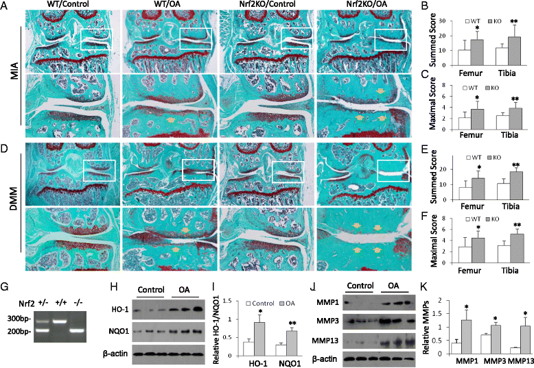
Fig. 1
Fig. 1
Nrf2-KO mice display more severe articular cartilage damage in osteoarthritis (OA) animal models. a Nrf2-KO and wild-type (WT) mice were subject to monosodium iodoacetate (MIA) OA. Mouse joint sections were stained with Safranin O/Fast Green. Representative images are shown (n = 10 mice per group). Lower panels show amplifications of upper panel insets. Yellow arrows point to the damage-affected cartilage areas. b Summed scores of femur and tibia damage in WT and Nrf2-KO mice in the MIA model. *p < 0.05, **p < 0.01. c Maximum scores of femur and tibia damage from WT and Nrf2-KO mice of MIA model. *p < 0.05, ** p < 0.01. d Nrf2-KO and WT mice were subject to DMM surgery. Mouse joint sections were stained with Safranin O/Fast Green and the representative images were shown (n = 10 mice per group). Lower panels were the amplifications of windows from upper panels. The yellow arrows indicate the damage-affected cartilage areas. e Summed scores of femur and tibia damage from WT and Nrf2-KO mice of DMM model. *p < 0.05, **p < 0.01. f Maximum scores of femur and tibia damage from WT and Nrf2-KO mice of DMM model. *p < 0.05, ** p < 0.01. g PCR genotyping of WT and Nrf2-KO mice on mouse tail DNA. h Western blot analysis of heme oxygenase 1 (HO-1) and NAD(P)H:quinine oxidoreductase 1 (NQO1) from mouse joint tissues of WT and Nrf2-KO mice (three mice in each group). i Quantitative analysis of HO1 and NQO1 protein levels. *p < 0.05, **p < 0.01. j Western blot analysis of matrix metalloproteinases MMP-1, MMP-3, and MMP-13 from the joint tissues of WT and Nrf2-KO mice (three mice in each group). k Quantitative analysis of MMP-1, MMP-3, and MMP-13 protein levels. *p < 0.05. KO knockout, Nrf2 nuclear factor (erythroid-derived 2)-like 2
Fig. 2
Fig. 2
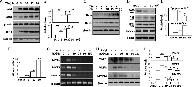
Trichostatin A (TSA) activates nuclear factor (erythroid-derived 2)-like 2 (Nrf2) and suppresses matrix metalloproteinase (MMP) expression in human chondrocytes. a SW1353 chondrosarcoma cells were treated with various doses of TSA for 16 h. Heme oxygenase 1 (HO-1), NAD(P)H:quinine oxidoreductase 1 (NQO1), and acetylated histone 3 (ac-H3) were assayed by Western blotting. β-actin and total histone 3 were used as loading controls. b Quantitative analysis of HO-1 and NQO1 protein levels. *p < 0.05 compared with control. c SW1353 chondrosarcoma cells were treated with TSA (10 nM) for various times. HO-1 and NQO1 were assayed by Western blotting. d SW1353 chondrosarcoma cells were treated with TSA (10 nM) for 4 h. The cell cytoplasmic and nuclear fractions were isolated and assayed for Nrf2 by Western blotting. β-actin, total Nrf2, and lamin B from total cell lysates served as controls. e Quantitative analysis of cytoplasmic and nuclear Nrf2 from (d). *p < 0.05 compared with no-treatment controls. f HEK293 cells were transiently transfected with HO-1 promoter reporter plasmid and a Renilla luciferase plasmid for 24 h. The cells were then treated with 10 nM and 30 nM of TSA for 24 h. The luciferase activity was measured. The reporter luciferase activities were normalized to Renilla luciferase activity. *p < 0.05, **p < 0.01 compared with no-treatment control. g SW1353 chondrosarcoma cells were treated with increasing concentrations of TSA for 1 h and then stimulated with interleukin (IL)-1β (5 ng/ml) for 16 h. MMP-1, MMP-3, and MMP-13 were assayed by RT-PCR. h MMP-1, MMP-3, and MMP-13 were assayed by Western blotting. i Quantitative analysis of MMP-1, MMP-3, and MMP-13 levels from (h). *p < 0.05 compared with IL-1β treatment only. All the Western blotting and RT-PCR results are representative of, and the statistical analyses were performed with results from, at least three independent experiments
Fig. 3
Fig. 3
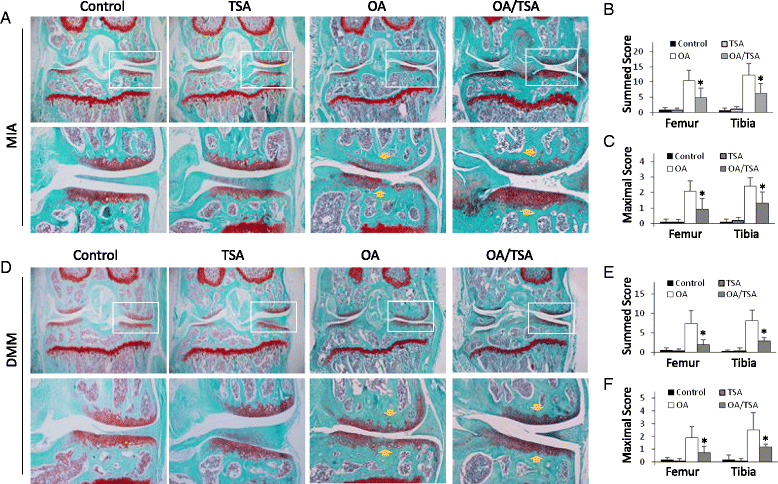
Trichostatin A (TSA) attenuates cartilage damage in monosodium iodoacetate (MIA) osteoarthritis (OA) and destabilization of the medial meniscus (DMM) OA models. a Histology of joints in the MIA OA model. Osteoarthritis was induced in mice by MIA articular injection. TSA was delivered via subcutaneous injection (2.0 mg/kg/day). Three weeks later, the joint sections were stained with Safranin O/Fast Green. Representative images of mouse joints are shown (n = 6 mice per group). Lower panels show amplifications of upper panel insets. Yellow arrows indicate the damage-affected cartilage areas. b Summed scores for femur and tibia. *p < 0.05. c Maximum scores for femur and tibia. *p < 0.05. d Histology of joints in the DMM model. Wild-type and nuclear factor (erythroid-derived 2)-like 2–knockout mice were subjected to DMM. TSA was administrated daily (2.0 mg/kg/day) for 6 weeks. The mouse joints were stained with Safranin O/Fast Green. Representative images are shown (n = 6 mice per group). Lower panels show amplifications of upper panel insets. Yellow arrows indicate the damage-affected cartilage areas. e Summed scores for femur and tibia. *p < 0.05. f Maximum scores for femur and tibia. *p < 0.05
Fig. 4
Fig. 4
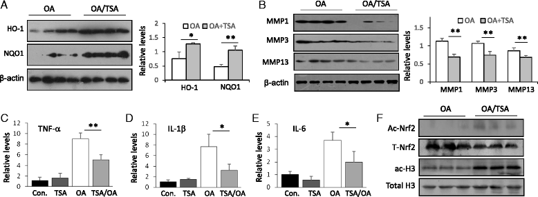
Trichostatin A (TSA) improves the expression of osteoarthritis (OA)-associated proteins and the induction of inflammatory cytokines in monosodium iodoacetate (MIA) OA mice. a Western blot analysis of heme oxygenase 1 (HO-1) and NAD(P)H:quinine oxidoreductase 1 (NQO1) from joints of MIA-OA or TSA-treated MIA-OA wild-type (WT) mice. The quantification was done on the right side. *p < 0.05, **p < 0.01 compared with MIA-OA mice. b Western blot analysis of matrix metalloproteinases MMP-1, MMP-3, and MMP-13 from joints of MIA-OA and TSA-treated MIA-OA WT mice. The quantification was done on the right side. **p < 0.01 compared with MIA-OA mice. Quantitative real-time polymerase chain reaction analysis of tumor necrosis factor (TNF)-α (c), interleukin (IL)-1β (d), and IL-6 (e) from joint tissues of MIA-OA or TSA-treated MIA-OA WT mice. *p < 0.05, **p < 0.01 compared with MIA-OA mice. f Western blot analysis of acetylated nuclear factor (erythroid-derived 2)-like 2 (Ac-Nrf2) and acetylated histone 3 (ac-H3) from MIA-OA or TSA-treated MIA-OA WT mice (three mice in each group). Total Nrf2 and histone 3 served as internal controls. All the Western blot analysis results are representative of, and the statistical analyses were performed with results from, at least six animals
Fig. 5
Fig. 5
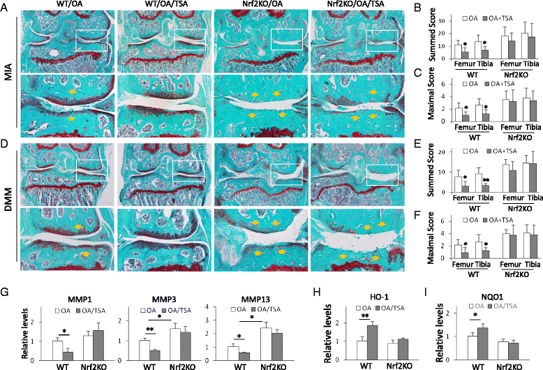
Trichostatin A (TSA) protection of osteoarthritis (OA) requires nuclear factor (erythroid-derived 2)-like 2 (Nrf2). a Wild-type (WT) and Nrf2-knockout (Nrf2-KO) mice were subjected to monosodium iodoacetate (MIA)-induced OA or MIA-induced OA with or without TSA treatment (n = 10 in each group). Mouse joint sections were stained with Safranin O/Fast Green. Representative images are shown. Lower panels show amplifications of upper panel insets. Yellow arrows indicate the damage-affected cartilage areas. b Summed scores of femur and tibia damage in WT and Nrf2-KO mice with MIA-induced OA. *p < 0.05. c Maximum scores representing femur and tibia damage in WT and Nrf2-KO mice with MIA-induced OA. *p < 0.05. d WT and Nrf2-KO mice were subjected to destabilization of the medial meniscus (DMM)-induced OA or to DMM-induced OA with or without TSA treatment (n = 10 in each group). Mouse joint tissue sections were stained with Safranin O/Fast Green. Representative images are shown. Lower panels show amplifications of upper panel insets. Yellow arrows indicate the damage-affected cartilage areas. e Summed scores of femur and tibia damage in WT and Nrf2-KO mice with DMM-induced OA. *p < 0.05, **p < 0.01 compared with OA group. f Maximum scores of femur and tibia damage in WT and Nrf2-KO mice with DMM-induced OA. *p < 0.05. g Quantitative real-time polymerase chain reaction (qRT-PCR) analysis of matrix metalloproteinases MMP-1, MMP-3, and MMP-13 from joints of WT and Nrf2-KO mice with MIA-induced OA or TSA-treated MIA-induced OA. *p < 0.05, **p < 0.01 compared with MIA-OA mice. qRT-PCR analysis of HO-1 (h) and NQO1 (i) from joints of WT and Nrf2-KO mice with MIA-OA or TSA-treated OA. *p < 0.05, **p < 0.01 compared with MIA-OA mice

References

    1. Bijlsma JW, Berenbaum F, Lafeber FP. Osteoarthritis: an update with relevance for clinical practice. Lancet. 2011;377:2115–26. doi: 10.1016/S0140-6736(11)60243-2. - DOI - PubMed
    1. Sellam J, Berenbaum F. Is osteoarthritis a metabolic disease? Joint Bone Spine. 2013;80:568–73. doi: 10.1016/j.jbspin.2013.09.007. - DOI - PubMed
    1. Su SC, Tanimoto K, Tanne Y, Kunimatsu R, Hirose N, Mitsuyoshi T, et al. Celecoxib exerts protective effects on extracellular matrix metabolism of mandibular condylar chondrocytes under excessive mechanical stress. Osteoarthritis Cartilage. 2014;22:845–51. doi: 10.1016/j.joca.2014.03.011. - DOI - PubMed
    1. de Vries HE, Witte M, Hondius D, Rozemuller AJ, Drukarch B, Hoozemans J, et al. Nrf2-induced antioxidant protection: a promising target to counteract ROS-mediated damage in neurodegenerative disease? Free Radic Biol Med. 2008;45:1375–83. doi: 10.1016/j.freeradbiomed.2008.09.001. - DOI - PubMed
    1. Zhao J, Zhang B, Li S, Zeng L, Chen Y, Fang J. Mangiferin increases Nrf2 protein stability by inhibiting its ubiquitination and degradation in human HL60 myeloid leukemia cells. Int J Mol Med. 2014;33:1348–54. - PubMed

"V体育平台登录" Publication types

MeSH terms

LinkOut - more resources