Skip to main page content
U.S. flag

An official website of the United States government

Dot gov

The . gov means it’s official. Federal government websites often end in VSports app下载. gov or . mil. Before sharing sensitive information, make sure you’re on a federal government site. .

Https

The site is secure. The https:// ensures that you are connecting to the official website and that any information you provide is encrypted and transmitted securely. V体育官网.

. 2015 Sep 15;6(27):24393-403.
doi: 10.18632/oncotarget.5162.

Heme oxygenase-1 accelerates erastin-induced ferroptotic cell death

Affiliations

Heme oxygenase-1 accelerates erastin-induced ferroptotic cell death (VSports手机版)

VSports注册入口 - Min-Young Kwon et al. Oncotarget. .

VSports在线直播 - Abstract

The oncogenic RAS-selective lethal small molecule Erastin triggers a unique iron-dependent form of nonapoptotic cell death termed ferroptosis. Ferroptosis is dependent upon the production of intracellular iron-dependent reactive oxygen species (ROS), but not other metals. However, key regulators remain unknown. The heme oxygenase (HO) is a major intracellular source of iron. In this study, the role of heme oxygenase in Erastin-triggered ferroptotic cancer cell death has been investigated VSports手机版. Zinc protoporphyrin IX (ZnPP), a HO-1 inhibitor, prevented Erastin-triggered ferroptotic cancer cell death. Furthermore, Erastin induced the protein and mRNA levels of HO-1 in HT-1080 fibrosarcoma cells. HO-1+/+ and HO-1-/- fibroblast, HO-1 overexpression, and chycloheximide-treated experiments revealed that the expression of HO-1 has a decisive effects in Erastin-triggered cell death. Hemin and CO-releasing molecules (CORM) promote Erastin-induced ferroptotic cell death, not by biliverdin and bilirubin. In addition, hemin and CORM accelerate the HO-1 expression in the presence of Erastin and increase membranous lipid peroxidation. Thus, HO-1 is an essential enzyme for iron-dependent lipid peroxidation during ferroptotic cell death. .

Keywords: free radicals; heme oxygenase-1; iron; oncogene; oncology V体育安卓版. .


Conflict of interest statement

CONFLICTS OF INTEREST

The authors declare no conflict of interest.

Figures
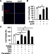
Figure 1
Figure 1. HO-1 inhibitor suppresses Erastin-induced ferroptotic death
A. HT-1080 fibrosacoma cells were treated vehicle or Erastin (10 μM) in the presence or absence of ZnPP (zinc protoporphyrin, 10 μM). After 12 hours treatment, cell nucleus was stained to detect live cells using Hoechst 33342. B. The number of live cells was measured and represented as a graph. Values are mean ± SD, n = 7. *p < 0.05 vs vehicle, †p < 0.05 vs Erastin. C. Cell viability of HT-1080 fibrosacoma cells was measured at 12 hours after vehicle, Erastin (10 μM), or Erastin plus ZnPP, DFO (deferoxamine, 100 μM), NAC (N-acetylcysteine, 10 mM), or ferrostatin-1 (0.5 μM) administration using Ez-Cytox Cell Viability Assay Kit. *p < 0.05 vs vehicle, †p < 0.05 vs Erastin. Values are mean ± SD, n = 12.
Figure 2
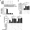
Figure 2. Erastin induces HO-1 expression in HT-1080 fibrosacoma cells
A. Western blotting for HO-1 was performed 4, 8, and 12 hours after vehicle (V), or Erastin (1 μMor 10 μM) in HT-1080 fibrosacoma cells. β-actin was used as controls for normalization. This represents a representative blot of three independent experiments. The fold change in protein levels were quantitated as signal intensity corrected for loading in vehicle or Erastin treated cells. *p < 0.05 vs vehicle. Values are mean ± SD, n = 3. Quantitative real-time PCR was performed to assess mRNA levels of HO-1 B. and HO-2 C. in HT-1080 fibrosacoma cells. *p < 0.05 vs vehicle. Quantitative real-time PCR was performed to assess mRNA levels of HO-1 D. and HO-2 E. 12 hours after vehicle, Erastin, or Erastin plus DFO, NAC, or ferrostatin-1 administration in HT-1080 fibrosacoma cells. *p < 0.05 vs vehicle, †p < 0.05 vs Erastin. Values are mean ± SD, n = 3.
Figure 3
Figure 3. The expression of HO-1 is an important for Erastin-induced ferroptotic cell death
A. Cell viability was measured 12 hours after vehicle or Erastin (1 μMor 10 μM) administration in mouse HO-1+/+ and HO-1−/− fibroblasts using Ez-Cytox Cell Viability Assay Kit. *p < 0.05 HO-1−/− vs HO-1+/+. Values are mean ± SD, n = 12. B. HT-1080 fibrosacoma cells were stably transfected with control vector (CON) or human HO-1 cDNA (HO-1). These groups of cells were exposed to vehicle or Erastin (10 μM). Cell viability was measured 12 hours after treatment in control vector (CON) or human HO-1 overexpressed HT-1080 fibrosacoma cells using Ez-Cytox Cell viability Assay Kit. *p < 0.05 HO-1 overexpressed vs control vector expressed HT-1080 fibrosacoma cells in the presence of Erastin. Values are mean ± SD, n = 12. C. The overexpression of human HO-1 was detected using western blotting for HO-1. D. CHX was treated in HT-1080 fibrosacoma cells for 12 hours in the absence or presence of Erastin. The expression of HO-1 was detected using Western blotting analysis and β-actin was used for loading control. E. Cell viability was measured 12 hours after vehicle or Erastin (10 μM) administration in the absence or presence of CHX. *p < 0.05 vs Erastin. Values are mean ± SD, n = 9. F. Cell viability was measured 12 hours after vehicle, Erastin (10 μM), or Erastin plus DFO, NAC, ferrostatin-1 administration in mouse HO-1+/+ and HO-1−/− fibroblasts using Ez-Cytox Cell Viability Assay Kit. *p < 0.05 HO-1−/− vs HO-1+/+. Values are mean ± SD, n = 11. G. Cell viability was measured 12 hours after vehicle, Erastin (10 μM), or Erastin plus CHX in mouse HO-1+/+ and HO-1−/− fibroblasts using Ez-Cytox Cell Viability Assay Kit. *p < 0.05 HO-1−/− vs HO-1+/+, †p < 0.05 Erastin plus CHX vs Erastin. Values are mean ± SD, n = 12.
Figure 4
Figure 4. Hemin and CORM accelerate Erasitn-induced ferroptotic cell death in HT-1080 fibrosacoma cells
A. Cell viability was analyzed 8 hours after vehicle, Erastin (10 μM), and hemin (5 μM), CORM (10 μM), bilirubin (10 μM), or biliverdin (10 μM)plus Erastin (10 μM)administration in HT-1080 fibrosacoma cells using Ez-Cytox Cell viability Assay Kit. *p < 0.05 vs Erastin. Values are mean ± SD, n = 8. B. Cell viability was analyzed 8 hours after vehicle, Erastin (10 μM), and hemin (5 μM) or CORM (10 μM)administration in HT-1080 fibrosacoma cells using Ez-Cytox Cell viability Assay Kit. *p < 0.05 vs vehicle. Values are mean ± SD, n = 8. C. Cell viability was analyzed 8 hours after vehicle, Erastin (10 μM), or Erastin plus hemin (5 μM)or CORM (10 μM) in the absence or presence of ZnPP or DFOadministration in HT-1080 fibrosacoma cells using Ez-Cytox Cell viability Assay Kit. *p < 0.05 vs Erastin, †p < 0.05 vs Erastin plus hemin or CORM. Values are mean ± SD, n = 8. D. Total protein was harvested 8 hours after vehicle, Erastin, or Erastin plus hemin (5 μM), CORM (10 μM), bilirubin (10 μM), or biliverdin (10 μM) administration in HT-1080 fibrosacoma cells. Western blotting for HO-1 was performed and β-actin was used as controls for normalization. This represents a representative blot of three independent experiments. The fold change in protein levels were quantitated as signal intensity corrected for loading in vehicle or Erastin treated cells. *p < 0.05 vs vehicle. Values are mean ± SD, n = 3.
Figure 5
Figure 5. The effects of hemin and CORM on Erastin-induced ferroptotic cell death play through the induction of HO-1 expression
A. Total protein was harvested 8 hours after vehicle, Erastin (10 μM), and hemin (5 μM) or CORM (10 μM) plus Erastin administration in the absence or presence of CHX (1 μg/mL) in HT-1080 fibrosacoma cells. Western blotting for HO-1 was performed and β-actin was used as controls for normalization. This represents a representative blot of three independent experiments. The fold change in protein levels were quantitated as signal intensity corrected for loading in vehicle or Erastin treated cells. *p < 0.05 vs vehicle. Values are mean ± SD, n = 3. B. Cell viability was analyzed 8 hours after vehicle, Erastin (10 μM), or Erastin plus hemin (5 μM) or CORM (10 μM)administration in the absence or presence of CHX in HT-1080 cells. *p < 0.05 vehicle, †p < 0.05 vs Erastin. Values are mean ± SD, n = 8. C. Cell viability was analyzed 8 hours after vehicle, Erastin (10 μM), and Erastin plus hemin (5 μM) or CORM (10 μM)administration in the absence or presence of DFO, NAC, or ferrostatin-1 in HT-1080 fibrosacoma cells. *p < 0.05 vs Erastin, †p < 0.05 vs Erastin plus hemin or CORM. Values are mean ± SD, n = 12.
Figure 6
Figure 6. Hemin and CORM accelerate the accumulation of lipid peroxidation in the presence of Erastin
HT-1080 fibrosacoma cells were treated with vehicle, Erastin (10 μM), or hemin (5 μM), CORM (10 μM), or biliverdin (10 μM) in the presence of Erastin. Cytosolic ROS A. and lipid peroxidation B. were assayed by flow cytometry using the fluorescent probes CellROX® Deep Red (cytosolic ROS) and C11-BODIPY (lipid peroxidation), respectively. Values are mean ± SD, n = 6. *p < 0.05 vs Erastin. C. HT-1080 fibrosacoma cells were treated vehicle, Erastin, Erastin plus hemin, or CORM in the presence or absence of HO-1 inhibitor, ZnPP (zinc protoporphyrin, 10 μM). Lipid peroxidation was analyzed. Values are mean ± SD, n = 6. *p < 0.05 vs Erastin, †p < 0.05 vs Erastin, #p < 0.05 vs Erastin plus hemin or CORM. D. Lipid peroxidation was analyzed after vehicle (0.1% DMSO), Erastin, or Erastin plus hemin or CORM administration in the absence or presence of DFO or NAC in HT-1080 fibrosacoma cells. Values are mean ± SD, n = 9. *p < 0.05 vs Erastin, †p < 0.05 vs Erastin, #p < 0.05 vs Erastin plus hemin or CORM. E. Lipid peroxidation was analyzed after vehicle (0.1% DMSO), Erastin, or Erastin plus hemin or CORM administration in the absence or presence of ferrostatin-1 in HT-1080 fibrosacoma cells. Values are mean ± SD, n = 9. *p < 0.05 vs Erastin, †p < 0.05 vs Erastin, #p < 0.05 vs Erastin plus hemin or CORM.

References

    1. Fuchs Y, Steller H. Programmed cell death in animal development and disease. Cell. 2011;147:742–758. - PMC - PubMed
    1. Thompson CB. Apoptosis in the pathogenesis and treatment of disease. Science. 1995;267:1456–1462. - PubMed
    1. Vigil D, Cherfils J, Rossman KL, Der CJ. Ras superfamily GEFs and GAPs: validated and tractable targets for cancer therapy? Nat. Rev. Cancer. 2010;10:842–857. - PMC - PubMed
    1. Dolma S, Lessnick SL, Hahn WC, Stockwell BR. Identification of genotype-selective antitumor agents using synthetic lethal chemical screening in engineered human tumor cells. Cancer Cell. 2003;3:285–296. - PubMed
    1. Yang WS, Stockwell BR. Synthetic lethal screening identifies compounds activating iron-dependent, nonapoptotic cell death in oncogenic-RAS-harboring cancer cells. Chem Biol. 2008;15:234–245. - "V体育2025版" PMC - PubMed

MeSH terms