Skip to main page content
U.S. flag

An official website of the United States government

Dot gov

The . gov means it’s official. Federal government websites often end in . gov or VSports app下载. mil. Before sharing sensitive information, make sure you’re on a federal government site. .

Https

The site is secure V体育官网. The https:// ensures that you are connecting to the official website and that any information you provide is encrypted and transmitted securely. .

. 2015 Sep 14;28(3):357-69.
doi: 10.1016/j.ccell.2015.08.003.

"VSports app下载" Loss of ATRX Suppresses Resolution of Telomere Cohesion to Control Recombination in ALT Cancer Cells

Affiliations

"V体育安卓版" Loss of ATRX Suppresses Resolution of Telomere Cohesion to Control Recombination in ALT Cancer Cells

VSports在线直播 - Mahesh Ramamoorthy et al. Cancer Cell. .

Abstract

The chromatin-remodeler ATRX is frequently lost in cancer cells that use ALT (alternative lengthening of telomeres) for telomere maintenance, but its function in telomere recombination is unknown. Here we show that loss of ATRX suppresses the timely resolution of sister telomere cohesion that normally occurs prior to mitosis. In the absence of ATRX, the histone variant macroH2A1. 1 binds to the poly(ADP-ribose) polymerase tankyrase 1, preventing it from localizing to telomeres and resolving cohesion. The resulting persistent telomere cohesion promotes recombination between sister telomeres, while it suppresses inappropriate recombination between non-sisters. Forced resolution of sister telomere cohesion induces excessive recombination between non-homologs, genomic instability, and impaired cell growth, indicating the ATRX-macroH2A1. 1-tankyrase axis as a potential therapeutic target in ALT tumors VSports手机版. .


Figures

Figure 1
Figure 1
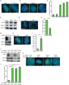
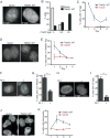
ATRX is required for resolution of sister telomere cohesion. (A, B) FISH analysis of non-ALT (A) and ALT (B) mitotic cells with a 16ptelo probe (green). (C) Quantification of the frequency of mitotic cells with cohered telomeres. Average of two independent experiments (n=50-99 cells each) ±SEM. (D) Immunoblot analysis of transfected U2OS cell extracts and (as a control for ATRX protein) HeLa cell extracts; * indicates the ATRX specific band. (E) FISH analysis of vector or ATRX-transfected U2OS mitotic cells with a 16ptelo probe (green). (F) Quantification of the frequency of mitotic cells with cohered telomeres. Average of two independent experiments (n=50-53 cells each) ±SEM. (G) Immunoblot analysis of siRNA-treated HeLa cell extracts. (H) FISH analysis of GFP or ATRX siRNA-treated HeLa mitotic cells with a 16ptelo probe (green). (I) Quantification of the frequency of mitotic cells with cohered telomeres. Average of three independent experiments (n=34-64 cells each) ±SD. (J) Immunoblot analysis of siRNA treated HeLa cell extracts. (K) FISH analysis of the indicated double siRNA-treated HeLa mitotic cells using a 16ptelo probe (green). (L) Quantification of the frequency of mitotic cells with cohered telomeres. Average of two independent experiments (n=49-51 cells each) ±SEM. (A, B, E, H, and K) DNA was stained with DAPI (blue). Scale bars, 5 μm. **p≤0.01, ***p≤0.001, students unpaired t-test. See also Figure S1.
Figure 2
Figure 2
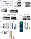
MacroH2A1.1 is a common partner of ATRX and tankyrase 1 that mediates telomere cohesion in ALT cells. (A) Schematic diagram of macroH2A1.1 as a common binding partner between ATRX and tankyrase 1 (TNKS1). (B) Immunoblot analysis of transfected HeLa cell extracts following immunoprecipitation with anti-myc antibody. (C) Immunoblot analysis of transfected U2OS cell extracts following immunoprecipitation with anti-flag antibody. (D) Immunoblot analysis of U2OS cell extracts following immunoprecipitation with anti-TNKS1 antibody. (E) Immunoblot analysis of the indicated cell extracts. (F) Immunoblot analysis of the indicated cell extracts following immunoprecipitation with IgG or anti-TNKS1 antibody. (G) Immunoblot analysis of transfected U2OS cells immunoprecipitated with anti-myc antibody. (H) Telomeric DNA ChIP analysis of HeLa cells transfected with vector, macroH2A1.1, or macroH2A1.2 using the indicated antibodies. (I) Quantification of the signal intensity of telomeric DNA immunoprecipitated by TNKS1 antibody. Average of two independent experiments ±SEM. (J) FISH analysis of vector, macroH2A1.1, or macroH2A1.2 transfected HeLa mitotic cells with a 16ptelo probe (green). DNA was stained with DAPI (blue). Scale bar, 5 μm. (K) Quantification of the frequency of mitotic cells with cohered telomeres. Average of two independent experiments, (n=41-53 cells each) ±SEM. (L) Immunoblot analysis of GM847 ALT cells following infection with GFP or macroH2A1 shRNA lentiviruses. (M) Quantification of the frequency of mitotic cells with cohered telomeres from 16p FISH analysis of mitotic cells. Average of two independent experiments (n=50 cells each) ±SEM. *p≤0.05, **p≤0.01, students unpaired t-test. See also Figure S2.
Figure 3
Figure 3
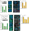
Resolution of telomere cohesion represses sister telomere recombination in ALT cells. (A) Immunoblot analysis of U2OS cells stably expressing vector, TNKS1.WT or TNKS1.PD. (B, C) FISH analysis of mitotic cells with a 16ptelo probe (B, green) and quantification of the frequency of mitotic cells with cohered telomeres (C) of U2OS cells stably expressing vector, TNKS1.WT or TNKS1.PD. DNA was stained with DAPI (blue). Scale bar, 5 μm. Average of two independent experiments (n=49-51 cells each) ±SEM. (D, E) CO-FISH analysis of metaphase spreads probed with TTAGGG (red) and CCCTAA (green) (D) and quantification of the frequency of T-SCE (E) from vector, TNKS1.WT, or TNKS1.PD expressing U2OS cells. DNA was stained with DAPI (blue). Scale bar, 10 μm. Inset scale bar, 2 μm. Average of two independent experiments (n=684-1338 chromosomes each) ±SEM. (F) immunoblot analysis of U2OS cells transiently transfected with GFP, TIN2, SA1, or SA2 siRNA. (G, H) FISH analysis of mitotic cells with a 16ptelo probe (green) (G) and quantification of the frequency of mitotic cells with cohered telomeres (n=50 cells) (H) of U2OS cells transiently transfected with GFP, TIN2, SA1, or SA2 siRNA. DNA was stained with DAPI (blue). Scale bar, 5 μm. (I, J) CO-FISH analysis of metaphase spreads probed with TTAGGG (red) and CCCTAA (green) (I) and quantification of the frequency of T-SCE (J) of U2OS cells transiently transfected with GFP, TIN2, SA1, or SA2 siRNA. DNA was stained with DAPI (blue). Scale bar, 10 μm. Inset scale bar, 1 μm. Average of two independent experiments (n=733-824 chromosomes each) ±SEM. *p≤0.05, **p≤0.01, ****p≤0.0001 students unpaired t-test.
Figure 4
Figure 4
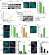
Resolution of telomere cohesion impairs cell growth and induces inter-telomere recombination in ALT cells. (A, B) Growth curves of U2OS (A) and HeLa (B) cells infected with vector, TNKS1.WT, or TNKS1.PD lentivirus. Average of three technical replicates ±SD. (C) Example of a TNKS1.WT overexpressing U2OS cell with three 16p loci assayed by FISH with a 16ptelo probe (green). DNA was stained with DAPI (blue). Scale bar, 5 μm. (D) Quantification of the frequency of mitotic cells with three 16p telomeric loci. Average of two independent experiments (n=49-51 cells each) ±SEM. *p≤0.05 students unpaired t-test. (E) Schematic representation of the tagged U2OS cell line. (F) Detection of lacO tags by immunofluorescence with anti-GFP antibody of F6B2(U2OS-3lacO)/GFPLacI cells expressing vector, TNKS1.WT, or TNKS1.PD. Scale bar, 5 μm. (G) Quantification of the frequency of cells with the indicated number of tags. Average of two independent experiments (n=25 cells each) ±SEM. See also Figure S3.
Figure 5
Figure 5
The macroH2A-binding domain of ATRX induces resolution of telomere cohesion and inter-telomere recombination in ALT cells. (A) Immunoblot analysis of transfected U2OS cell extracts following immunoprecipitation with anti-myc antibody. (B) FISH analysis of mitotic U2OS cells transfected with vector or ATRX.1-841 using a 16ptelo probe (green). DNA was stained with DAPI (blue). Scale bar, 5 μm. (C) Quantification of the frequency of mitotic cells with cohered telomeres. Average of two independent experiments (n=25-50 cells each) ±SEM. (D) Growth curves of U2OS cells infected with vector or ATRX.1-841 lentivirus. Average of three technical replicates ±SD. (E) Immunoblot analysis of transfected GM847 cell extracts following immunoprecipitation with anti-myc antibody. (F) FISH analysis of mitotic GM847 cells transfected with vector or ATRX.322-841 with a 16ptelo probe (green). DNA was stained with DAPI (blue). Scale bar, 5 μm. (G) Quantification of the frequency of mitotic cells with cohered telomeres. Average of two independent experiments (n=50 cells each) ±SEM. (H) Quantification of the frequency of T-SCE from CO-FISH analysis of metaphase spreads from U2OS cells stably expressing vector and ATRX.322-841. Average of two independent experiments (n=124-638 chromosomes each) ±SEM. (I) Example of an ATRX.322-841 overexpressing GM847 cell with four 16p loci assayed by FISH with a 16ptelo probe (green). DNA was stained with DAPI (blue). Scale bar, 5 μm. (J) Quantification of the frequency of mitotic cells with more than three 16p telomeric loci. Average of two independent experiments (n=50 cells each) ±SEM. (K) Dual FISH analysis of mitotic GM847 cells transfected with vector or ATRX.322-841 with a 13qtelo (green) and arm (red) probe. DNA was stained with DAPI (blue). Scale bar, 5 μm. (L) Quantification of the frequency of mitotic cells with more than two 13q telo and arm loci. Average of 3 independent experiments (n=11-26 cells each) ±SD. (M) Immunofluorescence analysis of analysis of lacO tags in F6B2(U2OS-3lacO) cells transfected with GFPLacI and ATRX.1-321 or ATRX.322-841. Scale bar, 5 μm. (N) Quantification of the frequency of cells with the indicated number of tags. Average of two independent experiments (n=25 cells each) ±SEM. *p≤0.05, **p≤0.01, ***p≤0.0001, ****p≤0.0001, students unpaired t-test. See also Figure S4.
Figure 6
Figure 6
Resolution of telomere cohesion leads to immediate, rampant recombination and genomic instability. (A, B) Detection of lacO tags by immunofluorescence with anti-GFP antibody (A) and quantification of the frequency of cells with the indicated number of tags (B) in F6B2(U2OS-3lacO)/GFPLacI cells following infection with vector or TNKS1.WT lentivirus on Day 1. Scale bar, 5 μm. Average of two independent experiments (n=25 cells each) ±SEM. (C) Quantification of the frequency of F6B2(U2OS-3lacO)/GFPLacI cells following infection with vector or TNKS1.WT lentivirus displaying more than 5 tags on days 1-4. Average of two independent experiments (n=25 cells each) ±SEM. (D, E) Detection of RAD51 foci by immunofluorescence analysis on Day 1 (D) and quantification of the frequency of cells displaying more than 10 RAD51 foci on days 1-4 (E) in U2OS cells following infection with vector or TNKS1.WT lentivirus. Scale bar, 10 μm. Average of two independent experiments (n=100 cells each) ±SEM. (F, G) Detection of RAD51 foci by immunofluorescence analysis (F) and quantification of the frequency of cells displaying more than 10 RAD51 foci (G) in U2OS cells on Day 1 following infection with TNKS1.WT lentivirus. Cells were treated with RAD51 inhibitor RI-1 for 8 hr prior to harvest. Scale bar, 10 μm. Average of two independent experiments (n=100 cells each) ±SEM. (H, I) Detection of lacO tags by immunofluorescence with anti-GFP antibody (H) and quantification of the frequency of cells with the indicated number of tags (I) in F6B2(U2OS-3lacO)/GFPLacI cells on Day 1 following infection with TNKS1.WT lentivirus. Cells were treated with RAD51 inhibitor RI-1 for 8 hr prior to harvest. Scale bar, 10 μm. Average of two independent experiments (n=25 cells each) ±SEM. *p≤0.05 students unpaired t-test. (J, K) Detection of micronuclei by immunofluorescence staining with DAPI on Day 2 (J) and quantification of the frequency of micronucleation events on days 1-4 (K) in U2OS cells following infection with vector or TNKS1.WT lentivirus. Scale bar, 10 μm. Average of two independent experiments (n=100 cells each) ±SEM. See also Figure S5.
Figure 7
Figure 7
Model for the role of persistent telomere cohesion in ALT. (A) Loss of ATRX in ALT frees macroH2A1.1 to bind and sequester tankyrase 1 thereby preventing resolution of sister telomere cohesion. When ATRX is reintroduced into ALT cells it binds macroH2A1.1, freeing tankyrase 1 to resolve telomere cohesion. (B) When telomeres are cohered the sister telomere is the favored copy template. Upon forced resolution of telomere cohesion in ALT cells (by introduction of ATRX, overexpression of tankyrase 1, or depletion of macroH2A1.1) any telomere can be the copy template.

Comment in (V体育官网)

"VSports最新版本" References

    1. Bailey SM, Cornforth MN, Kurimasa A, Chen DJ, Goodwin EH. Strand-specific postreplicative processing of mammalian telomeres. Science. 2001;293:2462–2465. - PubMed
    1. Bechter OE, Zou Y, Shay JW, Wright WE. Homologous recombination in human telomerase-positive and ALT cells occurs with the same frequency. EMBO Rep. 2003;4:1138–1143. - "V体育平台登录" PMC - PubMed
    1. Bisht KK, Daniloski Z, Smith S. SA1 binds directly to DNA through its unique AT-hook to promote sister chromatid cohesion at telomeres. J Cell Sci. 2013;126:3493–3503. - PMC (V体育平台登录) - PubMed
    1. Bisht KK, Dudognon C, Chang WG, Sokol ES, Ramirez A, Smith S. GDP-mannose-4,6-dehydratase is a cytosolic partner of tankyrase 1 that inhibits its poly(ADP-ribose) polymerase activity. Mol Cell Biol. 2012;32:3044–3053. - PMC - PubMed
    1. Bower K, Napier CE, Cole SL, Dagg RA, Lau LM, Duncan EL, Moy EL, Reddel RR. Loss of wild-type ATRX expression in somatic cell hybrids segregates with activation of Alternative Lengthening of Telomeres. PloS one. 2012;7:e50062. - V体育ios版 - PMC - PubMed

Publication types

MeSH terms

LinkOut - more resources