Skip to main page content (V体育平台登录)
U.S. flag

An official website of the United States government

Dot gov

The . gov means it’s official. Federal government websites often end in VSports app下载. gov or . mil. Before sharing sensitive information, make sure you’re on a federal government site. .

Https

The site is secure. The https:// ensures that you are connecting to the official website and that any information you provide is encrypted and transmitted securely V体育官网. .

. 2016 Jan;10(1):73-84.
doi: 10.1016/j.molonc.2015.08.003. Epub 2015 Aug 20.

Resistance to BRAF inhibitors induces glutamine dependency in melanoma cells

Affiliations

VSports最新版本 - Resistance to BRAF inhibitors induces glutamine dependency in melanoma cells

Franziska Baenke et al. Mol Oncol. 2016 Jan.

V体育安卓版 - Abstract

BRAF inhibitors can extend progression-free and overall survival in melanoma patients whose tumors harbor mutations in BRAF. However, the majority of patients eventually develop resistance to these drugs. Here we show that BRAF mutant melanoma cells that have developed acquired resistance to BRAF inhibitors display increased oxidative metabolism and increased dependency on mitochondria for survival. Intriguingly, the increased oxidative metabolism is associated with a switch from glucose to glutamine metabolism and an increased dependence on glutamine over glucose for proliferation. We show that the resistant cells are more sensitive to mitochondrial poisons and to inhibitors of glutaminolysis, suggesting that targeting specific metabolic pathways may offer exciting therapeutic opportunities to treat resistant tumors, or to delay emergence of resistance in the first-line setting. VSports手机版.

Keywords: BRAF; Glutaminolysis; Melanoma; Metabolism; Resistance V体育安卓版. .


Figures

Figure 1
Figure 1
BRAF inhibitor resistant melanoma cells exhibit increased mitochondrial mass and oxidative metabolism. (A) Photomicrographs showing mitochondrial morphology (Mitotracker Green) and nuclei (Hoechst, blue) in A375, Colo829, A375/R and Colo829/R cells. (B)PGC1α levels in A375, Colo829, A375/R and Colo829/R cells. Error bars represent SEM of at least two independent experiments with 3 biological replicates. ****p < 0.0001. (C) Photomicrographs showing mitochondrial morphology (Mitotracker Green) in A375, Colo829, A375/R and Colo829/R cells after silencing with siRNA control (siCtrl) or two different siRNA probes against PGC1α. (D–F)MT‐CO2(D), MT‐CYB(E), and MT‐ATP6/8(F) mRNA levels in A375, Colo829, A375/R and Colo829/R cells. Error bars represent SEM of at least two independent experiments with 3 biological replicates. *p < 0.05, **p < 0.01, ***p < 0.001 and ****p < 0.0001. (G) The box‐plot showing quantification of MT‐CO2 staining in 7 paired samples from patients before and after emergence of vemurafenib resistance; *p < 0.05, and test (Wilcoxon Signed‐Rank Test). (H) Oxygen consumption rate (OCR) changes in response to mitochondrial function modulators in A375, Colo829, A375/R and Colo829/R cells (n = 3). (I–J) Lactate secretion (I) and intracellular ATP levels (J) in A375, Colo829, A375/R and Colo829/R cells. Error bars represent SEM of at least two independent experiments with 3 biological replicates. **p < 0.01, ***p < 0.001, ****p < 0.0001. (K) Oxygen consumption rate (OCR) changes in response to mitochondrial function modulators in SKMEL5, SKMEL5/R, G361 and G361/R cells (n = 3). (L–M) Lactate secretion (L) and intracellular ATP levels (M) in SKMEL5, G361, SKMEL5/R and G361/R cells. Error bars represent SEM of at least two independent experiments with 3 biological replicates. n.s.: not significant.
Figure 2
Figure 2
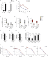
BRAF inhibitor resistant melanoma cells display decreased glycolytic flux. (A) Graph showing PGC1α mRNA levels in A375 and Colo829 cells grown for 5 days in medium containing glucose or galactose as the sole carbon source. Error bars represent SEM of at least two independent experiments with 3 biological replicates. ****p < 0.0001. (B) Graph showing A375 cell growth (sulforhodamine B) in glucose or galactose containing medium in the presence of increasing concentrations of PLX4720 for 72 h. (C–E) Graphs showing glucose (C), glucose‐6‐phosphate (D), and lactate (E) levels in A375, Colo829, A375/R and Colo829/R cells labeled with U‐13C6 glucose. The graphs show mean ± SD (n = 6) and similar results were observed in two independent experiments. Legend displays the different isotopomers. 13C1: one carbon atom labeled; 13C2 – 13C6: two to six carbon atoms labeled. Glucose and glucose‐6‐phosphate are 6‐carbon molecules hence 13C6 labeling (gray). Lactate is a 3‐carbon molecule hence the 13C3 labeling (red). Endogenous metabolites are unlabeled and shown in white. (F–H) Graphs showing glucose (F), pyruvate, (G) and lactate (H) consumption (extracellular exchange rate) in A375 and A375/R cells. Data are representative of three independent studies and displayed as mean ± SD of n = 6. (I) Growth (sulforhodamine B) of A375, A375/R, Colo829, Colo829/R, SKMEL5, SKMEL5/R, G361 and G361/R cells in medium containing 1 mM glucose for 72 h. Data are expressed relative to growth in medium containing 10 mM glucose (dotted line). Error bars represent SEM of at three independent experiments with 3 biological replicates. *p < 0.05, **p < 0.01, n.s.: non‐significant. (J) Growth of A375, A375/R, Colo829, Colo829/R, SKMEL5, SKMEL5/R, G361 and G361/R cells in the presence of increasing concentrations 2DG for 72 h (n = 3).
Figure 3
Figure 3
BRAF inhibitor resistant melanoma cells show increased glutamine metabolism. (A) Graph showing intracellular glutamine levels in A375, A375/R, Colo829 and Colo829/R cells labeled with U‐13C5 glutamine. Legend displays isotopomer distribution. 13C1: one carbon atom labeled; 13C2–13C5: two to five carbon atoms labeled. Glutamine is a 5‐carbon molecule hence 13C5 labeling (blue). Endogenous glutamine is unlabeled and shown in white. (B) Graph showing glutamine consumption (extracellular exchange rates) in A375 and A375/R cells. Data are representative of three independent studies and displayed as mean ± SD of n = 6. (C) Graph showing growth (sulforhodamine B) of A375, A375/R, Colo829, Colo829/R, SKMEL5, SKMEL5/R, G361 and G361/R cells in medium containing 0.5 mM glutamine for 72 h. Data are presented relative to medium containing 2 mM glutamine. Error bars represent SEM of three independent experiments with 3 biological replicates. *p < 0.05, ns: non‐significant. (D) Graph showing intracellular glutamine levels in A375, A375/R, Colo829 and Colo829/R cells labeled with U‐13C5 glutamine. Data are a representative experiment of mean ± SD of n = 6. Similar results were observed in two independent experiments. Legend displays isotopomer distribution. 13C1: one carbon atom labeled; 13C2 to 13C5: two to five carbon atoms labeled. Glutamate is a 5‐carbon molecule hence 13C5 labeling (blue). Endogenous glutamate is unlabeled and shown in white. (E) Graph showing GLS mRNA levels in A375, A375/R, Colo829, Colo829/R, SKMEL5, SKMEL5/R, G361 and G361/R cells. Three independent experiments with n = 3 were performed and results display mean ± SEM. *p < 0.05, **p < 0.01, and ****p <0.0001. (F) Ratio of intracellular citrate (Cit+2) to pyruvate (Pyr+3) in A375 and A375/R cells.
Figure 4
Figure 4
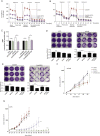
Glutaminolysis inhibition diminishes oxidative metabolism and cell viability of BRAF inhibitor resistant melanoma cells. (A, B) Oxygen consumption rate (OCR) changes in response to mitochondrial function modulators in A375 and A375/R cells (A), and in Colo829 and Colo829/R cells (B), treated with DMSO (control) or BPTES (2 μM) for 18 h. Raw values were normalized to protein content (n = 3). (C) Graph showing ATP levels in A375, A375/R, Colo829 and Colo829/R cells treated with DMSO (control) or BPTES (2 μM). Error bars represent SEM of at least two independent experiments with three biological replicates each. *p < 0.05, **p < 0.01, n.s.: not significant. (D, E) Long term (10 days) growth of A375 and A375/R cells (D) and in Colo829 and Colo829/R (E) cells in the presence of DMSO (control), BPTES (2 μM), dimethyl‐α‐KG (dmKM; 5 mM) or both. The graphs below the images show quantification by crystal violet. Error bars represent SEM of at least two independent experiments with three biological replicates each; ****p <0.0001 and n.s.: not significant. (F) Graph showing growth of A375/R xenografts in nude mice (n = 8 per group) treated with PLX4720 (45 mpk, p.o. daily) or BPTES (12.5 mpk, IP every second day) or both. Mann–Whitney‐U test *p < 0.05. (G) Graph showing growth of A375 xenografts in nude mice (n = 8 per group) treated with PLX4720 (45 mpk, p.o. daily) or BPTES (12.5 mpk IP every second day) or both. Mann–Whitney‐U test ****p < 0.0001.

"V体育官网" References

    1. Cantor, J.R. , Sabatini, D.M. , 2012. Cancer cell metabolism: one hallmark, many faces. Cancer Discov. 2, 881–898. - PMC - PubMed
    1. Chaneton, B. , Hillmann, P. , Zheng, L. , Martin, A.C. , Maddocks, O.D. , Chokkathukalam, A. , Coyle, J.E. , Jankevics, A. , Holding, F.P. , Vousden, K.H. , Frezza, C. , O'Reilly, M. , Gottlieb, E. , 2012. Serine is a natural ligand and allosteric activator of pyruvate kinase M2. Nature. 491, 458–462. - VSports在线直播 - PMC - PubMed
    1. Corazao-Rozas, P. , Guerreschi, P. , Jendoubi, M. , Andre, F. , Jonneaux, A. , Scalbert, C. , Garcon, G. , Malet-Martino, M. , Balayssac, S. , Rocchi, S. , Savina, A. , Formstecher, P. , Mortier, L. , Kluza, J. , Marchetti, P. , 2013. Mitochondrial oxidative stress is the Achille's heel of melanoma cells resistant to Braf-mutant inhibitor. Oncotarget. 4, 1986–1998. - PMC - PubMed
    1. Dang, L. , White, D.W. , Gross, S. , Bennett, B.D. , Bittinger, M.A. , Driggers, E.M. , Fantin, V.R. , Jang, H.G. , Jin, S. , Keenan, M.C. , Marks, K.M. , Prins, R.M. , Ward, P.S. , Yen, K.E. , Liau, L.M. , Rabinowitz, J.D. , Cantley, L.C. , Thompson, C.B. , Vander Heiden, M.G. , Su, S.M. , 2009. Cancer-associated IDH1 mutations produce 2-hydroxyglutarate. Nature. 462, 739–744. - PMC - PubMed
    1. Davies, H. , Bignell, G.R. , Cox, C. , Stephens, P. , Edkins, S. , Clegg, S. , Teague, J. , Woffendin, H. , Garnett, M.J. , Bottomley, W. , Davis, N. , Dicks, E. , Ewing, R. , Floyd, Y. , Gray, K. , Hall, S. , Hawes, R. , Hughes, J. , Kosmidou, V. , Menzies, A. , Mould, C. , Parker, A. , Stevens, C. , Watt, S. , Hooper, S. , Wilson, R. , Jayatilake, H. , Gusterson, B.A. , Cooper, C. , Shipley, J. , Hargrave, D. , Pritchard-Jones, K. , Maitland, N. , Chenevix-Trench, G. , Riggins, G.J. , Bigner, D.D. , Palmieri, G. , Cossu, A. , Flanagan, A. , Nicholson, A. , Ho, J.W. , Leung, S.Y. , Yuen, S.T. , Weber, B.L. , Seigler, H.F. , Darrow, T.L. , Paterson, H. , Marais, R. , Marshall, C.J. , Wooster, R. , Stratton, M.R. , Futreal, P.A. , 2002. Mutations of the BRAF gene in human cancer. Nature. 417, 949–954. - "V体育官网" PubMed

Publication types