Skip to main page content
U.S. flag

An official website of the United States government

Dot gov

The VSports app下载. gov means it’s official. Federal government websites often end in . gov or . mil. Before sharing sensitive information, make sure you’re on a federal government site. .

Https

The site is secure V体育官网. The https:// ensures that you are connecting to the official website and that any information you provide is encrypted and transmitted securely. .

. 2015 Sep 10;6(9):e1879.
doi: 10.1038/cddis.2015.248.

ER stress induces NLRP3 inflammasome activation and hepatocyte death

Affiliations

"V体育官网入口" ER stress induces NLRP3 inflammasome activation and hepatocyte death

C Lebeaupin et al. Cell Death Dis. .

"VSports最新版本" Abstract

The incidence of chronic liver disease is constantly increasing, owing to the obesity epidemic. However, the causes and mechanisms of inflammation-mediated liver damage remain poorly understood. Endoplasmic reticulum (ER) stress is an initiator of cell death and inflammatory mechanisms. Although obesity induces ER stress, the interplay between hepatic ER stress, NLRP3 inflammasome activation and hepatocyte death signaling has not yet been explored during the etiology of chronic liver diseases. Steatosis is a common disorder affecting obese patients; moreover, 25% of these patients develop steatohepatitis with an inherent risk for progression to hepatocarcinoma. Increased plasma LPS levels have been detected in the serum of patients with steatohepatitis. We hypothesized that, as a consequence of increased plasma LPS, ER stress could be induced and lead to NLRP3 inflammasome activation and hepatocyte death associated with steatohepatitis progression. In livers from obese mice, administration of LPS or tunicamycin results in IRE1α and PERK activation, leading to the overexpression of CHOP VSports手机版. This, in turn, activates the NLRP3 inflammasome, subsequently initiating hepatocyte pyroptosis (caspase-1, -11, interleukin-1β secretion) and apoptosis (caspase-3, BH3-only proteins). In contrast, the LPS challenge is blocked by the ER stress inhibitor TUDCA, resulting in: CHOP downregulation, reduced caspase-1, caspase-11, caspase-3 activities, lowered interleukin-1β secretion and rescue from cell death. The central role of CHOP in mediating the activation of proinflammatory caspases and cell death was characterized by performing knockdown experiments in primary mouse hepatocytes. Finally, the analysis of human steatohepatitis liver biopsies showed a correlation between the upregulation of inflammasome and ER stress markers, as well as liver injury. We demonstrate here that ER stress leads to hepatic NLRP3 inflammasome pyroptotic death, thus contributing as a novel mechanism of inflammation-mediated liver injury in chronic liver diseases. Inhibition of ER-dependent inflammasome activation and cell death pathways may represent a potential therapeutic approach in chronic liver diseases. .


Figures
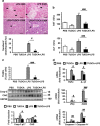
Figure 1
Figure 1
TUDCA protected against LPS-induced liver injury, apoptosis and inflammasome priming in ob/ob mice. TUDCA was injected intraperitoneally (500 μg/g) for 5 days. An LPS challenge (2 μg/g) was performed 6-h before killing. (a) Shown are photomicrographs of sections of murine liver stained with H&E (scale bar=50 μm at × 200 or 25 μm at × 400 magnification). The number of steatohepatitis foci (number of inflammatory foci in contact with ballooned hepatocytes, identified by arrows) was evaluated. (b) Serum AST and ALT transaminase levels were measured (n=9–12). (c) Apoptotic hepatocytes were visualized with TUNEL assay. The expression of active caspase-3 and CAD was evaluated in total liver lysates. Quantification was performed from the immunoblot analysis and expressed as fold induction (n=6). (d) Relative expression of hepatic TNFα, IL-1β, caspase-1 and caspase-11 mRNA (normalized to 36B4 mRNA). Data were expressed as fold induction (n=7). Data are expressed as mean±standard error of the mean. Statistical significance from controls is denoted by *,#P≤0.05, **,##P≤0.01, ***,###P≤0.001
Figure 2
Figure 2
TUDCA treatment prevented hepatic activation of the NLRP3 inflammasome in ob/ob mice challenged with LPS. (a) Analysis of whole-liver samples from PBS-, TUDCA-, LPS- and [TUDCA+LPS]-treated mice. Immunoblot analysis of active caspase-11, active caspase-1 and NLRP3 protein levels are shown (n=4–6). (b) Real-time quantitative PCR analysis was performed to compare relative hepatic levels of Nlrp3 and IL-18 mRNAs. (c) Plasma cytokine levels were quantified for IL-1β, TNFα and IFNγ (n=7–9). Statistical significance from controls is denoted by *,#P≤0.05, **,##P≤0.01, ***,###P≤0.001
Figure 3
Figure 3
TUDCA inhibited the LPS-induced IRE1α and PERK hepatic activities by favoring anti-apoptotic signaling pathways in ob/ob mice. The expression of ER stress (a and b) and apoptotic proteins, presented as Bcl-2 family pro-apoptotic/anti-apoptotic ratios (c and d), was compared by immunoblotting and quantified (n=6–9). Data are expressed as mean±standard error of the mean. Statistical significance from controls is denoted by *,#P≤0.05, **,##P≤0.01, ***,###P≤0.001
Figure 4
Figure 4
Challenge with tunicamycin in ob/ob mice increased apoptosis and activation of the NLRP3 inflammasome, and was associated with hepatic IRE1α and PERK activities. Mice were injected with TUNI (2 μg/g) 6 h before killing. (a) Serum AST (IU/l) levels were measured and the number of steatohepatitis foci was determined (n=5). (b) The number of apoptotic hepatocytes was monitored by TUNEL staining. Immunoblotting of active caspase-3, Puma and Bax was performed from whole-liver lysates. (c) Immunoblot analysis of the protein levels of active caspase-11, caspase-1 and IL-1β are shown (n=5). (d) Plasma levels of the cytokines IL-1β, IL-6 and MCP-1 are represented (n=5). (e) The expression of ER stress proteins was compared in TUNI- and PBS-injected mice (n=5). Data are expressed as mean±standard error of the mean. Statistical significance from controls is denoted by *P≤0.05, **P≤0.01
Figure 5
Figure 5
TUDCA inhibited CHOP-induced apoptosis and activation of the inflammasome in response to LPS and tunicamycin co-treatment in primary mouse hepatocytes. (a and b) Hepatocytes were pretreated for 48 h with 500 μg/ml TUDCA followed by culture for 24 h in normal medium (c) or 100 ng/ml LPS, 1 μg/ml TUNI or both (LPS+TUNI). After treatment, the percentage of viable cells quantified by MTT assay (a) (relative to control) and TUNEL-positive hepatocytes (b) was quantified (n=4–6). (c and d) Immunoblot analysis of CHOP, active caspase-11, caspase-1, pro-IL-1β and IL-1β protein levels assessed from hepatocytes pretreated either with TUDCA (c) or Chop siRNA (d) before stimulation (n=3). Data are expressed as mean±standard error of the mean. Statistical significance from controls is denoted by *,#P≤0.05, **P≤0.01
Figure 6
Figure 6
Increased expression of ER stress and inflammasome gene expression in human NASH. (a) The mRNA expression of Chop, Grp78, caspase-4, caspase-1 and IL-1β (normalized to RPLPO mRNA) was measured in the livers of massively obese patients without NAFLD (n=6), with steatosis (n=15) or with NASH (n=9). Statistical significance of controls (patients without NAFLD) was determined with the Mann–Whitney test. (b) Correlation between NLRP3 inflammasome components at the relative mRNA expression level is shown. Correlation was evaluated using the Spearman's rank correlation test. Statistical significance from controls is denoted by *P≤0.05, **,##P≤0.01, ***,###P≤0.001
Figure 7
Figure 7
ER stress activates NLRP3 inflammasome and hepatocyte death in liver disorders. Endotoxinemia (LPS), a pathological condition found in chronic liver diseases, overwhelms hepatic IRE1α and PERK activities, leading to the overexpression of CHOP, which regulates the expression of caspase-1, caspase-11 and Puma, and triggers hepatocyte pyroptosis and apoptosis. TUDCA increased the [GRP78/CHOP] ratio, promoting protection against ER-dependent NLRP3 inflammasome and cell death pathways. Using TUDCA, either alone or combined with hepatoprotective and anti-inflammatory interventions, could be a valid therapeutic strategy for the treatment of liver disorders

References (V体育平台登录)

    1. 1Henao-Mejia J, Elinav E, Jin C, Hao L, Mehal WZ, Strowig T et al. Inflammasome-mediated dysbiosis regulates progression of NAFLD and obesity. Nature 2012; 482: 179–185. - PMC - PubMed
    1. 2Angulo P. Nonalcoholic fatty liver disease. N Engl J Med 2002; 346: 1221–1231. - PubMed
    1. 3Ozcan U, Cao Q, Yilmaz E, Lee AH, Iwakoshi NN, Ozdelen E et al. Endoplasmic reticulum stress links obesity, insulin action, and type 2 diabetes. Science 2004; 306: 457–461. - PubMed
    1. 4Puri P, Mirshahi F, Cheung O, Natarajan R, Maher JW, Kellum JM et al. Activation and dysregulation of the unfolded protein response in nonalcoholic fatty liver disease. Gastroenterology 2008; 134: 568–576. - PubMed
    1. 5Xu C, Bailly-Maitre B, Reed JC. Endoplasmic reticulum stress: cell life and death decisions. J Clin Invest 2005; 115: 2656–2664. - "VSports注册入口" PMC - PubMed

"V体育平台登录" Publication types

MeSH terms

Substances