Skip to main page content
U.S. flag

An official website of the United States government

Dot gov

The . gov means it’s official. Federal government websites often end in . gov or . mil VSports app下载. Before sharing sensitive information, make sure you’re on a federal government site. .

Https

The site is secure. The https:// ensures that you are connecting to the official website and that any information you provide is encrypted and transmitted securely. V体育官网.

. 2015 Oct;11(10):765-71.
doi: 10.1038/nchembio.1891. Epub 2015 Aug 24.

Human calprotectin is an iron-sequestering host-defense protein

Affiliations

Human calprotectin is an iron-sequestering host-defense protein

Toshiki G Nakashige et al. Nat Chem Biol. 2015 Oct.

Abstract

Human calprotectin (CP) is a metal-chelating antimicrobial protein of the innate immune response. The current working model states that CP sequesters manganese and zinc from pathogens. We report the discovery that CP chelates iron and deprives bacteria of this essential nutrient. Elemental analysis of CP-treated growth medium establishes that CP reduces the concentrations of manganese, iron and zinc. Microbial growth studies reveal that iron depletion by CP contributes to the growth inhibition of bacterial pathogens. Biochemical investigations demonstrate that CP coordinates Fe(II) at an unusual hexahistidine motif, and the Mössbauer spectrum of (57)Fe(II)-bound CP is consistent with coordination of high-spin Fe(II) at this site (δ = 1. 20 mm/s, ΔEQ = 1 VSports手机版. 78 mm/s). In the presence of Ca(II), CP turns on its iron-sequestering function and exhibits subpicomolar affinity for Fe(II). Our findings expand the biological coordination chemistry of iron and support a previously unappreciated role for CP in mammalian iron homeostasis. .


Figures

Figure 1
Figure 1. CP houses two transition-metal binding sites at the S100A8/S100A9 interface
(a) CP heterodimer from the structure of the Mn(II)-, Ca(II)-, and Na(I)-bound heterotetramer (PDB: 4XJK). S100A8 is shown in green. S100A9 is shown in blue. The calcium ions are shown as yellow spheres. The sodium ions are shown as purple spheres. The manganese ion is shown as a magenta sphere. The amino acid residues at the metal binding sites are shown as orange sticks. (b) The His3Asp motif (site 1) is comprised of (A8)His83, (A8)His87, (A9)His20, and (A9)Asp30. (c) The His6 motif (site 2) is comprised of (A8)His17, (A8)His27, (A9)His91, (A9)His95, (A9)His103, and (A9)His105.
Figure 2
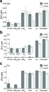
Figure 2. CP depleted metals from bacterial growth medium
Metal analysis of Mn (a), Zn (b), and Fe (c) concentrations in CP-treated and untreated growth medium (250 µg/mL CP, ~3 mM BME) in the absence (white bars) and presence (gray bars) of a 2-mM Ca(II) supplement (mean ± SEM, n ≥ 3).
Figure 3
Figure 3. Metal supplementation growth studies
Growth curves of Gram-negative bacteria Acinetobacter baumannii (a), laboratory and enterohemorrhagic strains of Escherichia coli (b,c), Klebsiella pneumoniae (d), and Pseudomonas aeruginosa (e), and Gram-positive bacteria Bacillus cereus (f) and Staphylococcus aureus (g,h) cultured in medium (+Ca(II), +BME) treated with CP-Ser (250 µg/mL) and supplemented with 0.15 µM Mn(II), 3 µM Fe(II), and/or 5 µM Zn(II) (mean ± SEM, n ≥ 3).
Figure 4
Figure 4. The antimicrobial activity of CP against E. coli, S. aureus, and L. plantarum
(a,b) Antibacterial activity of CP-Ser, ΔHis3Asp, and ΔHis4 preincubated with 0.9 equiv of Fe(II) against E. coli and S. aureus (t = 20 h, mean ± SEM, n = 6). (c,d) Antibacterial activity of CP against L. plantarum in the absence and presence of 2-mM Ca(II) and 3-mM BME supplements (t = 20 h, mean ± SEM, n = 3). (e) Growth of L. plantarum in medium treated with CP-Ser (500 µg/mL) supplemented with 10 µM Mn(II) and 10 µM Zn(II) (mean ± SEM, n = 3).
Figure 5
Figure 5. Inhibition of bacterial iron acquisition by CP
Uptake of radiolabeled 55Fe by E. coli (a) and P. aeruginosa (b) treated with 500 µg/mL CP-Ser (squares) or ΔΔ (diamonds) compared with untreated bacteria (circles). The upper panels show the amount of 55Fe uptake per mL of bacterial culture over the 2-h experiment. The lower panels indicate the corresponding OD600 values of the cultures at each time point. The mean ± SEM values for 55Fe and OD600 are shown (n = 4).
Figure 6
Figure 6. Hexahistidine Fe(II) coordination by CP
(a–c) Analytical SEC chromatograms of CP-Ser (400 µM) incubated with no metal, 5 equiv of Fe(II), and 5 equiv of Fe(III), and quantification of Fe and CP in eluent fractions. (d–f) Analytical SEC chromatograms of CP (20 µM) incubated with 10 equiv of Fe(II). Chromatograms for CP αβ (no metal) and α2β2 (+Ca(II)) are provided for reference. Peak intensities are normalized to 1. (g) The 4.2-K/53-mT Mössbauer spectrum of 57Fe(II)-bound CP-Ser (black vertical bars). Simulation using the parameters described in the main text is shown as the blue line, and the Mössbauer spectrum of Fe(II) sulfate in 50 mM Tris, pH 7.5 recorded under the same conditions is shown as the red line.
Figure 7
Figure 7. CP binds Fe(II) with remarkably high affinity in a Ca(II)-dependent manner
(a) ZP1 (1 µM) emission response to 3.5 µM Fe(II) in the absence (circles) and presence (squares) of 4 µM CP-Ser with addition of Ca(II). The red markers represent the 200-µM Ca(II) samples spiked with 4 µM Fe(II) (mean, n = 2). (b) ZP1 (1 µM) emission response to Fe(II) in the presence of 4 µM CP with or without 50 equiv of Ca(II) (mean ± SDM, n = 3). (c) Room-temperature EPR spectra of Mn(II) (black), CP-Ser in the presence of 50 equiv of Ca(II) and 0.9 equiv of Mn(II) (red), and CP-Ser in the presence of 50 equiv of Ca(II) and 0.9 equiv of Mn(II) with the addition of 0.9 equiv of Fe(II) (blue). (d) The concentration of free Mn(II) determined by room-temperature EPR (mean ± SDM, n = 3). (e) The concentration of unbound Fe(II) in ΔHis3Asp samples containing 0.9 equiv of Fe(II) after addition of 0.9 equiv of Zn(II) with and without 50 equiv of Ca(II) (mean ± SDM, n = 3). Control samples containing 25 µM protein, 0.9 equiv of Fe(II), and/or 50 equiv of Ca(II) are described in the right panel.

Comment in

References

    1. Bertini I, Gray HB, Stiefel EI, Valentine JS. Biological inorganic chemistry: structure and reactivity. University Science Books; 2007.
    1. Weinberg ED. Nutritional immunity: host’s attempt to withhold iron from microbial invaders. J. Am. Med. Assoc. 1975;231:39–41. - PubMed
    1. Fischbach MA, Lin H, Liu DR, Walsh CT. How pathogenic bacteria evade mammalian sabotage in the battle for iron. Nat. Chem. Biol. 2006;2:132–138. - PubMed
    1. Hood MI, Skaar EP. Nutritional immunity: transition metals at the pathogen-host interface. Nat. Rev. Microbiol. 2012;10:525–537. - "V体育2025版" PMC - PubMed
    1. Cassat JE, Skaar EP. Iron in infection and immunity. Cell Host Microbe. 2013;13:509–519. - PMC - PubMed

Methods-only references

    1. Carter P. Spectrophotometric determination of serum iron at the submicrogram level with a new reagent (ferrozine) Anal. Biochem. 1971;40:450–458. - PubMed

Publication types

MeSH terms