Skip to main page content
U.S. flag

An official website of the United States government

Dot gov

The . gov means it’s official. Federal government websites often end in VSports app下载. gov or . mil. Before sharing sensitive information, make sure you’re on a federal government site. .

Https

The site is secure. The https:// ensures that you are connecting to the official website and that any information you provide is encrypted and transmitted securely V体育官网. .

. 2015 Oct;34(4):1692-700.
doi: 10.3892/or.2015.4177. Epub 2015 Aug 5.

Nutlin-3 sensitizes nasopharyngeal carcinoma cells to cisplatin-induced cytotoxicity

Affiliations

Nutlin-3 sensitizes nasopharyngeal carcinoma cells to cisplatin-induced cytotoxicity

V体育2025版 - Yee-Lin Voon et al. Oncol Rep. 2015 Oct.

Abstract

The small-molecule inhibitor of p53-Mdm2 interaction, Nutlin-3, is known to be effective against cancers expressing wild-type (wt) p53. p53 mutations are rare in nasopharyngeal carcinoma (NPC), hence targeting disruption of p53-Mdm2 interaction to reactivate p53 may offer a promising therapeutic strategy for NPC. In the present study, the effects of Nutlin-3 alone or in combination with cisplatin, a standard chemotherapeutic agent, were tested on C666-1 cells, an Epstein-Barr virus (EBV)-positive NPC cell line bearing wt p53. Treatment with Nutlin-3 activated the p53 pathway and sensitized NPC cells to the cytotoxic effects of cisplatin VSports手机版. The combined treatment also markedly suppressed soft agar colony growth formation and increased apoptosis of NPC cells. The effect of Nutlin-3 on NPC cells was inhibited by knockdown of p53, suggesting that its effect was p53-dependent. Extended treatment with increasing concentrations of Nutlin-3 did not result in emergence of p53 mutations in the C666-1 cells. Collectively, the present study revealed supportive evidence of the effectiveness of combining cisplatin and Nutlin-3 as a potential therapy against NPC. .


Figures

Figure 1
Figure 1
Growth-inhibitory effects of cisplatin and Nutlin-3 on NPC (C666–1) and NPE (NP69, NP460) cells. Cells were treated with increasing concentrations of (A) cisplatin (0–66.64 µM) or (B) Nutlin-3 (0–40 µM) for 72 h. Cell viability was determined by MTS viability assay. The average percentage of cell viability relative to the untreated and 0.1% DMSO-treated controls are shown.
Figure 2
Figure 2
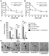
Nutlin-3 sensitizes C666-1 cells to the cytotoxic effect of cisplatin. The cell viability in response to (A) cisplatin alone (0–100 µM) and in combination with 10 µM Nutlin-3, and (B) Nutlin-3 alone (0–30 µM) and in combination with 33.33 µM cisplatin were assayed by MTS viability assay following 48 h of treatment. The graphs shown are representative average percentages of cell viability relative to the untreated and 0.1% DMSO-treated controls of three independent experiments with nine data points. (C) Effect of Nutlin-3 alone and in combination with cisplatin on colony formation of C666-1 cells. The plot shown is representative of two independent experiments carried out in triplicate. The untreated and 0.1% DMSO-treated cells were included as controls for cisplatin and Nutlin-3 treatments, respectively. Statistical analysis using the paired Student's t-test was performed to compare the difference in number of colonies scored on day 7, 14 and 21. *P<0.05 and **P<0.005 compared to the control. (D) Morphological appearances of C666-1 cell colonies in soft agar cultures. The image of the colonies was captured on day 21 using an Olympus stereomicroscope at a magnification of ×2.5 (scale bar, 100 µm).
Figure 3
Figure 3
Nutlin-3 activates the p53 pathway, upregulating p53, p21 and Mdm2 in C666-1 cells in a p53-dependent manner. (A) The expression of p53-related proteins in cancer cells bearing wt p53 (C666-1 and HCT116) and mutant p53 (HK1 and MDA-MB-231) after being treated with 10 µM Nutlin-3 (+) or 0.1% DMSO (−) for 24 h are shown. β-actin is included as loading control. Nutlin-3 induced the upregulation of p53, p21 and Mdm2 proteins in the C666-1 and HCT116 cells bearing wt p53. (B) Establishment of p53 knockdown. p53-knockdown C666-1 cell sublines (lenti-shΔp53 C666-1) were generated using the lentiviral-based shRNA constructs p53si-E1, -E2, -C12 or -D3 in parallel with the control vector and NS. The efficiency of knockdown was verified at 96 h post transduction. The plot shown is representative of three independent experiments. The mean percentage of the relative p53 expression level was obtained by densitometric analysis when normalized to β-actin. Among the 4 constructs, the p53 protein level was most significantly reduced by p53si-D3 or p53si-E2. (C) The effects of Nutlin-3 on the expression of p53, p21 and Mdm2 proteins in the lenti-shΔp53 C666-1 cells. The lenti-shΔp53 C666-1 and control cells were treated with 10 µM Nutlin-3 for 24 h prior to analysis. The attenuation of the activation of the p53 pathway was evident in cells transduced with p53si-D3 or p53si-E2 compared to the controls. (D) Growth inhibitory effect of Nutlin-3 on lenti-shΔp53 C666-1 cells. Cell viability responses to 0–40 µM Nutlin-3 after a 72-h treatment was quantified by MTS viability assay. The average percentage of cell viability relative to the 0.1% DMSO-treated vehicle control is shown for the lenti-shΔp53 C666-1 cells and controls. The plot shown is representative of mean values of three independent experiments with nine data points. *P<0.05 and **P<0.005 compared to the vector. Vector, vector-pLKO; NS, non-specific.
Figure 4
Figure 4
Nutlin-3 sensitizes C666-1 cells to cisplatin-induced apoptosis. (A) High content analysis of Annexin V-FITC/PI-stained cells to measure apoptosis induced by cisplatin and/or Nutlin-3 in C666-1 cells. The cells were treated with 33.33 µM cisplatin and/or 10 µM Nutlin-3 for 48 and 72 h prior to analysis. The untreated and 0.1% DMSO-treated cells served as non-treated controls for cisplatin and Nutlin-3, respectively. Student's t-test was performed to compare the difference in the apoptotic cell score. *P<0.05 and **P<0.005 compared to the control. (B) Representative high content images of C666-1 cells stained with Annexin V-FITC for apoptosis (green), PI for viability (red) and Hoechst 33342 as counterstaining (blue) are shown. Imaging was conducted at 72 h post-treatment at magnification of x20. Annexin V(+)/PI(−) and Annexin V(+)/PI(+) cells were defined as apoptotic cells. (C) The effects of Nutlin-3 on the expression of apoptosis-related proteins in cisplatin-treated C666-1 cells. C666-1 cells were treated with cisplatin and/or Nutlin-3 (+) or 0.1% DMSO (−) for 24 h prior to analysis. The levels of p53 target apoptosis-related proteins are shown. β-actin was probed to monitor protein loading. Treatment of C666-1 cells with Nutlin-3 resulted in induction of p53 target apoptosis-related proteins p21, Mdm2, BAX and PUMA as compared to the vehicle control. FITC, fluorescein isothiocyanate; PI, propidium iodide.
Figure 5
Figure 5
Extended treatment with Nutlin-3 results in reduced sensitivity without emergence of p53 mutations. (A) The diagram shown is the experimental design to establish Nutlin-3-adapted NPC cell sublines. Nutlin-3-adapted C666-1 cell sublines were established by treatment of C666-1 cells with 10 to 40 µM Nutlin-3 over a period of up to six months. The sublines tested for p53 mutations are labeled (★). (B) Sensitivity of Nutlin-3-adapted C666-1 sublines to Nutlin-3 indicated by the concentration that inhibits cell viability by 50% (IC50) following a 72-h treatment as quantified by MTS viability assay. The plot shown is representative mean IC values of three independent experiments carried out in triplicate. *P<0.05 and **P<0.005 compared to the parental control. ( ), Nutlin-3 adapted C666-1 sublines; R, resistance index indicates the degree of acquired resistance.

References

    1. Pathmanathan R, Prasad U, Chandrika G, Sadler R, Flynn K, Raab-Traub N. Undifferentiated, nonkeratinizing, and squamous cell carcinoma of the nasopharynx. Variants of Epstein-Barr virus-infected neoplasia. Am J Pathol. 1995;146:1355–1367. - PMC - PubMed
    1. Devi BC, Pisani P, Tang TS, Parkin DM. High incidence of nasopharyngeal carcinoma in native people of Sarawak, Borneo Island. Cancer Epidemiol Biomarkers Prev. 2004;13:482–486. - V体育官网入口 - PubMed
    1. Zeng MS, Zeng YX. Nasopharyngeal Cancer Medical Radiology. Springer Berlin; Heidelberg: 2010. Pathogenesis and etiology of nasopharyngeal carcinoma; pp. 9–25. - DOI
    1. Khoo AS, Pua KC. Nasopharyngeal Carcinoma: Keys for Translational Medicine and Biology. Landes Bioscience and Springer Science:Business Media; New York, NY: 2013. Diagnosis and clinical evaluation of nasopharyngeal carcinoma; pp. 1–9. - DOI
    1. Pua KC, Khoo AS, Yap YY, Subramaniam SK, Ong CA, Gopala Krishnan G, Shahid H, The Malaysian Nasopharyngeal Carcinoma Study Group Nasopharyngeal Carcinoma Database. Med J Malaysia. 2008;63(Suppl C):59–62. - "VSports app下载" PubMed

Publication types

VSports手机版 - MeSH terms