Skip to main page content
U.S. flag

An official website of the United States government

Dot gov

The VSports app下载. gov means it’s official. Federal government websites often end in . gov or . mil. Before sharing sensitive information, make sure you’re on a federal government site. .

Https

The site is secure. The https:// ensures that you are connecting to the official website and that any information you provide is encrypted and transmitted securely. V体育官网.

. 2015 Oct;43(10):1544-56.
doi: 10.1124/dmd.115.065276. Epub 2015 Jul 21.

Importance of Large Intestine in Regulating Bile Acids and Glucagon-Like Peptide-1 in Germ-Free Mice

Affiliations

Importance of Large Intestine in Regulating Bile Acids and Glucagon-Like Peptide-1 in Germ-Free Mice

Felcy Pavithra Selwyn (VSports手机版) et al. Drug Metab Dispos. 2015 Oct.

"V体育ios版" Abstract

It is known that 1) elevated serum bile acids (BAs) are associated with decreased body weight, 2) elevated glucagon-like peptide-1 (GLP-1) levels can decrease body weight, and 3) germ-free (GF) mice are resistant to diet-induced obesity. The purpose of this study was to test the hypothesis that a lack of intestinal microbiota results in more BAs in the body, resulting in increased BA-mediated transmembrane G protein-coupled receptor 5 (TGR5) signaling and increased serum GLP-1 as a mechanism of resistance of GF mice to diet-induced obesity VSports手机版. GF mice had 2- to 4-fold increased total BAs in the serum, liver, bile, and ileum. Fecal excretion of BAs was 63% less in GF mice. GF mice had decreased secondary BAs and increased taurine-conjugated BAs, as anticipated. Surprisingly, there was an increase in non-12α-OH BAs, namely, β-muricholic acid, ursodeoxycholic acid (UDCA), and their taurine conjugates, in GF mice. Further, in vitro experiments confirmed that UDCA is a primary BA in mice. There were minimal changes in the mRNA of farnesoid X receptor target genes in the ileum (Fibroblast growth factor 15, small heterodimer protein, and ileal bile acid-binding protein), in the liver (small heterodimer protein, liver receptor homolog-1, and cytochrome P450 7a1), and BA transporters (apical sodium dependent bile acid transporter, organic solute transporter α, and organic solute transporter β) in the ileum of GF mice. Surprisingly, there were marked increases in BA transporters in the large intestine. Increased GLP-1 levels and gallbladder size were observed in GF mice, suggesting activation of TGR5 signaling. In summary, the GF condition results in increased expression of BA transporters in the colon, resulting in 1) an increase in total BA concentrations in tissues, 2) a change in BA composition to favor an increase in non-12α-OH BAs, and 3) activation of TGR5 signaling with increased gallbladder size and GLP-1. .


Figures

Fig. 1.
Fig. 1.
Body and liver weight of GF mice. Data are presented as mean ± S.E.M., n = 10 per group. Asterisks (*) represent a statistically significant difference between CV and GF mice (P < 0.05) by Student’s t test. Dark blue and light blue bars represent CV and GF male mice, respectively, and red and pink bars represent CV and GF female mice, respectively. F, female; M, male.
Fig. 2.
Fig. 2.
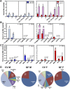
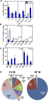
Concentrations of BAs in the serum of CV and GF mice. (A) Concentrations of total BAs (∑), total 1° (primary) and 2° (secondary) BAs, total 12α-OH BAs [CA, TCA, DCA, and taurine-conjugated deoxycholic acid (TDCA)] and non–12α-OH BAs (all other BAs), and total taurine-conjugated (T-Conj) and unconjugated (Unconj) BAs. (B) Concentrations of individual T-conj BAs. (C) Concentrations of individual Unconj BAs. Data are presented as mean ± S.E.M., n = 7–10 mice/group. (D) Proportion of individual BAs in the serum of CV and GF mice. Asterisks (*) represent statistically significant differences between CV and GF mice (P < 0.05) by Student’s t test. Dark blue and light blue bars represent CV and GF male mice, respectively, and red and pink bars represent CV and GF female mice, respectively. F, female; M, male.
Fig. 3.
Fig. 3.
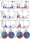
Concentrations of BAs in the livers of CV and GF mice. (A) Concentrations of total BAs (∑), total 1° (primary) and 2° (secondary) BAs, total 12α-OH BAs [CA, TCA, DCA, and taurine-conjugated deoxycholic acid (TDCA)] and non–12α-OH BAs (all other BAs), and total taurine-conjugated (T-Conj) and unconjugated (Unconj) BAs. (B) Concentrations of individual T-conj BAs. (C) Concentrations of individual Unconj BAs. Data are presented as mean ± S.E.M., n = 7–10 mice/group. (D) Proportion of individual BAs in the livers of CV and GF mice. Asterisks (*) represent statistically significant differences between CV and GF mice (P < 0.05) by Student’s t test. Dark blue and light blue bars represent CV and GF male mice, respectively, and red and pink bars represent CV and GF female mice, respectively. F, female; M, male.
Fig. 4.
Fig. 4.
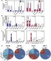
Biliary excretion of BAs in CV and GF mice. BAs in bile are expressed as nmol/kg body weight per min. (A) Concentrations of total BAs (∑), total 1° (primary) and 2° (secondary) BAs, total 12α-OH BAs [CA, TCA, DCA, and taurine-conjugated deoxycholic acid (TDCA)] and non–12α-OH BAs (all other BAs), and total taurine-conjugated (T-Conj) and unconjugated (Unconj) BAs. (B) Concentrations of individual T-conj BAs. (C) Concentrations of individual Unconj BAs. Bile was collected for 40 minutes from each mouse. Data are presented as mean ± S.E.M., n = 3–6 mice/group. (D) Proportion of individual BAs in the bile of CV and GF mice. Asterisks (*) represent statistically significant differences between CV and GF mice (P < 0.05) by Student’s t test. Dark blue and light blue bars represent CV and GF male mice, respectively, and red and pink bars represent CV and GF female mice, respectively. F, female; M, male.
Fig. 5.
Fig. 5.
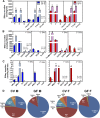
Concentrations of BAs in the ileal tissue of CV and GF mice. (A) Concentrations of total BAs (∑), total 1° (primary) and 2° (secondary) BAs, total 12α-OH BAs [CA, TCA, DCA, and taurine-conjugated deoxycholic acid (TDCA)] and non–12α-OH BAs (all other BAs), and total taurine-conjugated (T-Conj) and unconjugated (Unconj) BAs. (B) Concentrations of individual T-conj BAs. (C) Concentrations of individual Unconj BAs. Ileal tissue refers to the last one-third of the small intestine. Data are presented as mean ± S.E.M., n = 7–10 mice/group. (D) Proportion of individual BAs in the ileal tissue of CV and GF mice. Asterisks (*) represent statistically significant differences between CV and GF mice (P < 0.05) by Student’s t test. Dark blue and light blue bars represent CV and GF male mice, respectively, and red and pink bars represent CV and GF female mice, respectively. F, female; M, male.
Fig. 6.
Fig. 6.
Concentrations of BAs in the feces of CV and GF mice. BAs are expressed as nmols/gram of feces. (A) Concentrations of total BAs (∑), total 1° (primary) and 2° (secondary) BAs, total 12α-OH BAs [CA, TCA, DCA, and taurine-conjugated deoxycholic acid (TDCA)] and non–12α-OH BAs (all other BAs), and taurine-conjugated (total T-Conj) and unconjugated (Unconj) BAs. (B) Concentrations of individual T-conj BAs. (C) Concentrations of individual Unconj BAs. Mice were housed individually in metabolic chambers, and feces were collected over 24 hours. Data are presented as mean ± S.E.M., n = 4 mice/group. (D) Proportion of individual BAs in the feces of CV and GF mice. Asterisks (*) represent statistically significant differences between CV and GF mice (P < 0.05) by Student’s t test. Dark blue and light blue bars represent CV and GF male mice. M, male.
Fig. 7.
Fig. 7.
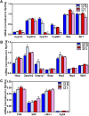
Gene expression in livers of CV and GF mice. (A) mRNA expression of genes involved in BA synthesis; (B) mRNA expression of genes involved in BA transport; and (C) mRNA expression of genes involved in FXR feedback regulation in the livers of male and female CV and GF mice. mRNA was quantified by beadplex assay; n = 6 per group. Data are presented as mean ± S.E.M. Asterisks (*) represent statistically significant differences between CV and GF mice (P < 0.05) by Student’s t test. Dark blue and light blue bars represent CV and GF male mice, respectively. Red and pink bars represent CV and GF females, respectively. F, female; M, male.
Fig. 8.
Fig. 8.
Gene expression in ileum of CV and GF mice. (A) mRNA expression of BA transporters in the ileal tissue of male and female CV and GF mice. (B) mRNA expression of genes involved in BA signaling in the ileum of male and female CV and GF mice. mRNA was quantified by beadplex assay; n = 6 per group. Data are presented as mean ± S.E.M. Asterisks (*) represent statistically significant differences between CV and GF mice (P < 0.05) by Student’s t test. Dark blue and light blue bars represent CV and GF male mice, respectively. Red and pink bars represent CV and GF females, respectively. F, female; M, male.
Fig. 9.
Fig. 9.
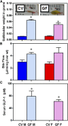
Targets of TGR5 signaling. (A) Gallbladder weights of CV and GF mice and (inset) representative pictures. (B) Bile flow in CV and GF mice. (C) Serum GLP-1 quantification by enzyme-linked immunosorbent assay. Data are presented as mean ± S.E.M.; n = 3–6 per group. Asterisks (*) represent statistically significant differences between CV and GF mice (P < 0.05) by Student’s t test. Dark blue and light blue bars represent CV and GF male mice, respectively. Red and pink bars represent CV and GF females, respectively. F, female; M, male.
Fig. 10.
Fig. 10.
Gene expression of BA-related genes across the intestinal sections. Total RNA was isolated from livers of adult male CV and GF C57BL/6 mice (n = 3 per group). The mRNA quantified by RNA-Seq as described in Materials and Methods. Asterisk (*) indicates differential expression determined using Cuffdiff (FDR-BH < 0.05). Dark blue and light blue bars represent CV and GF male mice, respectively. L.Int, large intestine (colon); M, male.

References

    1. Alnouti Y, Csanaky IL, Klaassen CD. (2008) Quantitative-profiling of bile acids and their conjugates in mouse liver, bile, plasma, and urine using LC-MS/MS. J Chromatogr B Analyt Technol Biomed Life Sci 873:209–217. - "V体育官网" PMC - PubMed
    1. Bäckhed F, Ding H, Wang T, Hooper LV, Koh GY, Nagy A, Semenkovich CF, Gordon JI. (2004) The gut microbiota as an environmental factor that regulates fat storage. Proc Natl Acad Sci USA 101:15718–15723. - PMC - PubMed
    1. Bäckhed F, Manchester JK, Semenkovich CF, Gordon JI. (2007) Mechanisms underlying the resistance to diet-induced obesity in germ-free mice. Proc Natl Acad Sci USA 104:979–984. - PMC - PubMed
    1. Chen L, McNulty J, Anderson D, Liu Y, Nystrom C, Bullard S, Collins J, Handlon AL, Klein R, Grimes A, et al. (2010) Cholestyramine reverses hyperglycemia and enhances glucose-stimulated glucagon-like peptide 1 release in Zucker diabetic fatty rats. J Pharmacol Exp Ther 334:164–170. - PubMed
    1. Chiang JY. (2004) Regulation of bile acid synthesis: pathways, nuclear receptors, and mechanisms. J Hepatol 40:539–551. - PubMed

"V体育安卓版" Publication types

"V体育官网" LinkOut - more resources